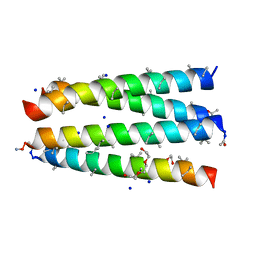
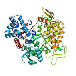
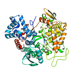
3CTO
 
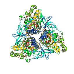
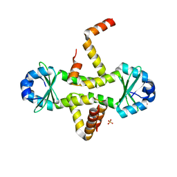 | | Crystal Structure of M. tuberculosis YefM antitoxin | | 分子名称: | SULFATE ION, Uncharacterized protein Rv3357/MT3465 | | 著者 | Kumar, P, Issac, B, Dodson, E.J, Turkenberg, J.P, Mande, S.C. | | 登録日 | 2008-04-14 | | 公開日 | 2008-12-02 | | 最終更新日 | 2024-03-20 | | 実験手法 | X-RAY DIFFRACTION (2.5 Å) | | 主引用文献 | Crystal structure of Mycobacterium tuberculosis YefM antitoxin reveals that it is not an intrinsically unstructured protein
J.Mol.Biol., 383, 2008
|
|
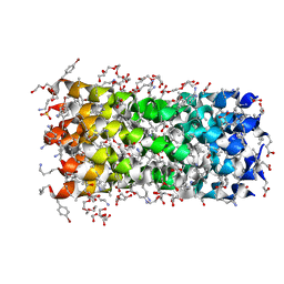
5WXU
 | | 11S globulin from Wrightia tinctoria reveals auxin binding site | | 分子名称: | 11S globulin, 1H-INDOL-3-YLACETIC ACID, CITRATE ANION, ... | | 著者 | Kumar, P, Kesari, P, Dhindwal, S, Kumar, P. | | 登録日 | 2017-01-09 | | 公開日 | 2018-05-23 | | 最終更新日 | 2024-10-16 | | 実験手法 | X-RAY DIFFRACTION (1.7 Å) | | 主引用文献 | A novel function for globulin in sequestering plant hormone: Crystal structure of Wrightia tinctoria 11S globulin in complex with auxin.
Sci Rep, 7, 2017
|
|
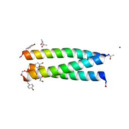
8BFD
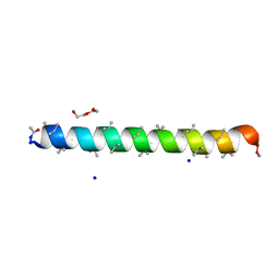 | | Racemic structure of PK-7 (310HD-U2U5) | | 分子名称: | 310HD-U2U5, D-310HD-U2U5, DI(HYDROXYETHYL)ETHER, ... | | 著者 | Kumar, P, Paterson, N.G, Woolfson, D.N. | | 登録日 | 2022-10-25 | | 公開日 | 2022-11-23 | | 実験手法 | X-RAY DIFFRACTION (2 Å) | | 主引用文献 | De novo design of discrete, stable 3 10 -helix peptide assemblies.
Nature, 607, 2022
|
|
8BFE
 | | A dimeric de novo coiled-coil assembly: PK-2 (CC-TypeN-LaUbUcLd) | | 分子名称: | (4S)-2-METHYL-2,4-PENTANEDIOL, 1,2-ETHANEDIOL, CC-TypeN-LaUbUcLd, ... | | 著者 | Kumar, P, Paterson, N.G, Woolfson, D.N. | | 登録日 | 2022-10-25 | | 公開日 | 2022-11-23 | | 実験手法 | X-RAY DIFFRACTION (2.1 Å) | | 主引用文献 | De novo design of discrete, stable 3 10 -helix peptide assemblies.
Nature, 607, 2022
|
|
2XSH
 | | CRYSTAL STRUCTURE OF P4 VARIANT OF BIPHENYL DIOXYGENASE FROM BURKHOLDERIA XENOVORANS LB400 IN COMPLEX WITH 2,6 DI CHLOROBIPHENYL | | 分子名称: | 2,6-DICHLOROBIPHENYL, BIPHENYL DIOXYGENASE SUBUNIT ALPHA, BIPHENYL DIOXYGENASE SUBUNIT BETA, ... | | 著者 | Kumar, P, Bolin, J.T. | | 登録日 | 2010-09-29 | | 公開日 | 2010-11-24 | | 最終更新日 | 2023-12-20 | | 実験手法 | X-RAY DIFFRACTION (2.29 Å) | | 主引用文献 | Structural Insight Into the Expanded Pcb-Degrading Abilities of a Biphenyl Dioxygenase Obtained by Directed Evolution.
J.Mol.Biol., 405, 2011
|
|
2XSO
 | |
2XR8
 | |
2YFL
 | |
2XRX
 | |
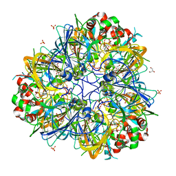
6O5U
 | | AAC-VIa bound to Kanamycin A | | 分子名称: | Aminoglycoside N(3)-acetyltransferase, KANAMYCIN A, MAGNESIUM ION | | 著者 | Kumar, P, Cuneo, M.J. | | 登録日 | 2019-03-04 | | 公開日 | 2019-09-25 | | 最終更新日 | 2023-10-11 | | 実験手法 | X-RAY DIFFRACTION (1.4 Å) | | 主引用文献 | Low-Barrier and Canonical Hydrogen Bonds Modulate Activity and Specificity of a Catalytic Triad.
Angew.Chem.Int.Ed.Engl., 58, 2019
|
|
3BVN
 | | High resolution crystal structure of HLA-B*1402 in complex with the latent membrane protein 2 peptide (LMP2) of Epstein-Barr virus | | 分子名称: | Beta-2-microglobulin, HLA class I histocompatibility antigen, B*1402 alpha chain, ... | | 著者 | Kumar, P, Vahedi-Faridi, A, Saenger, W, Uchanska-Ziegler, B, Ziegler, A. | | 登録日 | 2008-01-07 | | 公開日 | 2009-02-03 | | 最終更新日 | 2024-11-20 | | 実験手法 | X-RAY DIFFRACTION (2.55 Å) | | 主引用文献 | Structural basis for T cell alloreactivity among three HLA-B14 and HLA-B27 antigens
J.Biol.Chem., 284, 2009
|
|
5E33
 | | Structure of human DPP3 in complex with met-enkephalin | | 分子名称: | Dipeptidyl peptidase 3, MAGNESIUM ION, Met-enkephalin, ... | | 著者 | Kumar, P, Reithofer, V, Reisinger, M, Pavkov-Keller, T, Wallner, S, Macheroux, P, Gruber, K. | | 登録日 | 2015-10-01 | | 公開日 | 2016-04-13 | | 最終更新日 | 2024-11-13 | | 実験手法 | X-RAY DIFFRACTION (1.837 Å) | | 主引用文献 | Substrate complexes of human dipeptidyl peptidase III reveal the mechanism of enzyme inhibition.
Sci Rep, 6, 2016
|
|
8B45
 | | Structure of CC-Tri with Aib@b,c: CC-Tri-(UbUc)4 | | 分子名称: | 1,2-ETHANEDIOL, CC-Tri-(UbUc)4, SODIUM ION, ... | | 著者 | Kumar, P, Martin, F.J.O, Dawson, W.M, Zieleniewski, F, Woolfson, D.N. | | 登録日 | 2022-09-19 | | 公開日 | 2023-09-27 | | 最終更新日 | 2023-11-15 | | 実験手法 | X-RAY DIFFRACTION (1.6 Å) | | 主引用文献 | Structure of CC-Tri with Aib@b,c: CC-Tri-(UbUc)4
To Be Published
|
|
5E3A
 | | Structure of human DPP3 in complex with opioid peptide leu-enkephalin | | 分子名称: | Dipeptidyl peptidase 3, Leu-enkephalin, MAGNESIUM ION, ... | | 著者 | Kumar, P, Reithofer, V, Reisinger, M, Pavkov-Keller, T, Wallner, S, Macheroux, P, Gruber, K. | | 登録日 | 2015-10-02 | | 公開日 | 2016-04-13 | | 最終更新日 | 2024-11-06 | | 実験手法 | X-RAY DIFFRACTION (2.05 Å) | | 主引用文献 | Substrate complexes of human dipeptidyl peptidase III reveal the mechanism of enzyme inhibition.
Sci Rep, 6, 2016
|
|
5E3C
 | | Structure of human DPP3 in complex with hemorphin like opioid peptide IVYPW | | 分子名称: | Dipeptidyl peptidase 3, IVYPW, MAGNESIUM ION, ... | | 著者 | Kumar, P, Reithofer, V, Reisinger, M, Pavkov-Keller, T, Wallner, S, Macheroux, P, Gruber, K. | | 登録日 | 2015-10-02 | | 公開日 | 2016-04-13 | | 最終更新日 | 2024-01-10 | | 実験手法 | X-RAY DIFFRACTION (2.765 Å) | | 主引用文献 | Substrate complexes of human dipeptidyl peptidase III reveal the mechanism of enzyme inhibition.
Sci Rep, 6, 2016
|
|
5EHH
 | | Structure of human DPP3 in complex with endomorphin-2. | | 分子名称: | Dipeptidyl peptidase 3, Endomorphin-2, MAGNESIUM ION, ... | | 著者 | Kumar, P, Reithofer, V, Reisinger, M, Pavkov-Keller, T, Wallner, S, Macheroux, P, Gruber, K. | | 登録日 | 2015-10-28 | | 公開日 | 2016-04-13 | | 最終更新日 | 2024-11-13 | | 実験手法 | X-RAY DIFFRACTION (2.38 Å) | | 主引用文献 | Substrate complexes of human dipeptidyl peptidase III reveal the mechanism of enzyme inhibition.
Sci Rep, 6, 2016
|
|
5EGY
 | | Structure of ligand free human DPP3 in closed form. | | 分子名称: | Dipeptidyl peptidase 3, MAGNESIUM ION, ZINC ION | | 著者 | Kumar, P, Reithofer, V, Reisinger, M, Pavkov-Keller, T, Wallner, S, Macheroux, P, Gruber, K. | | 登録日 | 2015-10-27 | | 公開日 | 2016-04-13 | | 最終更新日 | 2024-11-06 | | 実験手法 | X-RAY DIFFRACTION (2.741 Å) | | 主引用文献 | Substrate complexes of human dipeptidyl peptidase III reveal the mechanism of enzyme inhibition.
Sci Rep, 6, 2016
|
|
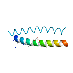
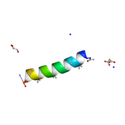
7QDI
 | | Structure of octameric left-handed 310-helix bundle: D-310HD | | 分子名称: | 1,2-ETHANEDIOL, 1-(2-METHOXY-ETHOXY)-2-{2-[2-(2-METHOXY-ETHOXY]-ETHOXY}-ETHANE, 2-(2-METHOXYETHOXY)ETHANOL, ... | | 著者 | Kumar, P, Paterson, N.G, Woolfson, D.N. | | 登録日 | 2021-11-27 | | 公開日 | 2022-04-27 | | 最終更新日 | 2022-07-27 | | 実験手法 | X-RAY DIFFRACTION (2.34 Å) | | 主引用文献 | De novo design of discrete, stable 3 10 -helix peptide assemblies.
Nature, 607, 2022
|
|
7QDK
 | | A trimeric de novo coiled-coil assembly: CC-TypeN-LaLd | | 分子名称: | (4S)-2-METHYL-2,4-PENTANEDIOL, CC-TypeN-LaLd, GLYCEROL, ... | | 著者 | Kumar, P, Paterson, N.G, Woolfson, D.N. | | 登録日 | 2021-11-27 | | 公開日 | 2022-04-27 | | 最終更新日 | 2022-07-27 | | 実験手法 | X-RAY DIFFRACTION (1.41 Å) | | 主引用文献 | De novo design of discrete, stable 3 10 -helix peptide assemblies.
Nature, 607, 2022
|
|
7QDJ
 | | Racemic structure of PK-10 and PK-11 | | 分子名称: | GLYCEROL, MALONATE ION, PK-10+PK-11, ... | | 著者 | Kumar, P, Paterson, N.G, Woolfson, D.N. | | 登録日 | 2021-11-27 | | 公開日 | 2022-04-27 | | 最終更新日 | 2022-07-27 | | 実験手法 | X-RAY DIFFRACTION (1.44 Å) | | 主引用文献 | De novo design of discrete, stable 3 10 -helix peptide assemblies.
Nature, 607, 2022
|
|
5E2Q
 | | Structure of human DPP3 in complex with angiotensin-II | | 分子名称: | Dipeptidyl peptidase 3, MAGNESIUM ION, POTASSIUM ION, ... | | 著者 | Kumar, P, Reisinger, M, Reithofer, V, Gruber, K. | | 登録日 | 2015-10-01 | | 公開日 | 2016-04-13 | | 最終更新日 | 2024-01-10 | | 実験手法 | X-RAY DIFFRACTION (2.404 Å) | | 主引用文献 | Substrate complexes of human dipeptidyl peptidase III reveal the mechanism of enzyme inhibition.
Sci Rep, 6, 2016
|
|
1I6B
 | |
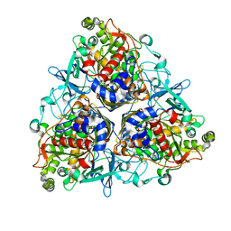
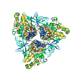
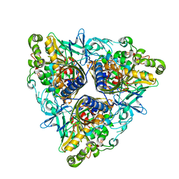
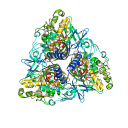
3LS6
 | | Crystal structure of 3,4-Dihydroxy-2-butanone 4-phosphate synthase in complex with sulfate and zinc | | 分子名称: | 3,4-Dihydroxy-2-butanone 4-phosphate synthase, GLYCEROL, MAGNESIUM ION, ... | | 著者 | Kumar, P, Karthikeyan, S. | | 登録日 | 2010-02-12 | | 公開日 | 2010-09-15 | | 最終更新日 | 2023-11-01 | | 実験手法 | X-RAY DIFFRACTION (1.86 Å) | | 主引用文献 | Potential anti-bacterial drug target: structural characterization of 3,4-dihydroxy-2-butanone-4-phosphate synthase from Salmonella typhimurium LT2.
Proteins, 78, 2010
|
|
3LQU
 | |
3LRJ
 | |


