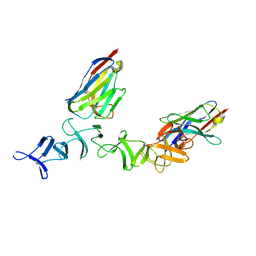
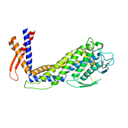
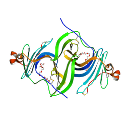
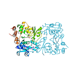
4NC0
 
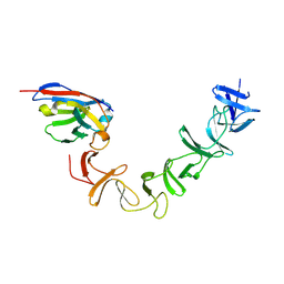 | | Crystal Structure of TcdA-A2 Bound to A26.8 VHH | | 分子名称: | A26.8 VHH, Cell wall-binding repeat protein | | 著者 | Murase, T, Eugenio, L, Schorr, M, Hussack, G, Tanha, J, Kitova, E.N, Klassen, J.S, Ng, K.K.S. | | 登録日 | 2013-10-23 | | 公開日 | 2013-12-11 | | 最終更新日 | 2023-09-20 | | 実験手法 | X-RAY DIFFRACTION (2.3 Å) | | 主引用文献 | Structural Basis for Antibody Recognition in the Receptor-binding Domains of Toxins A and B from Clostridium difficile.
J.Biol.Chem., 289, 2014
|
|
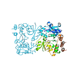
4NC2
 | | Crystal structure of TcdB-B1 bound to B39 VHH | | 分子名称: | B39 VHH, Toxin B | | 著者 | Murase, T, Eugenio, L, Schorr, M, Hussack, G, Tanha, J, Kitova, E.N, Klassen, J.S, Ng, K.K.S. | | 登録日 | 2013-10-23 | | 公開日 | 2013-12-11 | | 最終更新日 | 2023-09-20 | | 実験手法 | X-RAY DIFFRACTION (2.5 Å) | | 主引用文献 | Structural Basis for Antibody Recognition in the Receptor-binding Domains of Toxins A and B from Clostridium difficile.
J.Biol.Chem., 289, 2014
|
|
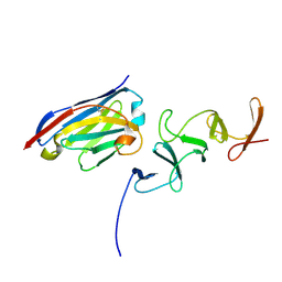
4NBX
 | | Crystal Structure of Clostridium difficile Toxin A fragment TcdA-A1 Bound to A20.1 VHH | | 分子名称: | A20.1 VHH, TcdA | | 著者 | Murase, T, Eugenio, L, Schorr, M, Hussack, G, Tanha, J, Kitova, E.N, Klassen, J.S, Ng, K.K.S. | | 登録日 | 2013-10-23 | | 公開日 | 2013-12-11 | | 最終更新日 | 2014-02-12 | | 実験手法 | X-RAY DIFFRACTION (1.75 Å) | | 主引用文献 | Structural Basis for Antibody Recognition in the Receptor-binding Domains of Toxins A and B from Clostridium difficile.
J.Biol.Chem., 289, 2014
|
|
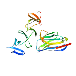
4NBZ
 | | Crystal Structure of TcdA-A1 Bound to A26.8 VHH | | 分子名称: | A26.8 VHH, TcdA | | 著者 | Murase, T, Eugenio, L, Schorr, M, Hussack, G, Tanha, J, Kitova, E.N, Klassen, J.S, Ng, K.K.S. | | 登録日 | 2013-10-23 | | 公開日 | 2013-12-11 | | 最終更新日 | 2023-09-20 | | 実験手法 | X-RAY DIFFRACTION (1.75 Å) | | 主引用文献 | Structural Basis for Antibody Recognition in the Receptor-binding Domains of Toxins A and B from Clostridium difficile.
J.Biol.Chem., 289, 2014
|
|
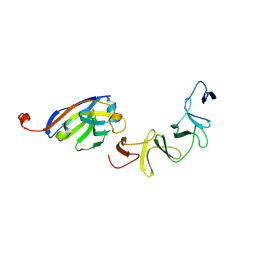
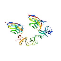
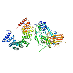
4NBY
 | | Crystal Structure of TcdA-A2 Bound to Two Molecules of A20.1 VHH | | 分子名称: | A20.1 VHH, Cell wall-binding repeat protein | | 著者 | Murase, T, Eugenio, L, Schorr, M, Hussack, G, Tanha, J, Kitova, E, Klassen, J.S, Ng, K.K.S. | | 登録日 | 2013-10-23 | | 公開日 | 2013-12-11 | | 最終更新日 | 2023-09-20 | | 実験手法 | X-RAY DIFFRACTION (2.08 Å) | | 主引用文献 | Structural Basis for Antibody Recognition in the Receptor-binding Domains of Toxins A and B from Clostridium difficile.
J.Biol.Chem., 289, 2014
|
|
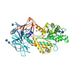
4NC1
 | | Crystal Structure of TcdA-A2 Bound to A20.1 VHH and A26.8 VHH | | 分子名称: | A20.1 VHH, A26.8 VHH, Cell wall-binding repeat protein | | 著者 | Murase, T, Eugenio, L, Schorr, M, Hussack, G, Tanha, J, Kitova, E.N, Klassen, J.S, Ng, K.K.S. | | 登録日 | 2013-10-23 | | 公開日 | 2013-12-11 | | 最終更新日 | 2023-09-20 | | 実験手法 | X-RAY DIFFRACTION (2.61 Å) | | 主引用文献 | Structural Basis for Antibody Recognition in the Receptor-binding Domains of Toxins A and B from Clostridium difficile.
J.Biol.Chem., 289, 2014
|
|
4CZS
 | | Discovery of Glycomimetic Ligands via Genetically-encoded Library of Phage displaying Mannose-peptides | | 分子名称: | 2-hydroxyethyl alpha-D-mannopyranoside, CALCIUM ION, Concanavalin V, ... | | 著者 | Ng, S, Lin, E, Tjhung, K.F, Gerlits, O, Sood, A, Kasper, B, Deng, L, Kitov, P.I, Matochko, W.L, Paschal, B.M, Noren, C.J, Klassen, J, Mahal, L.K, Coates, L, Woods, R.J, Derda, R. | | 登録日 | 2014-04-22 | | 公開日 | 2015-04-22 | | 最終更新日 | 2022-12-07 | | 実験手法 | X-RAY DIFFRACTION (1.73 Å) | | 主引用文献 | Genetically-Encoded Fragment-Based Discovery of Glycopeptide Ligands for Carbohydrate-Binding Proteins.
J.Am.Chem.Soc., 137, 2015
|
|
4O8V
 | | O-Acetyltransferase Domain of Pseudomonas putida AlgJ | | 分子名称: | Alginate biosynthesis protein AlgJ | | 著者 | Ricer, T, Little, D.J, Whitney, J.C, Robinson, H, Howell, P.L. | | 登録日 | 2013-12-30 | | 公開日 | 2014-10-01 | | 実験手法 | X-RAY DIFFRACTION (1.815 Å) | | 主引用文献 | P. aeruginosa SGNH Hydrolase-Like Proteins AlgJ and AlgX Have Similar Topology but Separate and Distinct Roles in Alginate Acetylation.
Plos Pathog., 10, 2014
|
|
7T8N
 | | Crystal structure of the PNAG binding module PgaA-TPR 220-359 | | 分子名称: | CHLORIDE ION, MAGNESIUM ION, Poly-beta-1,6-N-acetyl-D-glucosamine export protein | | 著者 | Pfoh, R, Little, D.J, Howell, P.L. | | 登録日 | 2021-12-16 | | 公開日 | 2022-08-03 | | 最終更新日 | 2024-02-28 | | 実験手法 | X-RAY DIFFRACTION (2.85 Å) | | 主引用文献 | The TPR domain of PgaA is a multifunctional scaffold that binds PNAG and modulates PgaB-dependent polymer processing.
Plos Pathog., 18, 2022
|
|
6GMM
 | | Crystal structure of Helicobacter pylori adhesin LabA | | 分子名称: | adhesin LabA | | 著者 | Paraskevopoulou, V, Overman, R.C, Stolnik, S, Winkler, S, Gellert, P, Falcone, F.H, Schimpl, M. | | 登録日 | 2018-05-27 | | 公開日 | 2019-12-18 | | 最終更新日 | 2024-01-17 | | 実験手法 | X-RAY DIFFRACTION (2.06 Å) | | 主引用文献 | Structural and binding characterization of the LacdiNAc-specific adhesin (LabA; HopD) exodomain from Helicobacter pylori
Current Research in Structural Biology, 2021
|
|
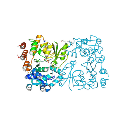
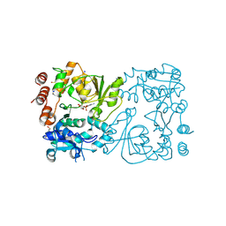
7ULA
 | | Structure of the Pseudomonas putida AlgKX modification and secretion complex | | 分子名称: | Alginate biosynthesis protein AlgK, Alginate biosynthesis protein AlgX, CHLORIDE ION, ... | | 著者 | Gheorghita, A.A, Li, E.Y, Pfoh, R, Howell, P.L. | | 登録日 | 2022-04-04 | | 公開日 | 2022-12-14 | | 最終更新日 | 2023-10-25 | | 実験手法 | X-RAY DIFFRACTION (2.46 Å) | | 主引用文献 | Structure of the AlgKX modification and secretion complex required for alginate production and biofilm attachment in Pseudomonas aeruginosa.
Nat Commun, 13, 2022
|
|
6NWZ
 | | Crystal structure of Agd3 a novel carbohydrate deacetylase | | 分子名称: | 2-acetamido-2-deoxy-beta-D-glucopyranose, 2-acetamido-2-deoxy-beta-D-glucopyranose-(1-4)-2-acetamido-2-deoxy-beta-D-glucopyranose, CHLORIDE ION, ... | | 著者 | Bamford, N.C, Howell, P.L. | | 登録日 | 2019-02-07 | | 公開日 | 2020-02-12 | | 最終更新日 | 2020-08-26 | | 実験手法 | X-RAY DIFFRACTION (2.6 Å) | | 主引用文献 | Structural and biochemical characterization of the exopolysaccharide deacetylase Agd3 required for Aspergillus fumigatus biofilm formation.
Nat Commun, 11, 2020
|
|
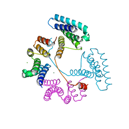
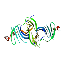
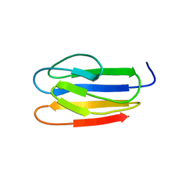
6MR1
 | | RbcS-like subdomain of CcmM | | 分子名称: | CHLORIDE ION, COBALT (II) ION, Carbon dioxide concentrating mechanism protein, ... | | 著者 | Ryan, P, Kimber, M.S. | | 登録日 | 2018-10-11 | | 公開日 | 2019-01-02 | | 最終更新日 | 2024-03-13 | | 実験手法 | X-RAY DIFFRACTION (1.35 Å) | | 主引用文献 | The small RbcS-like domains of the beta-carboxysome structural protein CcmM bind RubisCO at a site distinct from that binding the RbcS subunit.
J. Biol. Chem., 294, 2019
|
|
6E7O
 | | Crystal structure of deglycosylated human EPDR1 | | 分子名称: | Mammalian ependymin-related protein 1 | | 著者 | Wei, Y, Prive, G.G. | | 登録日 | 2018-07-27 | | 公開日 | 2019-01-23 | | 最終更新日 | 2020-01-08 | | 実験手法 | X-RAY DIFFRACTION (3 Å) | | 主引用文献 | Crystal structures of human lysosomal EPDR1 reveal homology with the superfamily of bacterial lipoprotein transporters.
Commun Biol, 2, 2019
|
|
6E8N
 | | Crystal structure of glycosylated human EPDR1 | | 分子名称: | Mammalian ependymin-related protein 1, NONAETHYLENE GLYCOL | | 著者 | Wei, Y, Prive, G.G. | | 登録日 | 2018-07-30 | | 公開日 | 2019-01-23 | | 最終更新日 | 2023-10-11 | | 実験手法 | X-RAY DIFFRACTION (3.2 Å) | | 主引用文献 | Crystal structures of human lysosomal EPDR1 reveal homology with the superfamily of bacterial lipoprotein transporters.
Commun Biol, 2, 2019
|
|
6CZT
 | |
6D10
 | |
8CSE
 | | WbbB in complex with alpha-Rha-(1-3)-beta-GlcNAc acceptor | | 分子名称: | CYTIDINE-5'-MONOPHOSPHATE, N-(8-hydroxyoctyl)-4-methoxybenzamide, N-acetyl glucosaminyl transferase, ... | | 著者 | Forrester, T.J.B, Kimber, M.S. | | 登録日 | 2022-05-12 | | 公開日 | 2022-11-09 | | 最終更新日 | 2023-11-15 | | 実験手法 | X-RAY DIFFRACTION (2.3 Å) | | 主引用文献 | The retaining beta-Kdo glycosyltransferase WbbB uses a double-displacement mechanism with an intermediate adduct rearrangement step.
Nat Commun, 13, 2022
|
|
8CSF
 | |
8CSB
 | | WbbB D232N in complex with CMP-beta-Kdo | | 分子名称: | CYTIDINE 5'-MONOPHOSPHATE 3-DEOXY-BETA-D-GULO-OCT-2-ULO-PYRANOSONIC ACID, CYTIDINE-5'-MONOPHOSPHATE, N-acetyl glucosaminyl transferase, ... | | 著者 | Forrester, T.J.B, Kimber, M.S. | | 登録日 | 2022-05-12 | | 公開日 | 2022-11-09 | | 最終更新日 | 2023-11-15 | | 実験手法 | X-RAY DIFFRACTION (2.25 Å) | | 主引用文献 | The retaining beta-Kdo glycosyltransferase WbbB uses a double-displacement mechanism with an intermediate adduct rearrangement step.
Nat Commun, 13, 2022
|
|
8CSD
 | | WbbB D232C Kdo adduct | | 分子名称: | 3-deoxy-alpha-D-manno-oct-2-ulopyranosonic acid, CHLORIDE ION, CYTIDINE-5'-MONOPHOSPHATE, ... | | 著者 | Forrester, T.J.B, Kimber, M.S. | | 登録日 | 2022-05-12 | | 公開日 | 2022-11-09 | | 最終更新日 | 2023-11-15 | | 実験手法 | X-RAY DIFFRACTION (1.95 Å) | | 主引用文献 | The retaining beta-Kdo glycosyltransferase WbbB uses a double-displacement mechanism with an intermediate adduct rearrangement step.
Nat Commun, 13, 2022
|
|
8CSC
 | | WbbB D232N-Kdo adduct | | 分子名称: | 3-deoxy-alpha-D-manno-oct-2-ulopyranosonic acid, CHLORIDE ION, CYTIDINE-5'-MONOPHOSPHATE, ... | | 著者 | Forrester, T.J.B, Kimber, M.S. | | 登録日 | 2022-05-12 | | 公開日 | 2022-11-09 | | 最終更新日 | 2023-11-15 | | 実験手法 | X-RAY DIFFRACTION (1.9 Å) | | 主引用文献 | The retaining beta-Kdo glycosyltransferase WbbB uses a double-displacement mechanism with an intermediate adduct rearrangement step.
Nat Commun, 13, 2022
|
|
4NK8
 | |
4NK6
 | |


