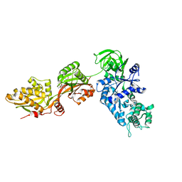
1N0V
 
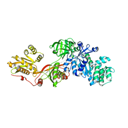 | | Crystal structure of elongation factor 2 | | 分子名称: | Elongation factor 2 | | 著者 | Joergensen, R, Ortiz, P.A, Carr-Schmid, A, Nissen, P, Kinzy, T.G, Andersen, G.R. | | 登録日 | 2002-10-15 | | 公開日 | 2002-11-27 | | 最終更新日 | 2024-04-03 | | 実験手法 | X-RAY DIFFRACTION (2.85 Å) | | 主引用文献 | Two crystal structures demonstrate large conformational changes in the eukaryotic ribosomal translocase.
Nat.Struct.Biol., 10, 2003
|
|
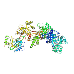
1ZM3
 | | Structure of the apo eEF2-ETA complex | | 分子名称: | Elongation factor 2, exotoxin A | | 著者 | Joergensen, R, Merrill, A.R, Yates, S.P, Marquez, V.E, Schwan, A.L, Boesen, T, Andersen, G.R. | | 登録日 | 2005-05-10 | | 公開日 | 2005-05-24 | | 最終更新日 | 2023-08-23 | | 実験手法 | X-RAY DIFFRACTION (3.07 Å) | | 主引用文献 | Exotoxin A-eEF2 complex structure indicates ADP ribosylation by ribosome mimicry.
Nature, 436, 2005
|
|
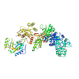
1ZM9
 | | Structure of eEF2-ETA in complex with PJ34 | | 分子名称: | Elongation factor 2, N~2~,N~2~-DIMETHYL-N~1~-(6-OXO-5,6-DIHYDROPHENANTHRIDIN-2-YL)GLYCINAMIDE, exotoxin A | | 著者 | Joergensen, R, Merrill, A.R, Yates, S.P, Marquez, V.E, Schwan, A.L, Boesen, T, Andersen, G.R. | | 登録日 | 2005-05-10 | | 公開日 | 2005-05-24 | | 最終更新日 | 2023-08-23 | | 実験手法 | X-RAY DIFFRACTION (2.8 Å) | | 主引用文献 | Exotoxin A-eEF2 complex structure indicates ADP ribosylation by ribosome mimicry.
Nature, 436, 2005
|
|
1ZM4
 | | Structure of the eEF2-ETA-bTAD complex | | 分子名称: | BETA-METHYLENE-THIAZOLE-4-CARBOXYAMIDE-ADENINE DINUCLEOTIDE, Elongation factor 2, exotoxin A | | 著者 | Joergensen, R, Merrill, A.R, Yates, S.P, Marquez, V.E, Schwan, A.L, Boesen, T, Andersen, G.R. | | 登録日 | 2005-05-10 | | 公開日 | 2005-05-24 | | 最終更新日 | 2023-08-23 | | 実験手法 | X-RAY DIFFRACTION (2.9 Å) | | 主引用文献 | Exotoxin A-eEF2 complex structure indicates ADP ribosylation by ribosome mimicry.
Nature, 436, 2005
|
|
1ZM2
 | | Structure of ADP-ribosylated eEF2 in complex with catalytic fragment of ETA | | 分子名称: | ADENOSINE-5-DIPHOSPHORIBOSE, Elongation factor 2, Exotoxin A | | 著者 | Joergensen, R, Merrill, A.R, Yates, S.P, Marquez, V.E, Schwan, A.L, Boesen, T, Andersen, G.R. | | 登録日 | 2005-05-10 | | 公開日 | 2005-05-24 | | 最終更新日 | 2023-08-23 | | 実験手法 | X-RAY DIFFRACTION (3.07 Å) | | 主引用文献 | Exotoxin A-eEF2 complex structure indicates ADP ribosylation by ribosome mimicry.
Nature, 436, 2005
|
|
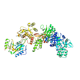
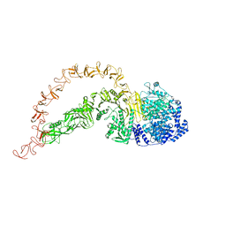
1N0U
 | | Crystal structure of yeast elongation factor 2 in complex with sordarin | | 分子名称: | Elongation factor 2, [1R-(1.ALPHA.,3A.BETA.,4.BETA.,4A.BETA.,7.BETA.,7A.ALPHA.,8A.BETA.)]8A-[(6-DEOXY-4-O-METHYL-BETA-D-ALTROPYRANOSYLOXY)METHYL]-4-FORMYL-4,4A,5,6,7,7A,8,8A-OCTAHYDRO-7-METHYL-3-(1-METHYLETHYL)-1,4-METHANO-S-INDACENE-3A(1H)-CARBOXYLIC ACID | | 著者 | Joergensen, R, Ortiz, P.A, Carr-Schmid, A, Nissen, P, Kinzy, T.G, Andersen, G.R. | | 登録日 | 2002-10-15 | | 公開日 | 2003-02-11 | | 最終更新日 | 2024-02-14 | | 実験手法 | X-RAY DIFFRACTION (2.12 Å) | | 主引用文献 | Two crystal structures demonstrate large conformational changes in the eukaryotic ribosomal translocase.
Nat. Struct. Biol., 10, 2003
|
|
1XK9
 | | Pseudomanas exotoxin A in complex with the PJ34 inhibitor | | 分子名称: | Exotoxin A, N~2~,N~2~-DIMETHYL-N~1~-(6-OXO-5,6-DIHYDROPHENANTHRIDIN-2-YL)GLYCINAMIDE | | 著者 | Yates, S.P, Taylor, P.J, Joergensen, R, Ferrraris, D, Zhang, J, Andersen, G.R, Merrill, A.R. | | 登録日 | 2004-09-28 | | 公開日 | 2005-05-17 | | 最終更新日 | 2023-10-25 | | 実験手法 | X-RAY DIFFRACTION (2.1 Å) | | 主引用文献 | Structure-function analysis of water-soluble inhibitors of the catalytic domain of exotoxin A from Pseudomonas aeruginosa
BIOCHEM.J., 385, 2005
|
|
7POG
 | | High-resolution structure of native toxin A from Clostridioides difficile | | 分子名称: | Toxin A, ZINC ION | | 著者 | Boesen, T, Joergensen, R, Aminzadeh, A, Engelbrecht Larsen, C. | | 登録日 | 2021-09-08 | | 公開日 | 2021-12-08 | | 最終更新日 | 2024-07-17 | | 実験手法 | ELECTRON MICROSCOPY (2.83 Å) | | 主引用文献 | High-resolution structure of native toxin A from Clostridioides difficile.
Embo Rep., 23, 2022
|
|


