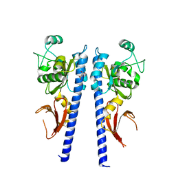
3G3Q
 
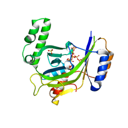 | |
3G3O
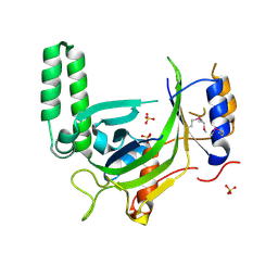 | |
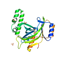
3G3U
 | |
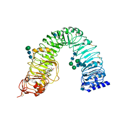
3G3R
 | |
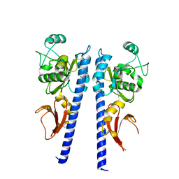
3RJ0
 | |
3RIZ
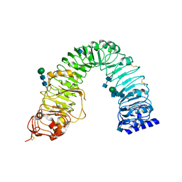 | | Crystal structure of the plant steroid receptor BRI1 ectodomain | | 分子名称: | 2-acetamido-2-deoxy-beta-D-glucopyranose, 2-acetamido-2-deoxy-beta-D-glucopyranose-(1-4)-2-acetamido-2-deoxy-beta-D-glucopyranose, Protein BRASSINOSTEROID INSENSITIVE 1, ... | | 著者 | Hothorn, M. | | 登録日 | 2011-04-14 | | 公開日 | 2011-06-22 | | 最終更新日 | 2020-07-29 | | 実験手法 | X-RAY DIFFRACTION (2.523 Å) | | 主引用文献 | Structural basis of steroid hormone perception by the receptor kinase BRI1.
Nature, 474, 2011
|
|
1RJ4
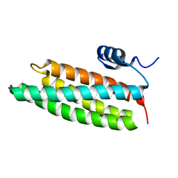
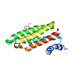 | | Structure of a Cell Wall Invertase Inhibitor from Tobacco in Complex with Cd2+ | | 分子名称: | 2-[BIS-(2-HYDROXY-ETHYL)-AMINO]-2-HYDROXYMETHYL-PROPANE-1,3-DIOL, CADMIUM ION, invertase inhibitor | | 著者 | Hothorn, M, D'Angelo, I, Marquez, J.A, Greiner, S, Scheffzek, K. | | 登録日 | 2003-11-18 | | 公開日 | 2004-02-03 | | 最終更新日 | 2011-07-13 | | 実験手法 | X-RAY DIFFRACTION (2 Å) | | 主引用文献 | The invertase inhibitor Nt-CIF from tobacco: a highly thermostable four-helix bundle with an unusual N-terminal extension
J.Mol.Biol., 335, 2004
|
|
1RJ1
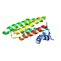 | | Crystal Structure of a Cell Wall Invertase Inhibitor from Tobacco | | 分子名称: | invertase inhibitor | | 著者 | Hothorn, M, D'Angelo, I, Marquez, J.A, Greiner, S, Scheffzek, K. | | 登録日 | 2003-11-18 | | 公開日 | 2004-02-03 | | 最終更新日 | 2023-10-25 | | 実験手法 | X-RAY DIFFRACTION (1.87 Å) | | 主引用文献 | The invertase inhibitor Nt-CIF from tobacco: a highly thermostable four-helix bundle with an unusual N-terminal extension
J.Mol.Biol., 335, 2004
|
|
3T4J
 | |
3T4Q
 | |
3T4O
 | |
3T4T
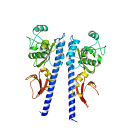 | |
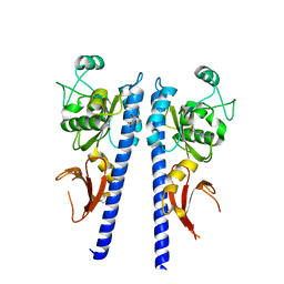
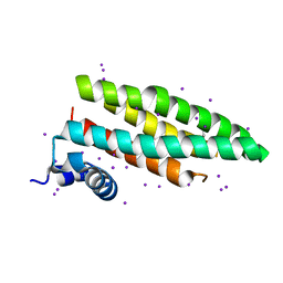
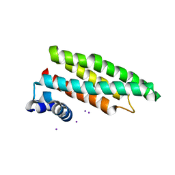
2XQR
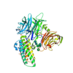 | | Crystal structure of plant cell wall invertase in complex with a specific protein inhibitor | | 分子名称: | 2-acetamido-2-deoxy-beta-D-glucopyranose, 2-acetamido-2-deoxy-beta-D-glucopyranose-(1-4)-2-acetamido-2-deoxy-beta-D-glucopyranose, 4-(2-HYDROXYETHYL)-1-PIPERAZINE ETHANESULFONIC ACID, ... | | 著者 | Hothorn, M, Van den Ende, W, Lammens, W, Rybin, V, Scheffzek, K. | | 登録日 | 2010-09-07 | | 公開日 | 2010-10-06 | | 最終更新日 | 2023-12-20 | | 実験手法 | X-RAY DIFFRACTION (2.58 Å) | | 主引用文献 | Structural Insights Into the Ph-Controlled Targeting of Plant Cell-Wall Invertase by a Specific Inhibitor Protein.
Proc.Natl.Acad.Sci.USA, 107, 2010
|
|
3T4L
 | |
3T4S
 | |
3T4K
 | |
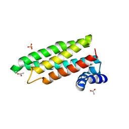
1X90
 | | Crystal structure of mutant form B of a pectin methylesterase inhibitor from Arabidopsis | | 分子名称: | invertase/pectin methylesterase inhibitor family protein | | 著者 | Hothorn, M, Wolf, S, Aloy, P, Greiner, S, Scheffzek, K. | | 登録日 | 2004-08-19 | | 公開日 | 2004-12-28 | | 最終更新日 | 2023-10-25 | | 実験手法 | X-RAY DIFFRACTION (2.68 Å) | | 主引用文献 | Structural insights into the target specificity of plant invertase and pectin methylesterase inhibitory proteins
Plant Cell, 16, 2004
|
|
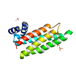
1X8Z
 | | Crystal structure of a pectin methylesterase inhibitor from Arabidopsis thaliana | | 分子名称: | invertase/pectin methylesterase inhibitor family protein | | 著者 | Hothorn, M, Wolf, S, Aloy, P, Greiner, S, Scheffzek, K. | | 登録日 | 2004-08-19 | | 公開日 | 2004-12-28 | | 最終更新日 | 2023-10-25 | | 実験手法 | X-RAY DIFFRACTION (2.86 Å) | | 主引用文献 | Structural insights into the target specificity of plant invertase and pectin methylesterase inhibitory proteins
Plant Cell, 16, 2004
|
|
1X91
 | | Crystal structure of mutant form A of a pectin methylesterase inhibitor from Arabidopsis | | 分子名称: | invertase/pectin methylesterase inhibitor family protein | | 著者 | Hothorn, M, Wolf, S, Aloy, P, Greiner, S, Scheffzek, K. | | 登録日 | 2004-08-19 | | 公開日 | 2004-12-28 | | 最終更新日 | 2023-10-25 | | 実験手法 | X-RAY DIFFRACTION (1.5 Å) | | 主引用文献 | Structural insights into the target specificity of plant invertase and pectin methylesterase inhibitory proteins
Plant Cell, 16, 2004
|
|
2CJ7
 | |
2CJ8
 | |
2CJ6
 | |
2CJ4
 | |
2CJ5
 | |
2GWD
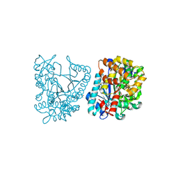 | | Crystal structure of plant glutamate cysteine ligase in complex with Mg2+ and L-glutamate | | 分子名称: | ACETATE ION, GLUTAMIC ACID, Glutamate cysteine ligase, ... | | 著者 | Hothorn, M, Wachter, A, Gromes, R, Stuwe, T, Rausch, T, Scheffzek, K. | | 登録日 | 2006-05-04 | | 公開日 | 2006-06-20 | | 最終更新日 | 2023-08-30 | | 実験手法 | X-RAY DIFFRACTION (2.09 Å) | | 主引用文献 | Structural basis for the redox control of plant glutamate cysteine ligase.
J.Biol.Chem., 281, 2006
|
|


