5L7P
 
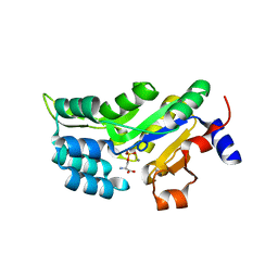
 | | In silico-powered specific incorporation of photocaged Dopa at multiple protein sites | | 分子名称: | (2~{S})-2-azanyl-3-[3-[(2-nitrophenyl)methoxy]-4-oxidanyl-phenyl]propanoic acid, CALCIUM ION, CHLORIDE ION, ... | | 著者 | Hauf, M, Richter, F, Schneider, T, Martins, B.M, Baumann, T, Durkin, P, Dobbek, H, Moeglich, A, Budisa, N. | | 登録日 | 2016-06-03 | | 公開日 | 2017-09-13 | | 最終更新日 | 2024-01-10 | | 実験手法 | X-RAY DIFFRACTION (1.9 Å) | | 主引用文献 | Photoactivatable Mussel-Based Underwater Adhesive Proteins by an Expanded Genetic Code.
Chembiochem, 18, 2017
|
|
8A21
 | |
8A1Z
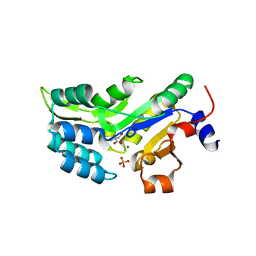
 | | Crystal structure of Phosphoserine phosphatase SerB from Mycobacterium avium in complex with 1-(2,4-dichlorophenyl)-3-hydroxyurea | | 分子名称: | 1-(2,4-dichlorophenyl)-3-oxidanyl-urea, CHLORIDE ION, MAGNESIUM ION, ... | | 著者 | Haufroid, M, Wouters, J. | | 登録日 | 2022-06-02 | | 公開日 | 2022-11-23 | | 最終更新日 | 2024-01-31 | | 実験手法 | X-RAY DIFFRACTION (2.28 Å) | | 主引用文献 | Targeting the phosphoserine phosphatase MtSerB2 for tuberculosis drug discovery, an hybrid knowledge based /fragment based approach.
Eur.J.Med.Chem., 245, 2022
|
|
6Q6J
 | | Human phosphoserine phosphatase with substrate analogue homo-cysteic acid | | 分子名称: | (2~{S})-2-azanyl-4-sulfo-butanoic acid, CALCIUM ION, CHLORIDE ION, ... | | 著者 | Wouters, J, Haufroid, M, Mirgaux, M. | | 登録日 | 2018-12-11 | | 公開日 | 2019-06-12 | | 最終更新日 | 2024-01-24 | | 実験手法 | X-RAY DIFFRACTION (1.985 Å) | | 主引用文献 | Crystal structures and snapshots along the reaction pathway of human phosphoserine phosphatase.
Acta Crystallogr D Struct Biol, 75, 2019
|
|
6HYJ
 | | PSPH Human phosphoserine phosphatase | | 分子名称: | CALCIUM ION, PHOSPHOSERINE, Phosphoserine phosphatase, ... | | 著者 | Wouters, J, Haufroid, M, Mirgaux, M. | | 登録日 | 2018-10-22 | | 公開日 | 2019-06-12 | | 最終更新日 | 2024-01-31 | | 実験手法 | X-RAY DIFFRACTION (1.929 Å) | | 主引用文献 | Crystal structures and snapshots along the reaction pathway of human phosphoserine phosphatase.
Acta Crystallogr D Struct Biol, 75, 2019
|
|
6HYY
 | | Human phosphoserine phosphatase with serine and phosphate | | 分子名称: | CALCIUM ION, MAGNESIUM ION, PHOSPHATE ION, ... | | 著者 | Wouters, J, Haufroid, M, Mirgaux, M. | | 登録日 | 2018-10-22 | | 公開日 | 2019-06-12 | | 最終更新日 | 2024-01-24 | | 実験手法 | X-RAY DIFFRACTION (1.566 Å) | | 主引用文献 | Crystal structures and snapshots along the reaction pathway of human phosphoserine phosphatase.
Acta Crystallogr D Struct Biol, 75, 2019
|
|
5NSF
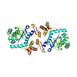 | | Structure of AzuAla | | 分子名称: | (2~{S})-2-azanyl-3-(2,6-dihydroazulen-1-yl)propanoic acid, CALCIUM ION, GLYCEROL, ... | | 著者 | Martins, B.M. | | 登録日 | 2017-04-26 | | 公開日 | 2019-01-16 | | 最終更新日 | 2024-01-17 | | 実験手法 | X-RAY DIFFRACTION (2.426 Å) | | 主引用文献 | Site-Resolved Observation of Vibrational Energy Transfer Using a Genetically Encoded Ultrafast Heater.
Angew. Chem. Int. Ed. Engl., 58, 2019
|
|
8B3X
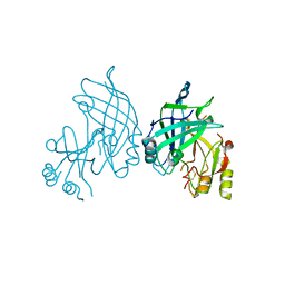 | | High resolution crystal structure of dimeric SUDV VP40 | | 分子名称: | Matrix protein VP40 | | 著者 | Werner, A.-D, Norris, M, Saphire, E.O, Becker, S. | | 登録日 | 2022-09-17 | | 公開日 | 2023-06-21 | | 最終更新日 | 2024-02-07 | | 実験手法 | X-RAY DIFFRACTION (1.531 Å) | | 主引用文献 | The C-terminus of Sudan ebolavirus VP40 contains a functionally important CX n C motif, a target for redox modifications.
Structure, 31, 2023
|
|
8B1P
 | |
8B1O
 | |


