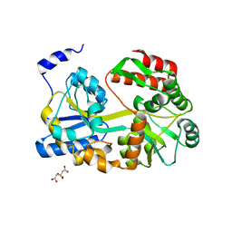
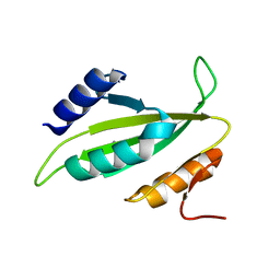
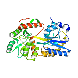
3WOA
 
 | |
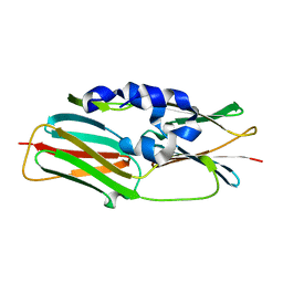
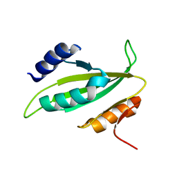
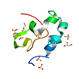
7VOS
 | | High-resolution neutron and X-ray joint refined structure of high-potential iron-sulfur protein in the oxidized state | | 分子名称: | AMMONIUM ION, GLYCEROL, High-potential iron-sulfur protein, ... | | 著者 | Hanazono, Y, Hirano, Y, Takeda, K, Kusaka, K, Tamada, T, Miki, K. | | 登録日 | 2021-10-14 | | 公開日 | 2022-06-01 | | 最終更新日 | 2024-04-03 | | 実験手法 | NEUTRON DIFFRACTION (0.66 Å), X-RAY DIFFRACTION | | 主引用文献 | Revisiting the concept of peptide bond planarity in an iron-sulfur protein by neutron structure analysis.
Sci Adv, 8, 2022
|
|
3W1Z
 | | Heat shock protein 16.0 from Schizosaccharomyces pombe | | 分子名称: | Heat shock protein 16 | | 著者 | Hanazono, Y, Takeda, K, Akiyama, N, Aikawa, Y, Miki, K. | | 登録日 | 2012-11-26 | | 公開日 | 2013-03-13 | | 最終更新日 | 2023-11-08 | | 実験手法 | X-RAY DIFFRACTION (2.401 Å) | | 主引用文献 | Nonequivalence Observed for the 16-Meric Structure of a Small Heat Shock Protein, SpHsp16.0, from Schizosaccharomyces pombe
Structure, 21, 2013
|
|
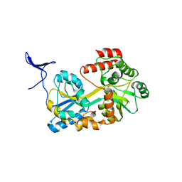
6AIR
 | | High resolution structure of perdeuterated high-potential iron-sulfur protein | | 分子名称: | GLYCEROL, High-potential iron-sulfur protein, IRON/SULFUR CLUSTER, ... | | 著者 | Hanazono, Y, Takeda, K, Miki, K. | | 登録日 | 2018-08-24 | | 公開日 | 2019-08-21 | | 最終更新日 | 2023-11-22 | | 実験手法 | X-RAY DIFFRACTION (0.85 Å) | | 主引用文献 | Characterization of perdeuterated high-potential iron-sulfur protein with high-resolution X-ray crystallography.
Proteins, 88, 2020
|
|
6AIQ
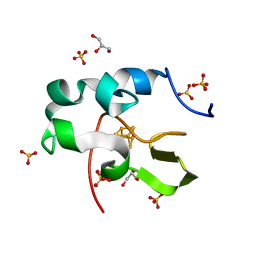 | | High resolution structure of recombinant high-potential iron-sulfur protein | | 分子名称: | GLYCEROL, High-potential iron-sulfur protein, IRON/SULFUR CLUSTER, ... | | 著者 | Hanazono, Y, Takeda, K, Miki, K. | | 登録日 | 2018-08-24 | | 公開日 | 2019-08-21 | | 最終更新日 | 2023-11-22 | | 実験手法 | X-RAY DIFFRACTION (0.85 Å) | | 主引用文献 | Characterization of perdeuterated high-potential iron-sulfur protein with high-resolution X-ray crystallography.
Proteins, 88, 2020
|
|
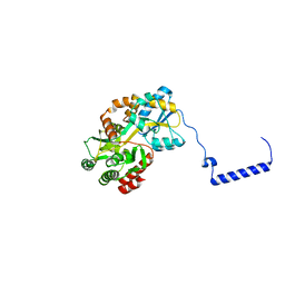
5ZCA
 | | Crystal structure of lambda repressor (1-20) fused with maltose-binding protein | | 分子名称: | CITRIC ACID, Repressor protein cI,Maltose-binding periplasmic protein, alpha-D-glucopyranose-(1-4)-alpha-D-glucopyranose | | 著者 | Hanazono, Y, Takeda, K, Miki, K. | | 登録日 | 2018-02-16 | | 公開日 | 2018-08-15 | | 最終更新日 | 2023-11-22 | | 実験手法 | X-RAY DIFFRACTION (1.801 Å) | | 主引用文献 | Co-translational folding of alpha-helical proteins: structural studies of intermediate-length variants of the lambda repressor
Febs Open Bio, 8, 2018
|
|
3VQL
 | |
3VQK
 | |
3VQM
 | |
5B3P
 | |
5B3Q
 | |
5B3Y
 | |
5B3W
 | | Crystal structure of hPin1 WW domain (5-15) fused with maltose-binding protein in C2221 form | | 分子名称: | CITRIC ACID, Peptidyl-prolyl cis-trans isomerase NIMA-interacting 1,Maltose-binding periplasmic protein, alpha-D-glucopyranose-(1-4)-alpha-D-glucopyranose | | 著者 | Hanazono, Y, Takeda, K, Miki, K. | | 登録日 | 2016-03-17 | | 公開日 | 2016-10-26 | | 最終更新日 | 2023-11-08 | | 実験手法 | X-RAY DIFFRACTION (2.4 Å) | | 主引用文献 | Structural studies of the N-terminal fragments of the WW domain: Insights into co-translational folding of a beta-sheet protein
Sci Rep, 6, 2016
|
|
5B3X
 | |
5BMY
 | |
5B3Z
 | |
5ZUI
 | | Crystal Structure of HSP104 from Chaetomium thermophilum | | 分子名称: | ADENOSINE-5'-DIPHOSPHATE, Heat Shock Protein 104, SULFATE ION | | 著者 | Hanazono, Y, Inoue, Y, Noguchi, K, Yohda, M, Shinohara, K, Takeda, K, Miki, K. | | 登録日 | 2018-05-07 | | 公開日 | 2019-06-19 | | 最終更新日 | 2023-11-22 | | 実験手法 | X-RAY DIFFRACTION (2.701 Å) | | 主引用文献 | Split conformation of Chaetomium thermophilum Hsp104 disaggregase.
Structure, 2021
|
|
6JGJ
 | | Crystal structure of the F99S/M153T/V163A/E222Q variant of GFP at 0.78 A | | 分子名称: | Green fluorescent protein, MAGNESIUM ION | | 著者 | Takaba, K, Tai, Y, Hanazono, Y, Miki, K, Takeda, K. | | 登録日 | 2019-02-14 | | 公開日 | 2019-04-17 | | 最終更新日 | 2023-11-22 | | 実験手法 | X-RAY DIFFRACTION (0.78 Å) | | 主引用文献 | Subatomic resolution X-ray structures of green fluorescent protein.
Iucrj, 6, 2019
|
|
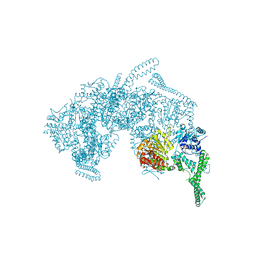
7CG3
 | | Staggered ring conformation of CtHsp104 (Hsp104 from Chaetomium Thermophilum) | | 分子名称: | Heat shock protein 104 | | 著者 | Inoue, Y, Hanazono, Y, Noi, K, Kawamoto, A, Kimatsuka, M, Harada, R, Takeda, K, Iwamasa, N, Shibata, K, Noguchi, K, Shigeta, Y, Namba, K, Ogura, T, Miki, K, Shinohara, K, Yohda, M. | | 登録日 | 2020-06-30 | | 公開日 | 2021-04-28 | | 最終更新日 | 2024-05-29 | | 実験手法 | ELECTRON MICROSCOPY (5.1 Å) | | 主引用文献 | Split conformation of Chaetomium thermophilum Hsp104 disaggregase.
Structure, 29, 2021
|
|
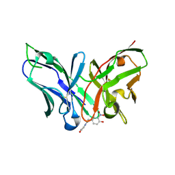
8YTN
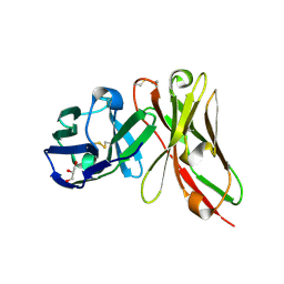 | | Single-chain Fv antibody of E11 | | 分子名称: | GLYCEROL, Single-chain Fv antibody of E11 | | 著者 | Yoshida, M, Hanazono, Y, Numoto, N, Ito, N, Oda, M. | | 登録日 | 2024-03-26 | | 公開日 | 2024-07-03 | | 最終更新日 | 2024-07-10 | | 実験手法 | X-RAY DIFFRACTION (1.72 Å) | | 主引用文献 | Affinity-matured antibody with a disulfide bond in H-CDR3 loop.
Arch.Biochem.Biophys., 758, 2024
|
|
8YTO
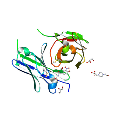 | | Single-chain Fv antibody of E11 complex with NP-glycine | | 分子名称: | 2-[2-(3-nitro-4-oxidanyl-phenyl)ethanoylamino]ethanoic acid, 4-(2-HYDROXYETHYL)-1-PIPERAZINE ETHANESULFONIC ACID, GLYCEROL, ... | | 著者 | Yoshida, M, Hanazono, Y, Numoto, N, Ito, N, Oda, M. | | 登録日 | 2024-03-26 | | 公開日 | 2024-07-03 | | 最終更新日 | 2024-07-10 | | 実験手法 | X-RAY DIFFRACTION (1.04 Å) | | 主引用文献 | Affinity-matured antibody with a disulfide bond in H-CDR3 loop.
Arch.Biochem.Biophys., 758, 2024
|
|
8YTP
 | | Single-chain Fv antibody of E11 complex with NP-glycine under reducing conditions | | 分子名称: | 2-[2-(3-nitro-4-oxidanyl-phenyl)ethanoylamino]ethanoic acid, Single-chain Fv antibody of E11 | | 著者 | Yoshida, M, Hanazono, Y, Numoto, N, Ito, N, Oda, M. | | 登録日 | 2024-03-26 | | 公開日 | 2024-07-03 | | 最終更新日 | 2024-07-10 | | 実験手法 | X-RAY DIFFRACTION (1.36 Å) | | 主引用文献 | Affinity-matured antibody with a disulfide bond in H-CDR3 loop.
Arch.Biochem.Biophys., 758, 2024
|
|
3AAC
 | | Small heat shock protein hsp14.0 with the mutations of I120F and I122F in the form II crystal | | 分子名称: | Putative uncharacterized protein ST1653 | | 著者 | Takeda, K, Hayashi, T, Abe, T, Hirano, Y, Hanazono, Y, Yohda, M, Miki, K. | | 登録日 | 2009-11-13 | | 公開日 | 2010-11-17 | | 最終更新日 | 2024-03-13 | | 実験手法 | X-RAY DIFFRACTION (2.4 Å) | | 主引用文献 | Dimer structure and conformational variability in the N-terminal region of an archaeal small heat shock protein, StHsp14.0
J.Struct.Biol., 174, 2011
|
|
3AAB
 | | Small heat shock protein hsp14.0 with the mutations of I120F and I122F in the form I crystal | | 分子名称: | GLYCEROL, ISOPROPYL ALCOHOL, Putative uncharacterized protein ST1653 | | 著者 | Takeda, K, Hayashi, T, Abe, T, Hirano, Y, Hanazono, Y, Yohda, M, Miki, K. | | 登録日 | 2009-11-13 | | 公開日 | 2010-11-17 | | 最終更新日 | 2024-03-13 | | 実験手法 | X-RAY DIFFRACTION (1.851 Å) | | 主引用文献 | Dimer structure and conformational variability in the N-terminal region of an archaeal small heat shock protein, StHsp14.0
J.Struct.Biol., 174, 2011
|
|
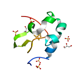
5WQR
 | | High resolution structure of high-potential iron-sulfur protein in the reduced state | | 分子名称: | GLYCEROL, High-potential iron-sulfur protein, IRON/SULFUR CLUSTER, ... | | 著者 | Ohno, H, Takeda, K, Niwa, S, Tsujinaka, T, Hanazono, Y, Hirano, Y, Miki, K. | | 登録日 | 2016-11-28 | | 公開日 | 2017-06-07 | | 最終更新日 | 2023-11-08 | | 実験手法 | X-RAY DIFFRACTION (0.8 Å) | | 主引用文献 | Crystallographic characterization of the high-potential iron-sulfur protein in the oxidized state at 0.8 angstrom resolution
PLoS ONE, 12, 2017
|
|


