2BAY
 
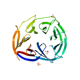
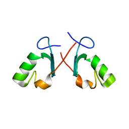 | | Crystal structure of the Prp19 U-box dimer | | 分子名称: | Pre-mRNA splicing factor PRP19 | | 著者 | Vander Kooi, C.W, Ohi, M.D, Rosenberg, J.A, Oldham, M.L, Newcomer, M.E, Gould, K.L, Chazin, W.J. | | 登録日 | 2005-10-15 | | 公開日 | 2006-01-10 | | 最終更新日 | 2024-02-14 | | 実験手法 | X-RAY DIFFRACTION (1.5 Å) | | 主引用文献 | The Prp19 U-box Crystal Structure Suggests a Common Dimeric Architecture for a Class of Oligomeric E3 Ubiquitin Ligases.
Biochemistry, 45, 2006
|
|
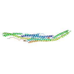
6XJ1
 | |
1PQN
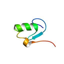
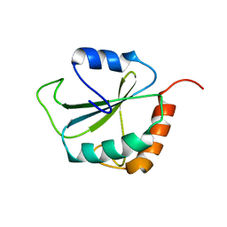 | | dominant negative human hDim1 (hDim1 1-128) | | 分子名称: | Spliceosomal U5 snRNP-specific 15 kDa protein | | 著者 | Zhang, Y.Z, Cheng, H, Gould, K.L, Golemis, E.A, Roder, H. | | 登録日 | 2003-06-18 | | 公開日 | 2003-08-26 | | 最終更新日 | 2024-05-22 | | 実験手法 | SOLUTION NMR | | 主引用文献 | Structure, stability and function of hDim1 investigated by NMR, circular
dichroism and mutational analysis
Biochemistry, 42, 2003
|
|
7P7G
 | | Crystal structure of phosphorylated pT220 Casein Kinase I delta (CK1d), conformation 2 and 3 | | 分子名称: | 1,2-ETHANEDIOL, ADENOSINE MONOPHOSPHATE, CITRIC ACID, ... | | 著者 | Chaikuad, A, Zhubi, R, Knapp, S, Structural Genomics Consortium (SGC) | | 登録日 | 2021-07-19 | | 公開日 | 2022-04-13 | | 最終更新日 | 2024-01-31 | | 実験手法 | X-RAY DIFFRACTION (1.7 Å) | | 主引用文献 | Kinase domain autophosphorylation rewires the activity and substrate specificity of CK1 enzymes.
Mol.Cell, 82, 2022
|
|
7P7H
 | |
7P7F
 | | Crystal structure of phosphorylated pT220 Casein Kinase I delta (CK1d), conformation 1 | | 分子名称: | 1,2-ETHANEDIOL, ADENOSINE, ADENOSINE MONOPHOSPHATE, ... | | 著者 | Chaikuad, A, Zhubi, R, Knapp, S, Structural Genomics Consortium (SGC) | | 登録日 | 2021-07-19 | | 公開日 | 2022-04-13 | | 最終更新日 | 2024-01-31 | | 実験手法 | X-RAY DIFFRACTION (1.96 Å) | | 主引用文献 | Kinase domain autophosphorylation rewires the activity and substrate specificity of CK1 enzymes.
Mol.Cell, 82, 2022
|
|
5C1F
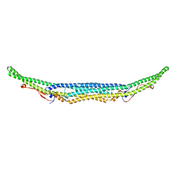 | | Structure of the Imp2 F-BAR domain | | 分子名称: | FORMIC ACID, Septation protein imp2 | | 著者 | Vander Kooi, C.W. | | 登録日 | 2015-06-13 | | 公開日 | 2016-01-27 | | 最終更新日 | 2019-12-25 | | 実験手法 | X-RAY DIFFRACTION (2.3551 Å) | | 主引用文献 | The Tubulation Activity of a Fission Yeast F-BAR Protein Is Dispensable for Its Function in Cytokinesis.
Cell Rep, 14, 2016
|
|
3LRV
 | |
1N87
 | |


