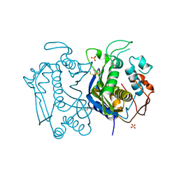
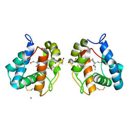
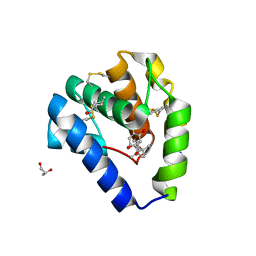
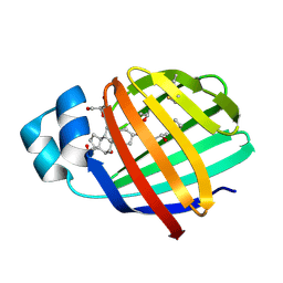
3EM0
 
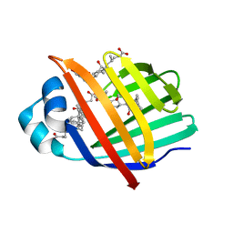 | | Crystal structure of Zebrafish Ileal Bile Acid-Bindin Protein complexed with cholic acid (crystal form B). | | 分子名称: | CHOLIC ACID, Ileal Bile Acid-Binding Protein | | 著者 | Capaldi, S, Saccomani, G, Fessas, D, Signorelli, M, Perduca, M, Monaco, H.L. | | 登録日 | 2008-09-23 | | 公開日 | 2009-01-13 | | 最終更新日 | 2023-09-06 | | 実験手法 | X-RAY DIFFRACTION (2.2 Å) | | 主引用文献 | The X-Ray structure of zebrafish (Danio rerio) ileal bile acid-binding protein reveals the presence of binding sites on the surface of the protein molecule.
J.Mol.Biol., 385, 2009
|
|
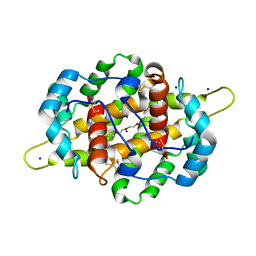
3ELZ
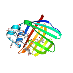 | | Crystal structure of Zebrafish Ileal Bile Acid-Bindin Protein complexed with cholic acid (crystal form A). | | 分子名称: | CHOLIC ACID, ileal Bile Acid-Binding Protein | | 著者 | Capaldi, S, Saccomani, G, Fessas, D, Signorelli, M, Perduca, M, Monaco, H.L. | | 登録日 | 2008-09-23 | | 公開日 | 2009-01-13 | | 最終更新日 | 2023-09-06 | | 実験手法 | X-RAY DIFFRACTION (2.2 Å) | | 主引用文献 | The X-Ray structure of zebrafish (Danio rerio) ileal bile acid-binding protein reveals the presence of binding sites on the surface of the protein molecule.
J.Mol.Biol., 385, 2009
|
|
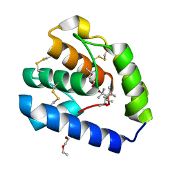
3ELX
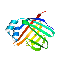 | | Crystal structure of apo Zebrafish Ileal Bile Acid-Binding Protein | | 分子名称: | 1,2-ETHANEDIOL, Ileal bile acid-binding protein | | 著者 | Capaldi, S, Saccomani, G, Fessas, D, Signorelli, M, Perduca, M, Monaco, H.L. | | 登録日 | 2008-09-23 | | 公開日 | 2009-01-13 | | 最終更新日 | 2023-09-06 | | 実験手法 | X-RAY DIFFRACTION (1.6 Å) | | 主引用文献 | The X-Ray structure of zebrafish (Danio rerio) ileal bile acid-binding protein reveals the presence of binding sites on the surface of the protein molecule.
J.Mol.Biol., 385, 2009
|
|
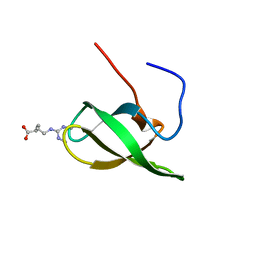
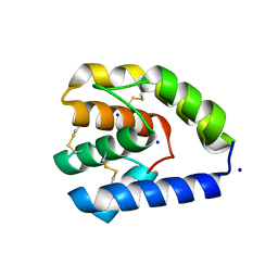
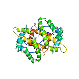
4A4F
 | | Solution structure of SPF30 Tudor domain in complex with symmetrically dimethylated arginine | | 分子名称: | N3, N4-DIMETHYLARGININE, SURVIVAL OF MOTOR NEURON-RELATED-SPLICING FACTOR 30 | | 著者 | Tripsianes, K, Madl, T, Machyna, M, Fessas, D, Englbrecht, C, Fischer, U, Neugebauer, K.M, Sattler, M. | | 登録日 | 2011-10-12 | | 公開日 | 2011-11-30 | | 最終更新日 | 2024-05-15 | | 実験手法 | SOLUTION NMR | | 主引用文献 | Structural Basis for Dimethyl-Arginine Recognition by the Tudor Domains of Human Smn and Spf30 Proteins
Nat.Struct.Mol.Biol., 18, 2011
|
|
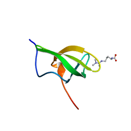
4A4E
 | | Solution structure of SMN Tudor domain in complex with symmetrically dimethylated arginine | | 分子名称: | N3, N4-DIMETHYLARGININE, SURVIVAL MOTOR NEURON PROTEIN | | 著者 | Tripsianes, K, Madl, T, Machyna, M, Fessas, D, Englbrecht, C, Fischer, U, Neugebauer, K.M, Sattler, M. | | 登録日 | 2011-10-12 | | 公開日 | 2011-11-30 | | 最終更新日 | 2024-06-19 | | 実験手法 | SOLUTION NMR | | 主引用文献 | Structural Basis for Dimethyl-Arginine Recognition by the Tudor Domains of Human Smn and Spf30 Proteins
Nat.Struct.Mol.Biol., 18, 2011
|
|
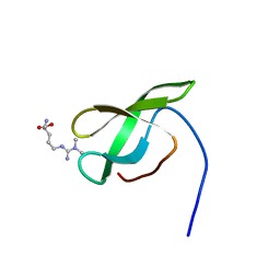
4A4H
 | | Solution structure of SPF30 Tudor domain in complex with asymmetrically dimethylated arginine | | 分子名称: | NG,NG-DIMETHYL-L-ARGININE, SURVIVAL OF MOTOR NEURON-RELATED-SPLICING FACTOR 30 | | 著者 | Tripsianes, K, Madl, T, Machyna, M, Fessas, D, Englbrecht, C, Fischer, U, Neugebauer, K.M, Sattler, M. | | 登録日 | 2011-10-12 | | 公開日 | 2011-11-30 | | 最終更新日 | 2023-11-15 | | 実験手法 | SOLUTION NMR | | 主引用文献 | Structural Basis for Dimethyl-Arginine Recognition by the Tudor Domains of Human Smn and Spf30 Proteins
Nat.Struct.Mol.Biol., 18, 2011
|
|
4A4G
 | | Solution structure of SMN Tudor domain in complex with asymmetrically dimethylated arginine | | 分子名称: | NG,NG-DIMETHYL-L-ARGININE, SURVIVAL MOTOR NEURON PROTEIN | | 著者 | Tripsianes, K, Madl, T, Machyna, M, Fessas, D, Englbrecht, C, Fischer, U, Neugebauer, K.M, Sattler, M. | | 登録日 | 2011-10-12 | | 公開日 | 2011-11-30 | | 最終更新日 | 2023-11-15 | | 実験手法 | SOLUTION NMR | | 主引用文献 | Structural Basis for Dimethyl-Arginine Recognition by the Tudor Domains of Human Smn and Spf30 Proteins
Nat.Struct.Mol.Biol., 18, 2011
|
|
3N5E
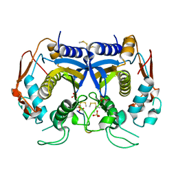 | | Crystal Structure of human thymidylate synthase bound to a peptide inhibitor | | 分子名称: | SULFATE ION, Synthetic peptide LR, Thymidylate synthase | | 著者 | Pozzi, C, Cardinale, D, Guaitoli, G, Tondi, D, Luciani, R, Myllykallio, H, Ferrari, S, Costi, M.P, Mangani, S. | | 登録日 | 2010-05-25 | | 公開日 | 2011-06-08 | | 最終更新日 | 2023-09-06 | | 実験手法 | X-RAY DIFFRACTION (2.26 Å) | | 主引用文献 | Protein-protein interface-binding peptides inhibit the cancer therapy target human thymidylate synthase.
Proc.Natl.Acad.Sci.USA, 108, 2011
|
|
3N5G
 | | Crystal Structure of histidine-tagged human thymidylate synthase | | 分子名称: | SULFATE ION, Thymidylate synthase | | 著者 | Pozzi, C, Cardinale, D, Guaitoli, G, Tondi, D, Luciani, R, Myllykallio, H, Ferrari, S, Costi, M.P, Mangani, S. | | 登録日 | 2010-05-25 | | 公開日 | 2011-06-08 | | 最終更新日 | 2023-09-06 | | 実験手法 | X-RAY DIFFRACTION (2.27 Å) | | 主引用文献 | Protein-protein interface-binding peptides inhibit the cancer therapy target human thymidylate synthase.
Proc.Natl.Acad.Sci.USA, 108, 2011
|
|
8C6G
 | |
8C68
 | |
8C6E
 | |
5EL2
 | |
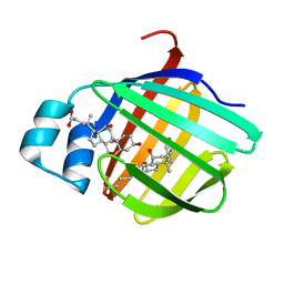
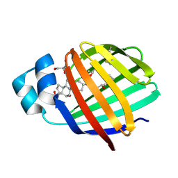
4IJ7
 | | Crystal structure of Odorant Binding Protein 48 from Anopheles gambiae (AgamOBP48) with PEG | | 分子名称: | 2,5,8,11,14,17,20,23,26,29,32,35,38,41,44,47,50,53,56,59,62,65,68,71,74,77,80-HEPTACOSAOXADOOCTACONTAN-82-OL, Odorant binding protein-8, SODIUM ION | | 著者 | Zographos, S.E, Tsitsanou, K.E, Drakou, C.E. | | 登録日 | 2012-12-21 | | 公開日 | 2013-10-16 | | 最終更新日 | 2023-09-20 | | 実験手法 | X-RAY DIFFRACTION (2.25 Å) | | 主引用文献 | Crystal and Solution Studies of the "Plus-C" Odorant-binding Protein 48 from Anopheles gambiae: CONTROL OF BINDING SPECIFICITY THROUGH THREE-DIMENSIONAL DOMAIN SWAPPING.
J.Biol.Chem., 288, 2013
|
|
8BXU
 | | Crystal structure of Odorant Binding Protein 5 from Anopheles gambiae (AgamOBP5) with MPD (2-Methyl-2,4-pentanediol) | | 分子名称: | (4S)-2-METHYL-2,4-PENTANEDIOL, 2-ETHOXYETHANOL, Odorant binding protein, ... | | 著者 | Liggri, P.G.V, Tsitsanou, K.E, Zographos, S.E. | | 登録日 | 2022-12-09 | | 公開日 | 2023-03-22 | | 最終更新日 | 2023-04-12 | | 実験手法 | X-RAY DIFFRACTION (1.35 Å) | | 主引用文献 | The structure of AgamOBP5 in complex with the natural insect repellents Carvacrol and Thymol: Crystallographic, fluorescence and thermodynamic binding studies.
Int.J.Biol.Macromol., 237, 2023
|
|
8BXV
 | | Crystal structure of Odorant Binding Protein 5 from Anopheles gambiae (AgamOBP5) with Thymol | | 分子名称: | (4S)-2-METHYL-2,4-PENTANEDIOL, 5-METHYL-2-(1-METHYLETHYL)PHENOL, DI(HYDROXYETHYL)ETHER, ... | | 著者 | Liggri, P.G.V, Tsitsanou, K.E, Zographos, S.E. | | 登録日 | 2022-12-10 | | 公開日 | 2023-03-22 | | 最終更新日 | 2023-04-12 | | 実験手法 | X-RAY DIFFRACTION (1.3 Å) | | 主引用文献 | The structure of AgamOBP5 in complex with the natural insect repellents Carvacrol and Thymol: Crystallographic, fluorescence and thermodynamic binding studies.
Int.J.Biol.Macromol., 237, 2023
|
|
8BXW
 | | Crystal structure of Odorant Binding Protein 5 from Anopheles gambiae (AgamOBP5) with Carvacrol | | 分子名称: | (4S)-2-METHYL-2,4-PENTANEDIOL, 1-BUTANOL, 2-methyl-5-propan-2-yl-phenol, ... | | 著者 | Liggri, P.G.V, Tsitsanou, K.E, Zographos, S.E. | | 登録日 | 2022-12-10 | | 公開日 | 2023-03-22 | | 最終更新日 | 2023-04-12 | | 実験手法 | X-RAY DIFFRACTION (1.3 Å) | | 主引用文献 | The structure of AgamOBP5 in complex with the natural insect repellents Carvacrol and Thymol: Crystallographic, fluorescence and thermodynamic binding studies.
Int.J.Biol.Macromol., 237, 2023
|
|
4KYN
 | |
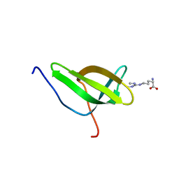
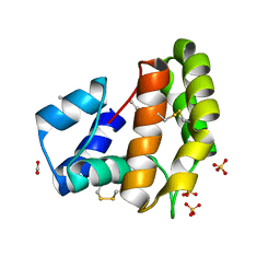
2QO5
 | | Crystal structure of the cysteine 91 threonine mutant of zebrafish liver bile acid-binding protein complexed with cholic acid | | 分子名称: | CHOLIC ACID, Liver-basic fatty acid binding protein | | 著者 | Capaldi, S, Saccomani, G, Perduca, M, Monaco, H.L. | | 登録日 | 2007-07-20 | | 公開日 | 2007-07-31 | | 最終更新日 | 2023-08-30 | | 実験手法 | X-RAY DIFFRACTION (1.5 Å) | | 主引用文献 | A Single Amino Acid Mutation in Zebrafish (Danio rerio) Liver Bile Acid-binding Protein Can Change the Stoichiometry of Ligand Binding.
J.Biol.Chem., 282, 2007
|
|
2QO6
 | | Crystal structure of the glycine 55 arginine mutant of zebrafish liver bile acid-binding protein complexed with cholic acid | | 分子名称: | CHOLIC ACID, GLYCEROL, ISOPROPYL ALCOHOL, ... | | 著者 | Capaldi, S, Saccomani, G, Perduca, M, Monaco, H.L. | | 登録日 | 2007-07-20 | | 公開日 | 2007-07-31 | | 最終更新日 | 2023-08-30 | | 実験手法 | X-RAY DIFFRACTION (1.9 Å) | | 主引用文献 | A Single Amino Acid Mutation in Zebrafish (Danio rerio) Liver Bile Acid-binding Protein Can Change the Stoichiometry of Ligand Binding.
J.Biol.Chem., 282, 2007
|
|
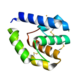
2QO4
 | | Crystal structure of zebrafish liver bile acid-binding protein complexed with cholic acid | | 分子名称: | CHOLIC ACID, GLYCEROL, ISOPROPYL ALCOHOL, ... | | 著者 | Capaldi, S, Saccomani, G, Perduca, M, Monaco, H.L. | | 登録日 | 2007-07-20 | | 公開日 | 2007-07-31 | | 最終更新日 | 2023-08-30 | | 実験手法 | X-RAY DIFFRACTION (1.5 Å) | | 主引用文献 | A Single Amino Acid Mutation in Zebrafish (Danio rerio) Liver Bile Acid-binding Protein Can Change the Stoichiometry of Ligand Binding.
J.Biol.Chem., 282, 2007
|
|


