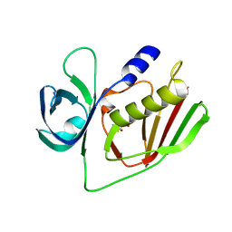
4O1N
 
 | |
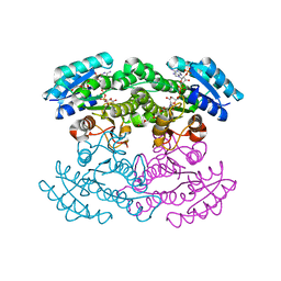
8G4L
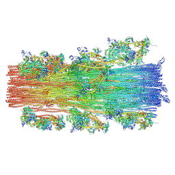 | | Cryo-EM structure of the human cardiac myosin filament | | 分子名称: | Myosin light chain 3, Myosin regulatory light chain 2, ventricular/cardiac muscle isoform, ... | | 著者 | Dutta, D, Nguyen, V, Padron, R, Craig, R. | | 登録日 | 2023-02-10 | | 公開日 | 2023-11-01 | | 最終更新日 | 2023-12-06 | | 実験手法 | ELECTRON MICROSCOPY (6.4 Å) | | 主引用文献 | Cryo-EM structure of the human cardiac myosin filament.
Nature, 623, 2023
|
|
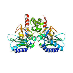
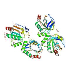
3Q6I
 | | Crystal structure of FabG4 and coenzyme binary complex | | 分子名称: | (2S,5R,8R,11S,14S,17S,21R)-5,8,11,14,17-PENTAMETHYL-4,7,10,13,16,19-HEXAOXADOCOSANE-2,21-DIOL, 3-oxoacyl-(Acyl-carrier-protein) reductase, NICOTINAMIDE-ADENINE-DINUCLEOTIDE | | 著者 | Dutta, D, Bhattacharyya, S, Das, A.K. | | 登録日 | 2011-01-01 | | 公開日 | 2012-01-11 | | 最終更新日 | 2023-11-01 | | 実験手法 | X-RAY DIFFRACTION (2.59 Å) | | 主引用文献 | Crystal structure of holoFabG4
To be Published
|
|
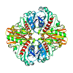
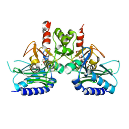
3M1L
 | | Crystal structure of a C-terminal trunacted mutant of a putative ketoacyl reductase (FabG4) from Mycobacterium tuberculosis H37Rv at 2.5 Angstrom resolution | | 分子名称: | 3-oxoacyl-(Acyl-carrier-protein) reductase, ACETATE ION | | 著者 | Dutta, D, Bhattacharyya, S, Saha, B, Das, A.K. | | 登録日 | 2010-03-05 | | 公開日 | 2010-12-22 | | 最終更新日 | 2024-03-20 | | 実験手法 | X-RAY DIFFRACTION (2.52 Å) | | 主引用文献 | Crystal structure of FabG4 from Mycobacterium tuberculosis reveals the importance of C-terminal residues in ketoreductase activity
J.Struct.Biol., 174, 2011
|
|
3SJ7
 | |
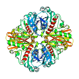
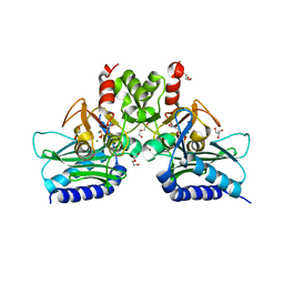
3V1U
 | | Crystal structure of a beta-ketoacyl reductase FabG4 from Mycobacterium tuberculosis H37Rv complexed with NAD+ and Hexanoyl-CoA at 2.5 Angstrom resolution | | 分子名称: | (2S,5R,8R,11S,14S,17S,21R)-5,8,11,14,17-PENTAMETHYL-4,7,10,13,16,19-HEXAOXADOCOSANE-2,21-DIOL, 3-oxoacyl-(Acyl-carrier-protein) reductase, HEXANOYL-COENZYME A, ... | | 著者 | Dutta, D, Bhattacharyya, S, Das, A.K. | | 登録日 | 2011-12-10 | | 公開日 | 2012-11-28 | | 最終更新日 | 2023-11-08 | | 実験手法 | X-RAY DIFFRACTION (2.5 Å) | | 主引用文献 | Crystal structure of hexanoyl-CoA bound to beta-ketoacyl reductase FabG4 of Mycobacterium tuberculosis
Biochem.J., 450, 2013
|
|
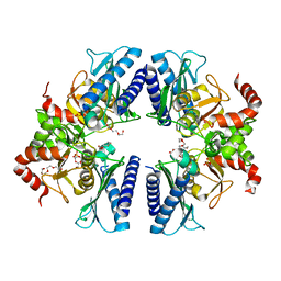
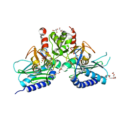
4FW8
 | | Crystal structure of FABG4 complexed with Coenzyme NADH | | 分子名称: | (2S,5R,8R,11S,14S,17S,21R)-5,8,11,14,17-PENTAMETHYL-4,7,10,13,16,19-HEXAOXADOCOSANE-2,21-DIOL, 1,4-DIHYDRONICOTINAMIDE ADENINE DINUCLEOTIDE, 3-oxoacyl-(Acyl-carrier-protein) reductase | | 著者 | Dutta, D, Bhattacharyya, S, Das, A.K. | | 登録日 | 2012-06-30 | | 公開日 | 2012-11-28 | | 最終更新日 | 2024-03-20 | | 実験手法 | X-RAY DIFFRACTION (2.79 Å) | | 主引用文献 | Crystal structure of hexanoyl-CoA bound to beta-ketoacyl reductase FabG4 of Mycobacterium tuberculosis
Biochem.J., 450, 2013
|
|
3V1T
 | |
5DAZ
 | |
5DZQ
 | | C3larvin toxin, an ADP-ribosyltransferase from Paenibacillus larvae, Orthorhombic Form | | 分子名称: | GLYCEROL, MAGNESIUM ION, Toxin-like protein | | 著者 | Dutta, D, Ravulapalli, R, Merrill, A.R. | | 登録日 | 2015-09-26 | | 公開日 | 2015-10-14 | | 最終更新日 | 2023-09-27 | | 実験手法 | X-RAY DIFFRACTION (1.892 Å) | | 主引用文献 | Structural variability of C3larvin toxin. Intrinsic dynamics of the alpha / beta fold of the C3-like group of mono-ADP-ribosyltransferase toxins.
J. Biomol. Struct. Dyn., 34, 2016
|
|
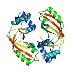
3NV9
 | | Crystal structure of Entamoeba histolytica Malic Enzyme | | 分子名称: | ACETATE ION, GLYCEROL, Malic enzyme, ... | | 著者 | Dutta, D, Chakrabarty, A, Ghosh, S.K, Das, A.K. | | 登録日 | 2010-07-08 | | 公開日 | 2011-07-20 | | 最終更新日 | 2024-03-20 | | 実験手法 | X-RAY DIFFRACTION (2.25 Å) | | 主引用文献 | Structural and functional characterization of Entamoeba histolytica Malic enzyme
To be Published
|
|
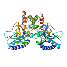
4RV2
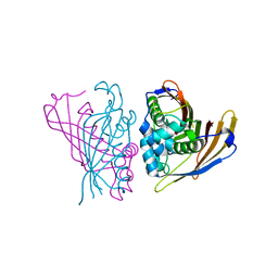 | | Crystal Structure of (3R)-hydroxyacyl-ACP dehydratase HadAB hetero-dimer from Mycobacterium smegmatis | | 分子名称: | MaoC family protein, SULFATE ION, UPF0336 protein MSMEG_1340/MSMEI_1302 | | 著者 | Biswas, R, Hazra, D, Dutta, D, Das, A.K. | | 登録日 | 2014-11-24 | | 公開日 | 2015-02-11 | | 最終更新日 | 2024-02-28 | | 実験手法 | X-RAY DIFFRACTION (2.7 Å) | | 主引用文献 | Crystal structure of dehydratase component HadAB complex of mycobacterial FAS-II pathway.
Biochem.Biophys.Res.Commun., 458, 2015
|
|
5I3S
 | |
5F24
 | | Crystal structure of dual specific IMPase/NADP phosphatase bound with D-inositol-1-phosphate | | 分子名称: | CALCIUM ION, CHLORIDE ION, D-MYO-INOSITOL-1-PHOSPHATE, ... | | 著者 | Bhattacharyya, S, Dutta, D, Ghosh, A.K, Das, A.K. | | 登録日 | 2015-12-01 | | 公開日 | 2015-12-23 | | 最終更新日 | 2023-11-08 | | 実験手法 | X-RAY DIFFRACTION (2.5 Å) | | 主引用文献 | Structural elucidation of the NADP(H) phosphatase activity of staphylococcal dual-specific IMPase/NADP(H) phosphatase
Acta Crystallogr D Struct Biol, 72, 2016
|
|
5EYH
 | | Crystal Structure of IMPase/NADP phosphatase complexed with NADP and Ca2+ at pH 7.0 | | 分子名称: | CALCIUM ION, GLYCEROL, Inositol monophosphatase, ... | | 著者 | Bhattacharyya, S, Dutta, D, Ghosh, A.K, Das, A.K. | | 登録日 | 2015-11-25 | | 公開日 | 2015-12-23 | | 最終更新日 | 2024-03-20 | | 実験手法 | X-RAY DIFFRACTION (2.5 Å) | | 主引用文献 | Structural elucidation of the NADP(H) phosphatase activity of staphylococcal dual-specific IMPase/NADP(H) phosphatase
Acta Crystallogr D Struct Biol, 72, 2016
|
|
5EYG
 | | Crystal structure of IMPase/NADP phosphatase complexed with NADP and Ca2+ | | 分子名称: | 2-(2-METHOXYETHOXY)ETHANOL, CALCIUM ION, CHLORIDE ION, ... | | 著者 | Bhattacharyya, S, Dutta, D, Ghosh, A.K, Das, A.K. | | 登録日 | 2015-11-25 | | 公開日 | 2015-12-23 | | 最終更新日 | 2023-11-08 | | 実験手法 | X-RAY DIFFRACTION (2.2 Å) | | 主引用文献 | Structural elucidation of the NADP(H) phosphatase activity of staphylococcal dual-specific IMPase/NADP(H) phosphatase
Acta Crystallogr D Struct Biol, 72, 2016
|
|
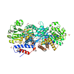
3HQ4
 | | Crystal Structure of C151S mutant of Glyceraldehyde-3-phosphate dehydrogenase 1 (GAPDH1) complexed with NAD from Staphylococcus aureus MRSA252 at 2.2 angstrom resolution | | 分子名称: | Glyceraldehyde-3-phosphate dehydrogenase 1, NICOTINAMIDE-ADENINE-DINUCLEOTIDE | | 著者 | Mukherjee, S, Dutta, D, Saha, B, Das, A.K. | | 登録日 | 2009-06-05 | | 公開日 | 2010-06-23 | | 最終更新日 | 2023-11-01 | | 実験手法 | X-RAY DIFFRACTION (2.2 Å) | | 主引用文献 | Crystal structure of glyceraldehyde-3-phosphate dehydrogenase 1 from methicillin-resistant Staphylococcus aureus MRSA252 provides novel insights into substrate binding and catalytic mechanism.
J.Mol.Biol., 401, 2010
|
|
3K9Q
 | | Crystal structure of C151G mutant of Glyceraldehyde 3-phosphate dehydrogenase 1 from Methicillin resistant Staphylococcus aureus (MRSA252) at 2.5 angstrom resolution | | 分子名称: | CHLORIDE ION, GLYCEROL, Glyceraldehyde-3-phosphate dehydrogenase 1, ... | | 著者 | Mukherjee, S, Dutta, D, Saha, B, Das, A.K. | | 登録日 | 2009-10-16 | | 公開日 | 2010-08-18 | | 最終更新日 | 2023-11-01 | | 実験手法 | X-RAY DIFFRACTION (2.5 Å) | | 主引用文献 | Crystal structure of glyceraldehyde-3-phosphate dehydrogenase 1 from methicillin-resistant Staphylococcus aureus MRSA252 provides novel insights into substrate binding and catalytic mechanism.
J.Mol.Biol., 401, 2010
|
|
3T0J
 | |
4PTK
 | | Crystal structure of Staphylococcal IMPase-I complex with 3Mg2+ and Phosphate | | 分子名称: | GLYCEROL, Inositol monophosphatase family protein, MAGNESIUM ION, ... | | 著者 | Dutta, A, Bhattacharyya, S, Dutta, D, Das, A.K. | | 登録日 | 2014-03-11 | | 公開日 | 2014-10-22 | | 最終更新日 | 2023-11-08 | | 実験手法 | X-RAY DIFFRACTION (2.503 Å) | | 主引用文献 | Structural elucidation of the binding site and mode of inhibition of Li(+) and Mg(2+) in inositol monophosphatase.
Febs J., 281, 2014
|
|
4I40
 | | crystal structure of Staphylococcal inositol monophosphatase-1: 50mM LiCl inhibited complex | | 分子名称: | GLYCEROL, Inositol monophosphatase family protein, MAGNESIUM ION, ... | | 著者 | Dutta, A, Bhattacharyya, S, Dutta, D, Das, A.K. | | 登録日 | 2012-11-27 | | 公開日 | 2013-11-27 | | 最終更新日 | 2023-11-08 | | 実験手法 | X-RAY DIFFRACTION (2.5 Å) | | 主引用文献 | Structural elucidation of the binding site and mode of inhibition of Li(+) and Mg(2+) in inositol monophosphatase.
Febs J., 281, 2014
|
|
4I3E
 | | Crystal structure of Staphylococcal IMPase - I complexed with products. | | 分子名称: | GLYCEROL, Inositol monophosphatase family protein, MAGNESIUM ION, ... | | 著者 | Bhattacharyya, S, Dutta, A, Dutta, D, Das, A.K. | | 登録日 | 2012-11-26 | | 公開日 | 2013-12-18 | | 最終更新日 | 2024-03-20 | | 実験手法 | X-RAY DIFFRACTION (2.6 Å) | | 主引用文献 | Crystal structure of Staphylococcal IMPase - I complexed with products.
To be Published
|
|
4I3Y
 | | Crystal structure of Staphylococcal inositol monophosphatase-1: 100 mM LiCl soaked inhibitory complex | | 分子名称: | GLYCEROL, Inositol monophosphatase family protein, MAGNESIUM ION, ... | | 著者 | Dutta, A, Bhattacharyya, S, Dutta, D, Das, A.K. | | 登録日 | 2012-11-26 | | 公開日 | 2013-11-27 | | 最終更新日 | 2023-11-08 | | 実験手法 | X-RAY DIFFRACTION (2.04 Å) | | 主引用文献 | Structural elucidation of the binding site and mode of inhibition of Li(+) and Mg(2+) in inositol monophosphatase.
Febs J., 281, 2014
|
|
4G61
 | | Crystal structure of IMPase/NADP phosphatase complexed with Mg2+ and phosphate | | 分子名称: | 1-HYDROXYSULFANYL-4-MERCAPTO-BUTANE-2,3-DIOL, 3,6,9,12,15,18-HEXAOXAICOSANE-1,20-DIOL, CHLORIDE ION, ... | | 著者 | Bhattacharyya, S, Dutta, D, Ghosh, A.K, Das, A.K. | | 登録日 | 2012-07-18 | | 公開日 | 2013-07-24 | | 最終更新日 | 2024-05-29 | | 実験手法 | X-RAY DIFFRACTION (2.3 Å) | | 主引用文献 | Structural elucidation of the binding site and mode of inhibition of Li(+) and Mg(2+) in inositol monophosphatase.
Febs J., 281, 2014
|
|
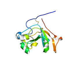
3P7X
 | | Crystal structure of an atypical two-cysteine peroxiredoxin (SAOUHSC_01822) from Staphylococcus aureus NCTC8325 | | 分子名称: | (2R,3S)-1,4-DIMERCAPTOBUTANE-2,3-DIOL, (2S,3S)-1,4-DIMERCAPTOBUTANE-2,3-DIOL, Probable thiol peroxidase, ... | | 著者 | Bhattacharyya, S, Dutta, D, Ghosh, A.K, Das, A.K. | | 登録日 | 2010-10-13 | | 公開日 | 2011-10-19 | | 最終更新日 | 2023-11-01 | | 実験手法 | X-RAY DIFFRACTION (1.96 Å) | | 主引用文献 | Crystal structure of an atypical two-cysteine peroxiredoxin (SAOUHSC_01822) from Staphylococcus aureus NCTC8325
To be Published
|
|


