1L5X
 
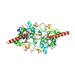 | | The 2.0-Angstrom resolution crystal structure of a survival protein E (SurE) homolog from Pyrobaculum aerophilum | | 分子名称: | ACETIC ACID, GLYCEROL, Survival protein E | | 著者 | Mura, C, Katz, J.E, Clarke, S.G, Eisenberg, D. | | 登録日 | 2002-03-08 | | 公開日 | 2003-02-25 | | 最終更新日 | 2011-07-13 | | 実験手法 | X-RAY DIFFRACTION (2 Å) | | 主引用文献 | Structure and Function of an Archaeal Homolog of Survival
Protein E (SurE-alpha): An Acid Phosphatase with Purine
Nucleotide Specificity
J.Mol.Biol., 326, 2003
|
|
6NB9
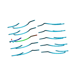 | | Amyloid-Beta (20-34) with L-isoaspartate 23 | | 分子名称: | Amyloid-beta A4 protein | | 著者 | Sawaya, M.R, Warmack, R.A, Boyer, D.R, Zee, C.T, Richards, L.S, Cascio, D, Gonen, T, Clarke, S.G, Eisenberg, D.S. | | 登録日 | 2018-12-06 | | 公開日 | 2019-08-07 | | 最終更新日 | 2022-09-07 | | 実験手法 | ELECTRON CRYSTALLOGRAPHY (1.05 Å) | | 主引用文献 | Structure of amyloid-beta (20-34) with Alzheimer's-associated isomerization at Asp23 reveals a distinct protofilament interface.
Nat Commun, 10, 2019
|
|
6OIZ
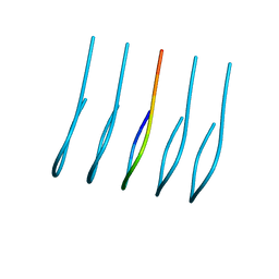 | | Amyloid-Beta (20-34) wild type | | 分子名称: | Amyloid beta A4 protein | | 著者 | Sawaya, M.R, Warmack, R.A, Zee, C.T, Gonen, T, Clarke, S.G, Eisenberg, D.S. | | 登録日 | 2019-04-10 | | 公開日 | 2019-08-07 | | 最終更新日 | 2024-05-15 | | 実験手法 | ELECTRON CRYSTALLOGRAPHY (1.1 Å) | | 主引用文献 | Structure of amyloid-beta (20-34) with Alzheimer's-associated isomerization at Asp23 reveals a distinct protofilament interface.
Nat Commun, 10, 2019
|
|
5EKU
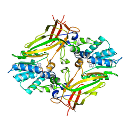 | |
6UMQ
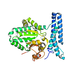 | | Structure of DUF89 | | 分子名称: | Damage-control phosphatase DUF89, MAGNESIUM ION | | 著者 | Perry, J.J, Kenjic, N, Dennis, T.N. | | 登録日 | 2019-10-10 | | 公開日 | 2020-07-29 | | 最終更新日 | 2023-10-11 | | 実験手法 | X-RAY DIFFRACTION (1.85 Å) | | 主引用文献 | Human ARMT1 structure and substrate specificity indicates that it is a DUF89 family damage-control phosphatase.
J.Struct.Biol., 212, 2020
|
|
6UMR
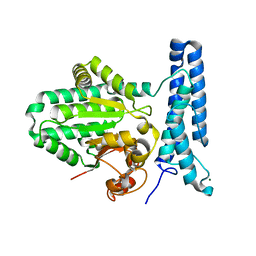 | | Structure of DUF89 - D291A mutant | | 分子名称: | Damage-control phosphatase DUF89, MAGNESIUM ION | | 著者 | Perry, J.J, Kenjic, N, Dennis, T.N. | | 登録日 | 2019-10-10 | | 公開日 | 2020-07-29 | | 最終更新日 | 2023-10-11 | | 実験手法 | X-RAY DIFFRACTION (2.21 Å) | | 主引用文献 | Human ARMT1 structure and substrate specificity indicates that it is a DUF89 family damage-control phosphatase.
J.Struct.Biol., 212, 2020
|
|
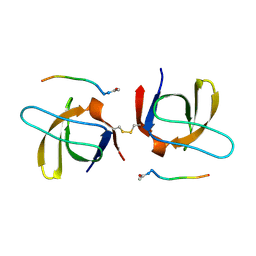
1SEM
 | |


