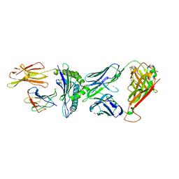
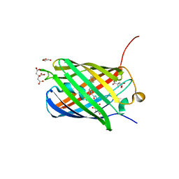
1XMZ
 
 | | Crystal structure of the dark state of kindling fluorescent protein kfp from anemonia sulcata | | 分子名称: | BETA-MERCAPTOETHANOL, GFP-like non-fluorescent chromoprotein FP595 chain 1, GFP-like non-fluorescent chromoprotein FP595 chain 2 | | 著者 | Quillin, M.L, Anstrom, D.M, Shu, X, O'Leary, S, Kallio, K, Chudakov, D.M, Remington, S.J. | | 登録日 | 2004-10-04 | | 公開日 | 2005-04-19 | | 最終更新日 | 2024-07-10 | | 実験手法 | X-RAY DIFFRACTION (1.38 Å) | | 主引用文献 | Kindling Fluorescent Protein from Anemonia sulcata: Dark-State Structure at 1.38 Resolution
Biochemistry, 44, 2005
|
|
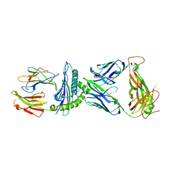
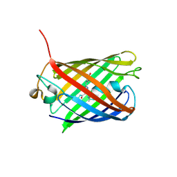
3O77
 | | The structure of Ca2+ Sensor (Case-16) | | 分子名称: | CALCIUM ION, CHLORIDE ION, Myosin light chain kinase, ... | | 著者 | Leder, L, Stark, W, Freuler, F, Marsh, M, Meyerhofer, M, Stettler, T, Mayr, L.M, Britanova, O.V, Strukova, L.A, Chudakov, D.M. | | 登録日 | 2010-07-30 | | 公開日 | 2010-09-29 | | 最終更新日 | 2024-11-20 | | 実験手法 | X-RAY DIFFRACTION (2.35 Å) | | 主引用文献 | The structure of Ca2+ sensor Case16 reveals the mechanism of reaction to low Ca2+ concentrations
Sensors (Basel), 10, 2010
|
|
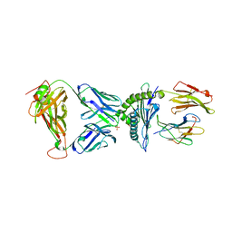
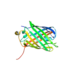
3O78
 | | The structure of Ca2+ Sensor (Case-12) | | 分子名称: | CALCIUM ION, Myosin light chain kinase, smooth muscle,Green fluorescent protein,Green fluorescent protein,Calmodulin-1 | | 著者 | Leder, L, Stark, W, Freuler, F, Marsh, M, Meyerhofer, M, Stettler, T, Mayr, L.M, Britanova, O.V, Strukova, L.A, Chudakov, D.M. | | 登録日 | 2010-07-30 | | 公開日 | 2010-09-29 | | 最終更新日 | 2024-11-06 | | 実験手法 | X-RAY DIFFRACTION (2.6 Å) | | 主引用文献 | The structure of Ca2+ sensor Case16 reveals the mechanism of reaction to low Ca2+ concentrations
Sensors (Basel), 10, 2010
|
|
2G6X
 | |
2G6Y
 | |
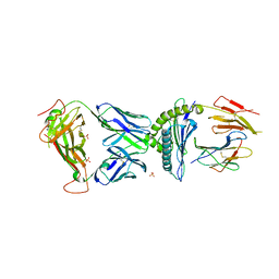
8CX4
 | | TCR-antigen complex AS8.4-YEIH-HLA*B27 | | 分子名称: | AS8.4a, AS8.4b, Beta-2-microglobulin, ... | | 著者 | Yang, X, Jude, K.M, Garcia, K.C. | | 登録日 | 2022-05-19 | | 公開日 | 2022-12-07 | | 最終更新日 | 2024-11-06 | | 実験手法 | X-RAY DIFFRACTION (2.2 Å) | | 主引用文献 | Autoimmunity-associated T cell receptors recognize HLA-B*27-bound peptides.
Nature, 612, 2022
|
|
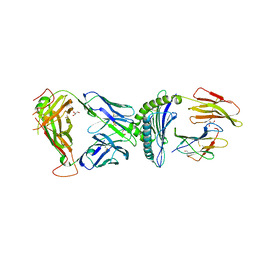
7N2O
 | | AS4.2-YEIH-HLA*B27 | | 分子名称: | 2-acetamido-2-deoxy-beta-D-glucopyranose, Beta-2-microglobulin, GLYCEROL, ... | | 著者 | Yang, X, Jude, K.M, Garcia, K.C. | | 登録日 | 2021-05-29 | | 公開日 | 2022-12-07 | | 最終更新日 | 2024-11-06 | | 実験手法 | X-RAY DIFFRACTION (2.3 Å) | | 主引用文献 | Autoimmunity-associated T cell receptors recognize HLA-B*27-bound peptides.
Nature, 612, 2022
|
|
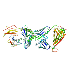
7N2Q
 | | AS4.3-YEIH-HLA*B27 | | 分子名称: | 2-acetamido-2-deoxy-beta-D-glucopyranose, AS4.3 T cell receptor alpha chain, AS4.3 T cell receptor beta chain, ... | | 著者 | Yang, X, Jude, K.M, Garcia, K.C. | | 登録日 | 2021-05-29 | | 公開日 | 2022-12-07 | | 最終更新日 | 2024-11-20 | | 実験手法 | X-RAY DIFFRACTION (2.7 Å) | | 主引用文献 | Autoimmunity-associated T cell receptors recognize HLA-B*27-bound peptides.
Nature, 612, 2022
|
|
7N2S
 | | AS3.1-PRPF3-HLA*B27 | | 分子名称: | 2-acetamido-2-deoxy-beta-D-glucopyranose, Beta-2-microglobulin, GLYCEROL, ... | | 著者 | Yang, X, Jude, K.M, Garcia, K.C. | | 登録日 | 2021-05-29 | | 公開日 | 2022-12-07 | | 最終更新日 | 2024-11-13 | | 実験手法 | X-RAY DIFFRACTION (2.37 Å) | | 主引用文献 | Autoimmunity-associated T cell receptors recognize HLA-B*27-bound peptides.
Nature, 612, 2022
|
|
7N2R
 | | AS4.3-PRPF3-HLA*B27 | | 分子名称: | 1,2-ETHANEDIOL, 2-acetamido-2-deoxy-beta-D-glucopyranose, AS4.3 T cell receptor alpha chain, ... | | 著者 | Yang, X, Jude, K.M, Garcia, K.C. | | 登録日 | 2021-05-29 | | 公開日 | 2022-12-07 | | 最終更新日 | 2024-11-13 | | 実験手法 | X-RAY DIFFRACTION (2.28 Å) | | 主引用文献 | Autoimmunity-associated T cell receptors recognize HLA-B*27-bound peptides.
Nature, 612, 2022
|
|
7N2N
 | | TCR-antigen complex AS4.2-PRPF3-HLA*B27 | | 分子名称: | 2-acetamido-2-deoxy-beta-D-glucopyranose, Beta-2-microglobulin, GLYCEROL, ... | | 著者 | Yang, X, Jude, K.M, Garcia, K.C. | | 登録日 | 2021-05-29 | | 公開日 | 2022-12-07 | | 最終更新日 | 2024-10-23 | | 実験手法 | X-RAY DIFFRACTION (2.6 Å) | | 主引用文献 | Autoimmunity-associated T cell receptors recognize HLA-B*27-bound peptides.
Nature, 612, 2022
|
|
7N2P
 | | AS4.3-RNASEH2b-HLA*B27 | | 分子名称: | 2-acetamido-2-deoxy-beta-D-glucopyranose, AS4.3 T cell receptor alpha chain, AS4.3 T cell receptor beta chain, ... | | 著者 | Yang, X, Jude, K.M, Garcia, K.C. | | 登録日 | 2021-05-29 | | 公開日 | 2022-12-07 | | 最終更新日 | 2024-10-23 | | 実験手法 | X-RAY DIFFRACTION (2.5 Å) | | 主引用文献 | Autoimmunity-associated T cell receptors recognize HLA-B*27-bound peptides.
Nature, 612, 2022
|
|
6U1A
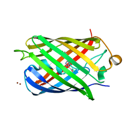 | | Crystal Structure of Fluorescent Protein FusionRed | | 分子名称: | CALCIUM ION, NICKEL (II) ION, Red fluorescent protein | | 著者 | Pletnev, S, Muslinkina, L, Pletneva, N, Pletnev, V.Z. | | 登録日 | 2019-08-15 | | 公開日 | 2020-04-22 | | 最終更新日 | 2023-11-15 | | 実験手法 | X-RAY DIFFRACTION (1.09 Å) | | 主引用文献 | Two independent routes of post-translational chemistry in fluorescent protein FusionRed.
Int.J.Biol.Macromol., 155, 2020
|
|
3BX9
 | |
3BXB
 | |
3BXC
 | |
3BXA
 | |
3PIB
 | |
3PJ7
 | |
3PJB
 | |
3PJ5
 | |
3RWA
 | |
3RWT
 | |
4ZBL
 | |
2OGR
 | |


