7P8V
 
 | |
7OU2
 | | The structure of MutS bound to two molecules of ADP | | 分子名称: | ADENOSINE-5'-DIPHOSPHATE, DNA mismatch repair protein MutS | | 著者 | Lamers, M.H, Borsellini, A, Friedhoff, P, Kunetsky, V. | | 登録日 | 2021-06-11 | | 公開日 | 2022-01-12 | | 最終更新日 | 2022-10-26 | | 実験手法 | ELECTRON MICROSCOPY (4.8 Å) | | 主引用文献 | Cryogenic electron microscopy structures reveal how ATP and DNA binding in MutS coordinates sequential steps of DNA mismatch repair.
Nat.Struct.Mol.Biol., 29, 2022
|
|
7OTO
 | | The structure of MutS bound to two molecules of AMPPNP | | 分子名称: | DNA mismatch repair protein MutS, MAGNESIUM ION, PHOSPHOAMINOPHOSPHONIC ACID-ADENYLATE ESTER | | 著者 | Lamers, M.H, Borsellini, A, Friedhoff, P, Kunetsky, V. | | 登録日 | 2021-06-10 | | 公開日 | 2022-01-12 | | 最終更新日 | 2022-10-26 | | 実験手法 | ELECTRON MICROSCOPY (3.4 Å) | | 主引用文献 | Cryogenic electron microscopy structures reveal how ATP and DNA binding in MutS coordinates sequential steps of DNA mismatch repair.
Nat.Struct.Mol.Biol., 29, 2022
|
|
7OU0
 | | The structure of MutS bound to two molecules of ADP-Vanadate | | 分子名称: | ADENOSINE-5'-DIPHOSPHATE, DNA mismatch repair protein MutS, MAGNESIUM ION, ... | | 著者 | Lamers, M.H, Borsellini, A, Friedhoff, P, Kunetsky, V. | | 登録日 | 2021-06-10 | | 公開日 | 2022-01-12 | | 最終更新日 | 2022-10-26 | | 実験手法 | ELECTRON MICROSCOPY (3.8 Å) | | 主引用文献 | Cryogenic electron microscopy structures reveal how ATP and DNA binding in MutS coordinates sequential steps of DNA mismatch repair.
Nat.Struct.Mol.Biol., 29, 2022
|
|
7OU4
 | | The structure of MutS bound to one molecule of ATP and one molecule of ADP | | 分子名称: | ADENOSINE-5'-DIPHOSPHATE, ADENOSINE-5'-TRIPHOSPHATE, DNA mismatch repair protein MutS, ... | | 著者 | Lamers, M.H, Borsellini, A, Friedhoff, P, Kunetsky, V. | | 登録日 | 2021-06-11 | | 公開日 | 2022-01-12 | | 最終更新日 | 2022-10-26 | | 実験手法 | ELECTRON MICROSCOPY (3.2 Å) | | 主引用文献 | Cryogenic electron microscopy structures reveal how ATP and DNA binding in MutS coordinates sequential steps of DNA mismatch repair.
Nat.Struct.Mol.Biol., 29, 2022
|
|
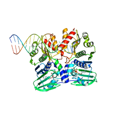
7PU7
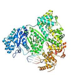 | | DNA polymerase from M. tuberculosis | | 分子名称: | DNA polymerase III subunit alpha, Template, ZINC ION, ... | | 著者 | Borsellini, A, Lamers, M.H. | | 登録日 | 2021-09-28 | | 公開日 | 2022-02-23 | | 最終更新日 | 2022-04-13 | | 実験手法 | ELECTRON MICROSCOPY (2.9 Å) | | 主引用文献 | DNA-Dependent Binding of Nargenicin to DnaE1 Inhibits Replication in Mycobacterium tuberculosis.
Acs Infect Dis., 8, 2022
|
|
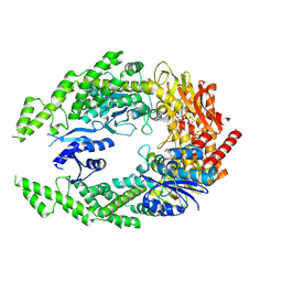
8OOY
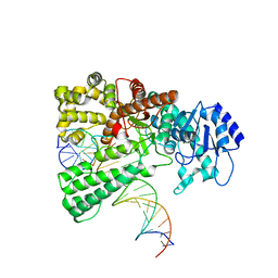 | | Pol I bound to extended and displaced DNA section - open conformation | | 分子名称: | DNA polymerase I, Displacing Primer, Extending Primer, ... | | 著者 | Botto, M, Borsellini, A, Lamers, M.H. | | 登録日 | 2023-04-06 | | 公開日 | 2023-08-09 | | 最終更新日 | 2023-11-01 | | 実験手法 | ELECTRON MICROSCOPY (4 Å) | | 主引用文献 | A four-point molecular handover during Okazaki maturation.
Nat.Struct.Mol.Biol., 30, 2023
|
|
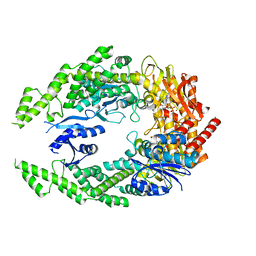
8OO6
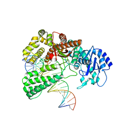 | | Pol I bound to extended and displaced DNA section - closed conformation | | 分子名称: | DNA polymerase I, Displaced primer, Extending Primer, ... | | 著者 | Botto, M, Borsellini, A, Lamers, M.H. | | 登録日 | 2023-04-04 | | 公開日 | 2023-08-09 | | 最終更新日 | 2023-11-01 | | 実験手法 | ELECTRON MICROSCOPY (4.3 Å) | | 主引用文献 | A four-point molecular handover during Okazaki maturation.
Nat.Struct.Mol.Biol., 30, 2023
|
|


