3T1U
 
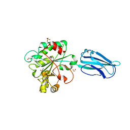
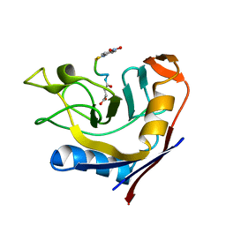 | | Crystal Structure of the complex of Cyclophilin-A enzyme from Azotobacter vinelandii with sucAFPFpNA peptide | | 分子名称: | Peptidyl-prolyl cis-trans isomerase, succinyl-Ala-Phe-Pro-Phe-p-nitroanilide | | 著者 | Karpusas, M, Christoforides, E, Bethanis, K, Dimou, M, Katinakis, P. | | 登録日 | 2011-07-22 | | 公開日 | 2012-03-07 | | 最終更新日 | 2023-09-13 | | 実験手法 | X-RAY DIFFRACTION (2 Å) | | 主引用文献 | Structure of a bacterial cytoplasmic cyclophilin A in complex with a tetrapeptide.
Acta Crystallogr.,Sect.F, 68, 2012
|
|
4HJ2
 | |
6TVL
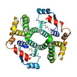
 | | Hen Egg White Lysozyme in complex with a "half sandwich"-type Ru(II) coordination compound | | 分子名称: | CHLORIDE ION, Lysozyme C, SODIUM ION, ... | | 著者 | Chiniadis, L, Giastas, P, Bratsos, I, Papakyriakou, A. | | 登録日 | 2020-01-10 | | 公開日 | 2020-04-29 | | 最終更新日 | 2024-01-24 | | 実験手法 | X-RAY DIFFRACTION (1.395 Å) | | 主引用文献 | High-resolution crystal structures of a "half sandwich"-type Ru(II) coordination compound bound to hen egg-white lysozyme and proteinase K.
J.Biol.Inorg.Chem., 25, 2020
|
|
6TXG
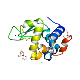
 | | Proteinase K in complex with a "half sandwich"-type Ru(II) coordination compound | | 分子名称: | 1,2-ETHANEDIOL, CALCIUM ION, NITRATE ION, ... | | 著者 | Chiniadis, L, Giastas, P, Bratsos, I, Papakyriakou, A. | | 登録日 | 2020-01-14 | | 公開日 | 2020-04-29 | | 最終更新日 | 2024-01-24 | | 実験手法 | X-RAY DIFFRACTION (1.372 Å) | | 主引用文献 | High-resolution crystal structures of a "half sandwich"-type Ru(II) coordination compound bound to hen egg-white lysozyme and proteinase K.
J.Biol.Inorg.Chem., 25, 2020
|
|
6GO1
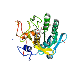
 | | Crystal Structure of a Bacillus anthracis peptidoglycan deacetylase | | 分子名称: | 1,2-ETHANEDIOL, ACETATE ION, Polysaccharide deacetylase-like protein, ... | | 著者 | Giastas, P, Andreou, A, Eliopoulos, E.E. | | 登録日 | 2018-06-01 | | 公開日 | 2019-04-10 | | 最終更新日 | 2024-01-17 | | 実験手法 | X-RAY DIFFRACTION (2.59 Å) | | 主引用文献 | The putative polysaccharide deacetylase Ba0331: cloning, expression, crystallization and structure determination.
Acta Crystallogr.,Sect.F, 75, 2019
|
|


