2BOS
 
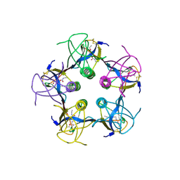
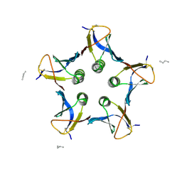 | | A MUTANT SHIGA-LIKE TOXIN IIE BOUND TO ITS RECEPTOR | | 分子名称: | N-BUTANE, PROTEIN (SHIGA-LIKE TOXIN IIE B SUBUNIT), alpha-D-galactopyranose-(1-4)-beta-D-galactopyranose, ... | | 著者 | Ling, H, Boodhoo, A, Armstrong, G.D, Clark, C.G, Brunton, J.L, Read, R.J. | | 登録日 | 1998-10-20 | | 公開日 | 1999-10-20 | | 最終更新日 | 2024-10-16 | | 実験手法 | X-RAY DIFFRACTION (2 Å) | | 主引用文献 | A mutant Shiga-like toxin IIe bound to its receptor Gb(3): structure of a group II Shiga-like toxin with altered binding specificity.
Structure Fold.Des., 8, 2000
|
|
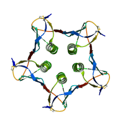
1BOS
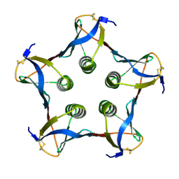
 | | SHIGA-LIKE TOXIN COMPLEXED WITH ITS RECEPTOR | | 分子名称: | SHIGA-LIKE TOXIN I B SUBUNIT, alpha-D-galactopyranose-(1-4)-beta-D-galactopyranose, alpha-D-galactopyranose-(1-4)-beta-D-galactopyranose-(1-4)-beta-D-glucopyranose, ... | | 著者 | Ling, H, Boodhoo, A, Hazes, B, Cummings, M.D, Armstrong, G.D, Brunton, J.L, Read, R.J. | | 登録日 | 1998-01-13 | | 公開日 | 1999-02-02 | | 最終更新日 | 2023-08-09 | | 実験手法 | X-RAY DIFFRACTION (2.8 Å) | | 主引用文献 | Structure of the shiga-like toxin I B-pentamer complexed with an analogue of its receptor Gb3.
Biochemistry, 37, 1998
|
|
1QOH
 | | A MUTANT SHIGA-LIKE TOXIN IIE | | 分子名称: | SHIGA-LIKE TOXIN IIE B SUBUNIT | | 著者 | Pannu, N.S, Boodhoo, A, Armstrong, G.D, Clark, C.G, Brunton, J.L, Read, R.J. | | 登録日 | 1999-11-08 | | 公開日 | 2000-07-03 | | 最終更新日 | 2023-12-13 | | 実験手法 | X-RAY DIFFRACTION (2.35 Å) | | 主引用文献 | A Mutant Shiga-Like Toxin Iie Bound to its Receptor Gb(3): Structure of a Group II Shiga-Like Toxin with Altered Binding Specificity
Structure, 8, 2000
|
|
1BCP
 | |
1PTO
 | |
1QNU
 | | Shiga-Like Toxin I B Subunit Complexed with the Bridged-Starfish Inhibitor | | 分子名称: | ETHYL-CARBAMIC ACID METHYL ESTER, METHYL-CARBAMIC ACID ETHYL ESTER, Shiga toxin 1 variant B subunit, ... | | 著者 | Pannu, N.S, Hayakawa, K, Read, R.J. | | 登録日 | 1999-10-21 | | 公開日 | 2000-04-11 | | 最終更新日 | 2023-12-13 | | 実験手法 | X-RAY DIFFRACTION (2.23 Å) | | 主引用文献 | Shiga-like toxins are neutralized by tailored multivalent carbohydrate ligands.
Nature, 403, 2000
|
|
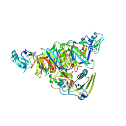
2GA4
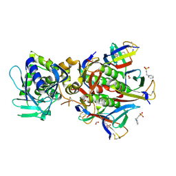 | | Stx2 with adenine | | 分子名称: | 1,2-ETHANEDIOL, 3-PYRIDINIUM-1-YLPROPANE-1-SULFONATE, ADENINE, ... | | 著者 | Fraser, M.E. | | 登録日 | 2006-03-07 | | 公開日 | 2006-07-11 | | 最終更新日 | 2017-10-18 | | 実験手法 | X-RAY DIFFRACTION (1.8 Å) | | 主引用文献 | Binding of adenine to Stx2, the protein toxin from Escherichia coli O157:H7.
Acta Crystallogr.,Sect.F, 62, 2006
|
|
1C48
 | | MUTATED SHIGA-LIKE TOXIN B SUBUNIT (G62T) | | 分子名称: | PROTEIN (SHIGA-LIKE TOXIN I B SUBUNIT) | | 著者 | Ling, H, Bast, D, Brunton, J.L, Read, R.J. | | 登録日 | 1999-08-11 | | 公開日 | 2000-08-16 | | 最終更新日 | 2023-08-09 | | 実験手法 | X-RAY DIFFRACTION (1.6 Å) | | 主引用文献 | Identification of the Primary Receptor Binding Site of Shiga-Like Toxin B Subunits: Structures of Mutated Shiga-Like Toxin I B-Pentamer with and without Bound Carbohydrate
To be Published
|
|
1C4Q
 | |
4OGM
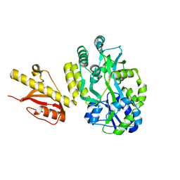 | | MBP-fusion protein of PilA1 residues 26-159 | | 分子名称: | Maltose ABC transporter periplasmic protein, pilin protein chimera, alpha-D-glucopyranose-(1-4)-alpha-D-glucopyranose | | 著者 | Piepenbrink, K.H, Sundberg, E.J. | | 登録日 | 2014-01-16 | | 公開日 | 2015-01-14 | | 最終更新日 | 2024-02-28 | | 実験手法 | X-RAY DIFFRACTION (2.234 Å) | | 主引用文献 | Structural and Evolutionary Analyses Show Unique Stabilization Strategies in the Type IV Pili of Clostridium difficile.
Structure, 23, 2015
|
|
4PE2
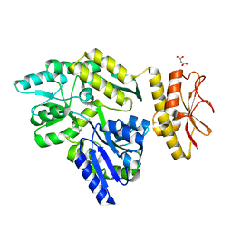 | | MBP PilA1 CD160 | | 分子名称: | MALONATE ION, Maltose ABC transporter periplasmic protein,Prepilin-type N-terminal cleavage/methylation domain protein, alpha-D-glucopyranose-(1-4)-alpha-D-glucopyranose | | 著者 | Piepenbrink, K.H, Sundberg, E.J. | | 登録日 | 2014-04-22 | | 公開日 | 2015-01-14 | | 最終更新日 | 2023-12-27 | | 実験手法 | X-RAY DIFFRACTION (1.724 Å) | | 主引用文献 | Structural and Evolutionary Analyses Show Unique Stabilization Strategies in the Type IV Pili of Clostridium difficile.
Structure, 23, 2015
|
|
4TSM
 | |
4IXJ
 | | The structure of PilJ, a Type IV pilin from Clostridium difficile | | 分子名称: | Fimbrial protein (Pilin), GLYCEROL, ZINC ION | | 著者 | Piepenbrink, K.H, Sundberg, E.J, Snyder, G.A. | | 登録日 | 2013-01-25 | | 公開日 | 2014-01-01 | | 最終更新日 | 2024-02-28 | | 実験手法 | X-RAY DIFFRACTION (1.983 Å) | | 主引用文献 | Structure of Clostridium difficile PilJ Exhibits Unprecedented Divergence from Known Type IV Pilins.
J.Biol.Chem., 289, 2014
|
|
1PRT
 | |


