4K02
 
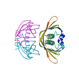 | | Crystal structure of AtDHNAT1, a 1,4-dihydroxy-2-naphthoyl-CoA thioesterase from Arabidopsis thaliana | | 分子名称: | 1,4-dihydroxy-2-naphthoyl-CoA thioesterase | | 著者 | Furt, F, Allen, W.J, Widhalm, J.R, Madzelan, P, Rizzo, R.C, Basset, G, Wilson, M.A. | | 登録日 | 2013-04-03 | | 公開日 | 2013-04-17 | | 最終更新日 | 2023-09-20 | | 実験手法 | X-RAY DIFFRACTION (1.9 Å) | | 主引用文献 | Functional convergence of structurally distinct thioesterases from cyanobacteria and plants involved in phylloquinone biosynthesis.
Acta Crystallogr.,Sect.D, 69, 2013
|
|
4K00
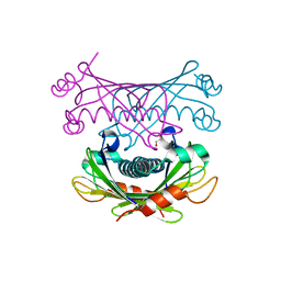 | | Crystal structure of Slr0204, a 1,4-dihydroxy-2-naphthoyl-CoA thioesterase from Synechocystis | | 分子名称: | 1,2-ETHANEDIOL, 1,4-dihydroxy-2-naphthoyl-CoA hydrolase | | 著者 | Furt, F, Allen, W.J, Widhalm, J.R, Madzelan, P, Rizzo, R.C, Basset, G, Wilson, M.A. | | 登録日 | 2013-04-03 | | 公開日 | 2013-04-17 | | 最終更新日 | 2023-09-20 | | 実験手法 | X-RAY DIFFRACTION (1.9 Å) | | 主引用文献 | Functional convergence of structurally distinct thioesterases from cyanobacteria and plants involved in phylloquinone biosynthesis.
Acta Crystallogr.,Sect.D, 69, 2013
|
|
4WGK
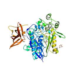 | | Crystal structure of human neutral ceramidase with Zn-bound phosphate | | 分子名称: | 1,2-ETHANEDIOL, 2-acetamido-2-deoxy-beta-D-glucopyranose, 2-acetamido-2-deoxy-beta-D-glucopyranose-(1-4)-2-acetamido-2-deoxy-beta-D-glucopyranose, ... | | 著者 | Airola, M.V, Pulkoski-Gross, M.J, Obeid, L.M, Hannun, Y.A. | | 登録日 | 2014-09-18 | | 公開日 | 2015-07-08 | | 最終更新日 | 2023-09-27 | | 実験手法 | X-RAY DIFFRACTION (2.582 Å) | | 主引用文献 | Structural Basis for Ceramide Recognition and Hydrolysis by Human Neutral Ceramidase.
Structure, 23, 2015
|
|
3OHN
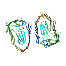 | |
3RFZ
 | | Crystal structure of the FimD usher bound to its cognate FimC:FimH substrate | | 分子名称: | Chaperone protein fimC, Outer membrane usher protein, type 1 fimbrial synthesis, ... | | 著者 | Phan, G, Remaut, H, Lebedev, A, Geibel, S, Waksman, G. | | 登録日 | 2011-04-07 | | 公開日 | 2011-06-01 | | 最終更新日 | 2012-03-28 | | 実験手法 | X-RAY DIFFRACTION (2.8 Å) | | 主引用文献 | Crystal structure of the FimD usher bound to its cognate FimC-FimH substrate.
Nature, 474, 2011
|
|


