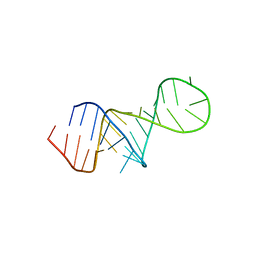
5UZF
 
 | | Insights into Watson-Crick/Hoogsteen Breathing Dynamics and Damage Repair from the Solution Structure and Dynamic Ensemble of DNA Duplexes containing m1A - A6-DNA structure | | 分子名称: | DNA (5'-D(*CP*GP*AP*TP*TP*TP*TP*TP*TP*GP*GP*C)-3'), DNA (5'-D(*GP*CP*CP*AP*AP*AP*AP*AP*AP*TP*CP*G)-3') | | 著者 | Sathyamoorthy, B, Shi, H, Xue, Y, Al-Hashimi, H.M. | | 登録日 | 2017-02-26 | | 公開日 | 2017-04-05 | | 最終更新日 | 2024-05-15 | | 実験手法 | SOLUTION NMR | | 主引用文献 | Insights into Watson-Crick/Hoogsteen breathing dynamics and damage repair from the solution structure and dynamic ensemble of DNA duplexes containing m1A.
Nucleic Acids Res., 45, 2017
|
|
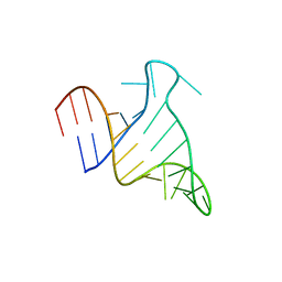
5UZD
 | | Insights into Watson-Crick/Hoogsteen Breathing Dynamics and Damage Repair from the Solution Structure and Dynamic Ensemble of DNA Duplexes containing m1A - A2-DNA structure | | 分子名称: | DNA (5'-D(*GP*CP*AP*TP*CP*GP*AP*TP*TP*GP*GP*C)-3'), DNA (5'-D(*GP*CP*CP*AP*AP*TP*CP*GP*AP*TP*GP*C)-3') | | 著者 | Sathyamoorthy, B, Shi, H, Xue, Y, Al-Hashimi, H.M. | | 登録日 | 2017-02-26 | | 公開日 | 2017-04-05 | | 最終更新日 | 2024-05-15 | | 実験手法 | SOLUTION NMR | | 主引用文献 | Insights into Watson-Crick/Hoogsteen breathing dynamics and damage repair from the solution structure and dynamic ensemble of DNA duplexes containing m1A.
Nucleic Acids Res., 45, 2017
|
|
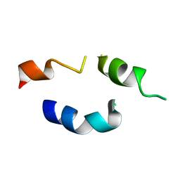
5UZI
 | | Insights into Watson-Crick/Hoogsteen Breathing Dynamics and Damage Repair from the Solution Structure and Dynamic Ensemble of DNA Duplexes containing m1A - A6-DNAm1A16 structure | | 分子名称: | DNA (5'-D(*CP*GP*AP*TP*TP*TP*TP*TP*TP*GP*GP*C)-3'), DNA (5'-D(*GP*CP*CP*(M1A)P*AP*AP*AP*AP*AP*TP*CP*G)-3') | | 著者 | Sathyamoorthy, B, Shi, H, Xue, Y, Al-Hashimi, H.M. | | 登録日 | 2017-02-26 | | 公開日 | 2017-04-05 | | 最終更新日 | 2024-05-15 | | 実験手法 | SOLUTION NMR | | 主引用文献 | Insights into Watson-Crick/Hoogsteen breathing dynamics and damage repair from the solution structure and dynamic ensemble of DNA duplexes containing m1A.
Nucleic Acids Res., 45, 2017
|
|
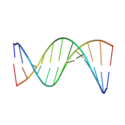
8THV
 | | FARFAR-NMR ensemble of HIV-1 TAR with apical loop capturing ground and excited conformational states | | 分子名称: | RNA (29-MER) | | 著者 | Roy, R, Geng, A, Shi, H, Merriman, D.K, Dethoff, E.A, Salmon, L, Al-Hashimi, H.M. | | 登録日 | 2023-07-18 | | 公開日 | 2023-08-02 | | 最終更新日 | 2024-05-15 | | 実験手法 | SOLUTION NMR | | 主引用文献 | Kinetic Resolution of the Atomic 3D Structures Formed by Ground and Excited Conformational States in an RNA Dynamic Ensemble.
J.Am.Chem.Soc., 145, 2023
|
|
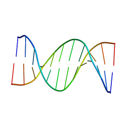
8U3M
 | | The FARFAR-MD-NMR ensemble of an HIV-1 TAR excited state | | 分子名称: | The excited state of HIV-1 transactivation response element (31-MER) | | 著者 | Geng, A, Ganser, L, Roy, R, Shi, H, Pratihar, S, Case, D.A, Al-Hashimi, H.M. | | 登録日 | 2023-09-07 | | 公開日 | 2023-10-04 | | 最終更新日 | 2024-05-15 | | 実験手法 | SOLUTION NMR | | 主引用文献 | An RNA excited conformational state at atomic resolution.
Nat Commun, 14, 2023
|
|
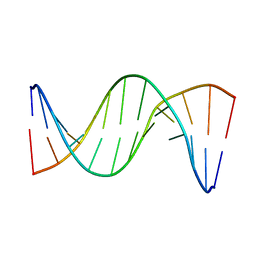
7JU1
 | | The FARFAR-NMR Ensemble of 29-mer HIV-1 Trans-activation Response Element RNA (N=20) | | 分子名称: | RNA (29-MER) | | 著者 | Shi, H, Rangadurai, A, Roy, R, Yesselman, J.D, Al-Hashimi, H.M. | | 登録日 | 2020-08-18 | | 公開日 | 2020-10-07 | | 最終更新日 | 2024-05-15 | | 実験手法 | SOLUTION NMR | | 主引用文献 | Rapid and accurate determination of atomistic RNA dynamic ensemble models using NMR and structure prediction
Nat Commun, 11, 2020
|
|
1FH1
 | |
6UEP
 | |
6UER
 | |
6UEQ
 | |
6M82
 | | Crystal structure of TylM1 Y14paF bound to SAH and dTDP-phenol | | 分子名称: | 1,2-ETHANEDIOL, 5'-O-[(S)-hydroxy{[(S)-hydroxy(phenoxy)phosphoryl]oxy}phosphoryl]thymidine, S-ADENOSYL-L-HOMOCYSTEINE, ... | | 著者 | Fick, R.J, McDole, B.G, Trievel, R.C. | | 登録日 | 2018-08-21 | | 公開日 | 2019-03-13 | | 最終更新日 | 2025-04-02 | | 実験手法 | X-RAY DIFFRACTION (1.3971 Å) | | 主引用文献 | Structural and Functional Characterization of Sulfonium Carbon-Oxygen Hydrogen Bonding in the Deoxyamino Sugar Methyltransferase TylM1.
Biochemistry, 58, 2019
|
|
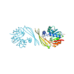
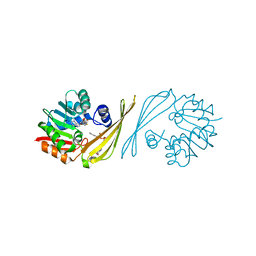
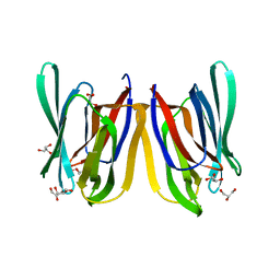
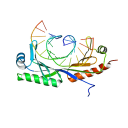
6NJQ
 | | Structure of TBP-Hoogsteen containing DNA complex | | 分子名称: | DNA (5'-D(*GP*CP*TP*AP*TP*AP*AP*AP*CP*GP*GP*GP*CP*A)-3'), DNA (5'-D(*TP*GP*CP*CP*CP*GP*TP*TP*TP*AP*TP*AP*GP*C)-3'), TATA-box-binding protein 1 | | 著者 | Schumacher, M.A, Stelling, A. | | 登録日 | 2019-01-04 | | 公開日 | 2019-10-30 | | 最終更新日 | 2023-10-11 | | 実験手法 | X-RAY DIFFRACTION (2.75 Å) | | 主引用文献 | Infrared Spectroscopic Observation of a G-C+Hoogsteen Base Pair in the DNA:TATA-Box Binding Protein Complex Under Solution Conditions.
Angew.Chem.Int.Ed.Engl., 58, 2019
|
|
6E4N
 | |
6E4O
 | |
6E4P
 | |
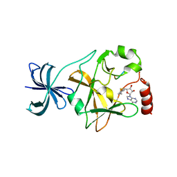
6M81
 | | Crystal structure of TylM1 Y14F bound to SAH and dTDP-phenol | | 分子名称: | 5'-O-[(S)-hydroxy{[(S)-hydroxy(phenoxy)phosphoryl]oxy}phosphoryl]thymidine, S-ADENOSYL-L-HOMOCYSTEINE, dTDP-3-amino-3,6-dideoxy-alpha-D-glucopyranose N,N-dimethyltransferase | | 著者 | Fick, R.J, McDole, B.G, Trievel, R.C. | | 登録日 | 2018-08-21 | | 公開日 | 2019-03-13 | | 最終更新日 | 2024-03-13 | | 実験手法 | X-RAY DIFFRACTION (1.782 Å) | | 主引用文献 | Structural and Functional Characterization of Sulfonium Carbon-Oxygen Hydrogen Bonding in the Deoxyamino Sugar Methyltransferase TylM1.
Biochemistry, 58, 2019
|
|
6M83
 | | Crystal structure of TylM1 S120A bound to SAH and dTDP-phenol | | 分子名称: | 5'-O-[(S)-hydroxy{[(S)-hydroxy(phenoxy)phosphoryl]oxy}phosphoryl]thymidine, CHLORIDE ION, S-ADENOSYL-L-HOMOCYSTEINE, ... | | 著者 | Fick, R.J, McDole, B.G, Trievel, R.C. | | 登録日 | 2018-08-21 | | 公開日 | 2019-03-13 | | 最終更新日 | 2024-03-13 | | 実験手法 | X-RAY DIFFRACTION (1.3685 Å) | | 主引用文献 | Structural and Functional Characterization of Sulfonium Carbon-Oxygen Hydrogen Bonding in the Deoxyamino Sugar Methyltransferase TylM1.
Biochemistry, 58, 2019
|
|
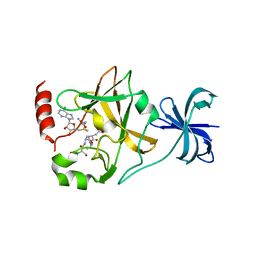
4J8O
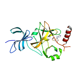 | | SET7/9 in complex with TAF10K189A peptide and AdoHcy | | 分子名称: | Histone-lysine N-methyltransferase SETD7, S-ADENOSYL-L-HOMOCYSTEINE, Transcription initiation factor TFIID subunit 10 | | 著者 | Horowitz, S, Trievel, R.C. | | 登録日 | 2013-02-14 | | 公開日 | 2014-01-08 | | 最終更新日 | 2024-10-30 | | 実験手法 | X-RAY DIFFRACTION (1.63 Å) | | 主引用文献 | Conservation and functional importance of carbon-oxygen hydrogen bonding in AdoMet-dependent methyltransferases.
J.Am.Chem.Soc., 135, 2013
|
|
4J7I
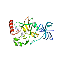 | | SET7/9Y335F in complex with TAF10 peptide and AdoHcy | | 分子名称: | Histone-lysine N-methyltransferase SETD7, S-ADENOSYL-L-HOMOCYSTEINE, Transcription initiation factor TFIID subunit 10 | | 著者 | Horowitz, S, Trievel, R.C. | | 登録日 | 2013-02-13 | | 公開日 | 2014-01-08 | | 最終更新日 | 2024-11-27 | | 実験手法 | X-RAY DIFFRACTION (2.56 Å) | | 主引用文献 | Conservation and functional importance of carbon-oxygen hydrogen bonding in AdoMet-dependent methyltransferases.
J.Am.Chem.Soc., 135, 2013
|
|
4J83
 | |
4J7F
 | |
4PIF
 | |
4PIU
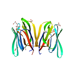 | |
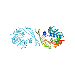
6UEO
 | | Structure of A. thaliana TBP-AC mismatch DNA site | | 分子名称: | DNA (5'-D(*GP*CP*TP*AP*TP*AP*AP*AP*AP*GP*GP*GP*CP*A)-3'), DNA (5'-D(*TP*GP*CP*CP*CP*CP*TP*TP*TP*AP*TP*AP*GP*C)-3'), TATA-box-binding protein 1 | | 著者 | Schumacher, M.A. | | 登録日 | 2019-09-22 | | 公開日 | 2020-09-02 | | 最終更新日 | 2023-10-11 | | 実験手法 | X-RAY DIFFRACTION (2 Å) | | 主引用文献 | DNA mismatches reveal conformational penalties in protein-DNA recognition.
Nature, 587, 2020
|
|
4PIK
 | |


