4WGK
 
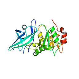
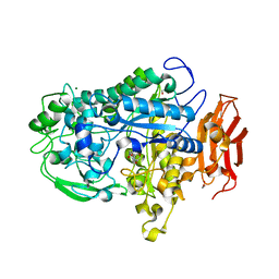
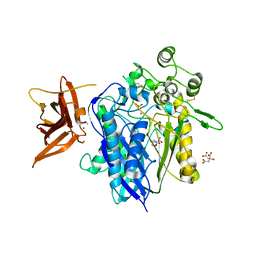 | | Crystal structure of human neutral ceramidase with Zn-bound phosphate | | 分子名称: | 1,2-ETHANEDIOL, 2-acetamido-2-deoxy-beta-D-glucopyranose, 2-acetamido-2-deoxy-beta-D-glucopyranose-(1-4)-2-acetamido-2-deoxy-beta-D-glucopyranose, ... | | 著者 | Airola, M.V, Pulkoski-Gross, M.J, Obeid, L.M, Hannun, Y.A. | | 登録日 | 2014-09-18 | | 公開日 | 2015-07-08 | | 最終更新日 | 2023-09-27 | | 実験手法 | X-RAY DIFFRACTION (2.582 Å) | | 主引用文献 | Structural Basis for Ceramide Recognition and Hydrolysis by Human Neutral Ceramidase.
Structure, 23, 2015
|
|
5UVG
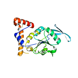
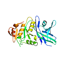
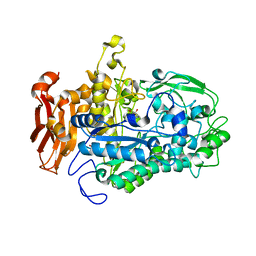
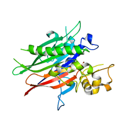 | | Crystal structure of the human neutral sphingomyelinase 2 (nSMase2) catalytic domain with insertion deleted and calcium bound | | 分子名称: | CALCIUM ION, Sphingomyelin phosphodiesterase 3,Sphingomyelin phosphodiesterase 3 | | 著者 | Airola, M.V, Guja, K.E, Garcia-Diaz, M, Hannun, Y.A. | | 登録日 | 2017-02-20 | | 公開日 | 2017-06-28 | | 最終更新日 | 2024-03-06 | | 実験手法 | X-RAY DIFFRACTION (1.849 Å) | | 主引用文献 | Structure of human nSMase2 reveals an interdomain allosteric activation mechanism for ceramide generation.
Proc. Natl. Acad. Sci. U.S.A., 114, 2017
|
|
3LNR
 | |
4HI4
 | |
4I44
 | |
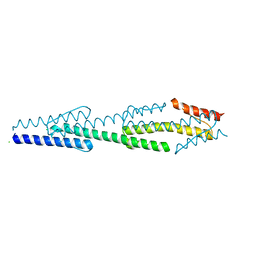
4I3M
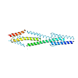
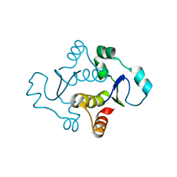
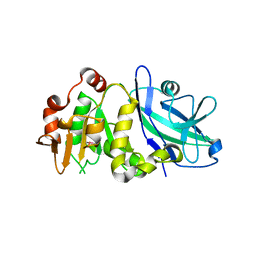
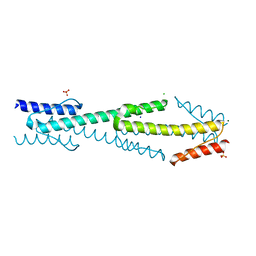 | | Aer2 poly-HAMP domains: L44H HAMP1 CW-lock mutant | | 分子名称: | Aerotaxis transducer Aer2, CHLORIDE ION, MAGNESIUM ION, ... | | 著者 | Airola, M.V, Sukomon, N, Crane, B.R. | | 登録日 | 2012-11-26 | | 公開日 | 2013-02-27 | | 最終更新日 | 2024-02-28 | | 実験手法 | X-RAY DIFFRACTION (1.95 Å) | | 主引用文献 | HAMP Domain Conformers That Propagate Opposite Signals in Bacterial Chemoreceptors.
Plos Biol., 11, 2013
|
|
8EXD
 | |
8UJL
 | |
8UJM
 | |
7KIL
 | |
7KIH
 | |
7KIQ
 | |
6TZZ
 | |
6TZY
 | |
6U8Z
 | |
7LPO
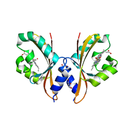
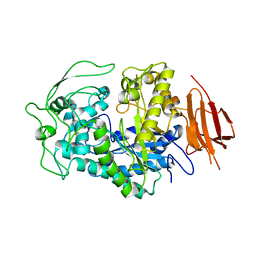
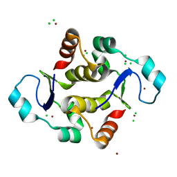
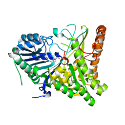
 | | Crystal structure of Cryptococcus neoformans sterylglucosidase 1 with tris | | 分子名称: | 2-AMINO-2-HYDROXYMETHYL-PROPANE-1,3-DIOL, Cytoplasmic protein, MAGNESIUM ION | | 著者 | Pereira de Sa, N, Del Poeta, M, Airola, M.V. | | 登録日 | 2021-02-12 | | 公開日 | 2021-09-01 | | 最終更新日 | 2024-05-22 | | 実験手法 | X-RAY DIFFRACTION (2.13 Å) | | 主引用文献 | Structure and inhibition of Cryptococcus neoformans sterylglucosidase to develop antifungal agents.
Nat Commun, 12, 2021
|
|
7LPQ
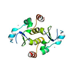
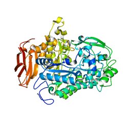
 | | Crystal structure of Cryptococcus neoformans sterylglucosidase 1 with hit 9 | | 分子名称: | Cytoplasmic protein, GLYCEROL, MAGNESIUM ION, ... | | 著者 | Pereira de Sa, N, Del Poeta, M, Airola, M.V. | | 登録日 | 2021-02-12 | | 公開日 | 2021-09-01 | | 最終更新日 | 2023-10-18 | | 実験手法 | X-RAY DIFFRACTION (2.32 Å) | | 主引用文献 | Structure and inhibition of Cryptococcus neoformans sterylglucosidase to develop antifungal agents.
Nat Commun, 12, 2021
|
|
7LPP
 | | Crystal structure of Cryptococcus neoformans sterylglucosidase 1 with hit 1 | | 分子名称: | 4-(hydroxymethyl)-1-[[2-(3-methoxyphenyl)-1,3-thiazol-5-yl]methyl]piperidin-4-ol, Cytoplasmic protein, GLYCEROL, ... | | 著者 | Pereira de Sa, N, Del Poeta, M, Airola, M.V. | | 登録日 | 2021-02-12 | | 公開日 | 2021-09-01 | | 最終更新日 | 2024-05-22 | | 実験手法 | X-RAY DIFFRACTION (2.85 Å) | | 主引用文献 | Structure and inhibition of Cryptococcus neoformans sterylglucosidase to develop antifungal agents.
Nat Commun, 12, 2021
|
|
7LHK
 | |


