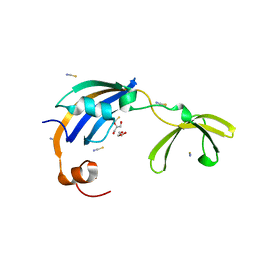
3LUO
 
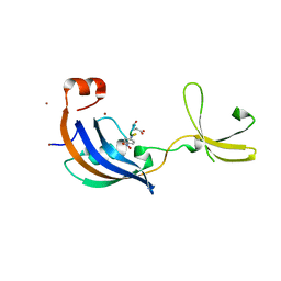 | | Crystal Structure and functional characterization of the thermophilic prolyl isomerase and chaperone SlyD | | 分子名称: | Peptidyl-prolyl cis-trans isomerase, Suc-Ala-Leu-Pro-Phe-pNA, ZINC ION | | 著者 | Loew, C, Neumann, P, Weininger, U, Stubbs, M.T, Balbach, J. | | 登録日 | 2010-02-18 | | 公開日 | 2010-03-31 | | 最終更新日 | 2023-11-01 | | 実験手法 | X-RAY DIFFRACTION (2.55 Å) | | 主引用文献 | Crystal Structure Determination and Functional Characterization of the Metallochaperone SlyD from Thermus thermophilus
J.Mol.Biol., 398, 2010
|
|
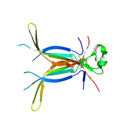
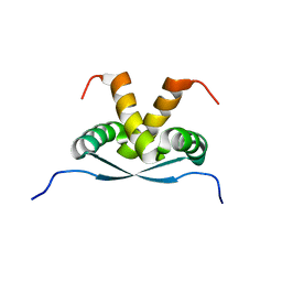
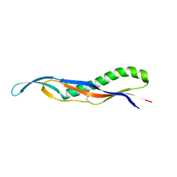
2RMY
 | |
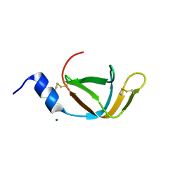
5AX2
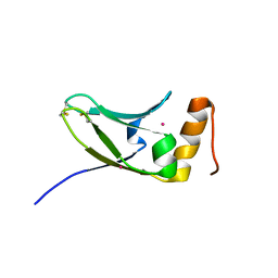 | | Crystal structure of S.cerevisiae Kti11p | | 分子名称: | CADMIUM ION, Diphthamide biosynthesis protein 3 | | 著者 | Kumar, A, Nagarathinam, K, Tanabe, M, Balbach, J. | | 登録日 | 2015-07-13 | | 公開日 | 2016-07-20 | | 最終更新日 | 2019-02-20 | | 実験手法 | X-RAY DIFFRACTION (2.4 Å) | | 主引用文献 | Hyperbolic Pressure-Temperature Phase Diagram of the Zinc-Finger Protein apoKti11 Detected by NMR Spectroscopy.
J Phys Chem B, 123, 2019
|
|
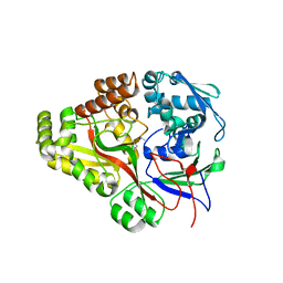
3CGN
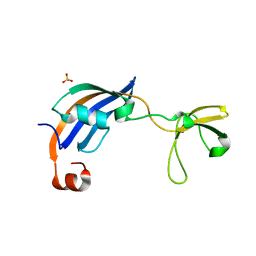 | | Crystal Structure of thermophilic SlyD | | 分子名称: | Peptidyl-prolyl cis-trans isomerase, SULFATE ION | | 著者 | Neumann, P, Loew, C, Stubbs, M.T, Balbach, J. | | 登録日 | 2008-03-06 | | 公開日 | 2009-03-10 | | 最終更新日 | 2023-11-01 | | 実験手法 | X-RAY DIFFRACTION (2.7 Å) | | 主引用文献 | Crystal Structure Determination and Functional Characterization of the Metallochaperone SlyD from Thermus thermophilus
J.Mol.Biol., 398, 2010
|
|
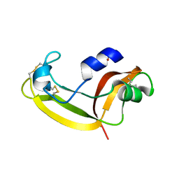
3CGM
 | | Crystal structure of thermophilic SlyD | | 分子名称: | GLYCEROL, NICKEL (II) ION, Peptidyl-prolyl cis-trans isomerase, ... | | 著者 | Loew, C, Neumann, P, Stubbs, M.T, Balbach, J. | | 登録日 | 2008-03-06 | | 公開日 | 2009-03-10 | | 最終更新日 | 2023-11-01 | | 実験手法 | X-RAY DIFFRACTION (2.41 Å) | | 主引用文献 | Crystal Structure Determination and Functional Characterization of the Metallochaperone SlyD from Thermus thermophilus
J.Mol.Biol., 398, 2010
|
|
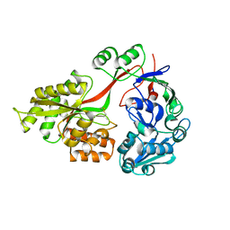
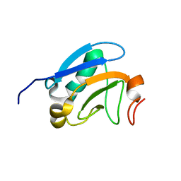
2RFM
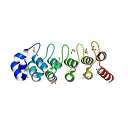 | | Structure of a Thermophilic Ankyrin Repeat Protein | | 分子名称: | 1,3-BUTANEDIOL, CHLORIDE ION, GLYCEROL, ... | | 著者 | Loew, C, Weininger, U, Neumann, P, Stubbs, M.T, Balbach, J. | | 登録日 | 2007-10-01 | | 公開日 | 2008-03-11 | | 最終更新日 | 2024-03-13 | | 実験手法 | X-RAY DIFFRACTION (1.65 Å) | | 主引用文献 | Structural insights into an equilibrium folding intermediate of an archaeal ankyrin repeat protein
Proc.Natl.Acad.Sci.Usa, 105, 2008
|
|
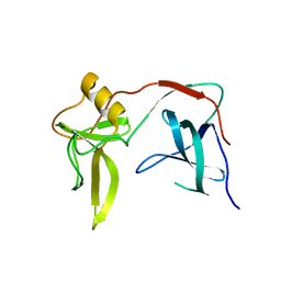
2RND
 | |
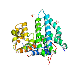
2F52
 | |
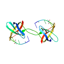
3E07
 | | Crystal structure of spatzle cystine knot | | 分子名称: | GLYCEROL, Protein spaetzle | | 著者 | Hoffmann, A, Funkner, A, Neumann, P, Juhnke, S, Walther, M, Schierhorn, A, Weininger, U, Balbach, J, Reuter, G, Stubbs, M.T. | | 登録日 | 2008-07-31 | | 公開日 | 2008-09-23 | | 最終更新日 | 2023-11-01 | | 実験手法 | X-RAY DIFFRACTION (2.4 Å) | | 主引用文献 | Biophysical Characterization of Refolded Drosophila Spatzle, a Cystine Knot Protein, Reveals Distinct Properties of Three Isoforms
J.Biol.Chem., 283, 2008
|
|
3TCH
 | | Crystal structure of E. coli OppA in an open conformation | | 分子名称: | Periplasmic oligopeptide-binding protein | | 著者 | Klepsch, M.M, Kovermann, M, Low, C, Balbach, J, de Gier, J.W, Slotboom, D.J, Berntsson, R.P.-A. | | 登録日 | 2011-08-09 | | 公開日 | 2011-10-12 | | 最終更新日 | 2012-01-11 | | 実験手法 | X-RAY DIFFRACTION (1.98 Å) | | 主引用文献 | Escherichia coli peptide binding protein OppA has a preference for positively charged peptides.
J.Mol.Biol., 414, 2011
|
|
3TCG
 | | Crystal structure of E. coli OppA complexed with the tripeptide KGE | | 分子名称: | KGE Peptide, Periplasmic oligopeptide-binding protein | | 著者 | Klepsch, M.M, Kovermann, M, Low, C, Balbach, J, de Gier, J.W, Slotboom, D.J, Berntsson, R.P.-A. | | 登録日 | 2011-08-09 | | 公開日 | 2011-10-12 | | 最終更新日 | 2017-10-25 | | 実験手法 | X-RAY DIFFRACTION (2 Å) | | 主引用文献 | Escherichia coli peptide binding protein OppA has a preference for positively charged peptides.
J.Mol.Biol., 414, 2011
|
|
3HG6
 | | Crystal Structure of the Recombinant Onconase from Rana pipiens | | 分子名称: | GLYCEROL, Onconase, SULFATE ION | | 著者 | Camara-Artigas, A, Gavira, J.A, Casares-Atienza, S, Weininger, U, Balbach, J, Garcia-Mira, M.M. | | 登録日 | 2009-05-13 | | 公開日 | 2010-05-19 | | 最終更新日 | 2023-11-08 | | 実験手法 | X-RAY DIFFRACTION (1.7 Å) | | 主引用文献 | Three-state thermal unfolding of onconase.
Biophys.Chem., 159, 2011
|
|
3TCF
 | | Crystal structure of E. coli OppA complexed with endogenous ligands | | 分子名称: | Endogenous peptide, Periplasmic oligopeptide-binding protein | | 著者 | Klepsch, M.M, Kovermann, M, Low, C, Balbach, J, de Gier, J.W, Slotboom, D.J, Berntsson, R.P.-A. | | 登録日 | 2011-08-09 | | 公開日 | 2011-10-12 | | 最終更新日 | 2017-10-25 | | 実験手法 | X-RAY DIFFRACTION (2 Å) | | 主引用文献 | Escherichia coli peptide binding protein OppA has a preference for positively charged peptides.
J.Mol.Biol., 414, 2011
|
|
2KB6
 | | Solution structure of onconase C87A/C104A | | 分子名称: | Protein P-30 | | 著者 | Weininger, U, Schulenburg, C, Arnold, U, Ulbrich-Hofmann, R, Balbach, J. | | 登録日 | 2008-11-21 | | 公開日 | 2009-11-24 | | 最終更新日 | 2021-11-10 | | 実験手法 | SOLUTION NMR | | 主引用文献 | Impact of the C-terminal disulfide bond on the folding and stability of onconase.
Chembiochem, 11, 2010
|
|
2K9I
 | |
2M1M
 | | Solution structure of the PsIAA4 oligomerization domain reveals interaction modes for transcription factors in early auxin response | | 分子名称: | Auxin-induced protein IAA4 | | 著者 | Kovermann, M, Dinesh, D.C, Gopalswamy, M, Abel, S, Balbach, J. | | 登録日 | 2012-12-03 | | 公開日 | 2013-12-11 | | 最終更新日 | 2024-05-01 | | 実験手法 | SOLUTION NMR | | 主引用文献 | Solution structure of the PsIAA4 oligomerization domain reveals interaction modes for transcription factors in early auxin response.
Proc.Natl.Acad.Sci.USA, 112, 2015
|
|
2K8I
 | | Solution structure of E.Coli SlyD | | 分子名称: | Peptidyl-prolyl cis-trans isomerase | | 著者 | Weininger, U, Balbach, J. | | 登録日 | 2008-09-11 | | 公開日 | 2009-03-24 | | 最終更新日 | 2024-05-01 | | 実験手法 | SOLUTION NMR | | 主引用文献 | NMR solution structure of SlyD from Escherichia coli: spatial separation of prolyl isomerase and chaperone function.
J.Mol.Biol., 387, 2009
|
|
2M2A
 | |
4EO0
 | | crystal structure of the pilus binding domain of the filamentous phage IKe | | 分子名称: | Attachment protein G3P | | 著者 | Jakob, R.P, Geitner, A.J, Weininger, U, Balbach, J, Dobbek, H, Schmid, F.X. | | 登録日 | 2012-04-13 | | 公開日 | 2012-05-30 | | 最終更新日 | 2017-10-25 | | 実験手法 | X-RAY DIFFRACTION (1.61 Å) | | 主引用文献 | Structural and energetic basis of infection by the filamentous bacteriophage IKe.
Mol.Microbiol., 84, 2012
|
|
4EO1
 | | crystal structure of the TolA binding domain from the filamentous phage IKe | | 分子名称: | Attachment protein G3P, MAGNESIUM ION | | 著者 | Jakob, R.P, Geitner, A.J, Weininger, U, Balbach, J, Dobbek, H, Schmid, F.X. | | 登録日 | 2012-04-13 | | 公開日 | 2012-05-30 | | 最終更新日 | 2023-09-13 | | 実験手法 | X-RAY DIFFRACTION (1.8 Å) | | 主引用文献 | Structural and energetic basis of infection by the filamentous bacteriophage IKe.
Mol.Microbiol., 84, 2012
|
|
3KNQ
 | |
4NY3
 | | Human PTPA in complex with peptide | | 分子名称: | GLYCEROL, SULFATE ION, Serine/threonine-protein phosphatase 2A activator, ... | | 著者 | Loew, C, Quistgaard, E.M, Nordlund, P. | | 登録日 | 2013-12-10 | | 公開日 | 2014-07-23 | | 最終更新日 | 2024-02-28 | | 実験手法 | X-RAY DIFFRACTION (1.797 Å) | | 主引用文献 | Structural basis for PTPA interaction with the invariant C-terminal tail of PP2A.
Biol.Chem., 395, 2014
|
|
3PF4
 | | Crystal structure of Bs-CspB in complex with r(GUCUUUA) | | 分子名称: | Cold shock protein cspB, MAGNESIUM ION, SODIUM ION, ... | | 著者 | Sachs, R, Max, K.E.A, Heinemann, U. | | 登録日 | 2010-10-27 | | 公開日 | 2011-09-21 | | 最終更新日 | 2023-09-06 | | 実験手法 | X-RAY DIFFRACTION (1.38 Å) | | 主引用文献 | RNA single strands bind to a conserved surface of the major cold shock protein in crystals and solution.
Rna, 18, 2012
|
|
2HAX
 | |
2X9A
 | | crystal structure of g3p from phage IF1 in complex with its coreceptor, the C-terminal domain of TolA | | 分子名称: | ATTACHMENT PROTEIN G3P, MEMBRANE SPANNING PROTEIN, REQUIRED FOR OUTER MEMBRANE INTEGRITY | | 著者 | Lorenz, S.H, Jakob, R.P, Dobbek, H, Schmid, F.X. | | 登録日 | 2010-03-15 | | 公開日 | 2010-12-01 | | 最終更新日 | 2023-12-20 | | 実験手法 | X-RAY DIFFRACTION (2.47 Å) | | 主引用文献 | The Filamentous Phages Fd and If1 Use Different Mechanisms to Infect Escherichia Coli.
J.Mol.Biol., 405, 2011
|
|


