3HWK
 
 | |
8UAI
 | |
3ICO
 | |
7K7Q
 | |
7K7O
 | |
6IV3
 | |
4NFN
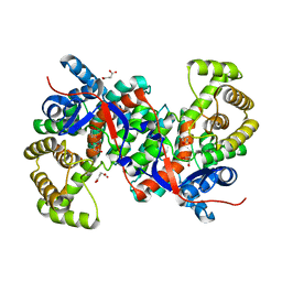
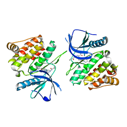
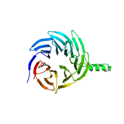
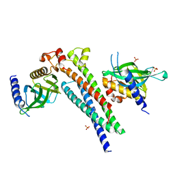
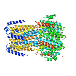
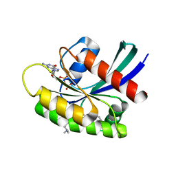
 | | Human tau tubulin kinase 1 (TTBK1) complexed with 3-({5-[(4-amino-4-methylpiperidin-1-yl)methyl]pyrrolo[2,1-f][1,2,4]triazin-4-yl}amino)-5-bromophenol | | 分子名称: | 3-({5-[(4-amino-4-methylpiperidin-1-yl)methyl]pyrrolo[2,1-f][1,2,4]triazin-4-yl}amino)-5-bromophenol, SULFATE ION, Tau-tubulin kinase 1 | | 著者 | Sheriff, S. | | 登録日 | 2013-10-31 | | 公開日 | 2014-02-05 | | 最終更新日 | 2023-09-20 | | 実験手法 | X-RAY DIFFRACTION (1.42 Å) | | 主引用文献 | The structure of human tau-tubulin kinase 1 both in the apo form and in complex with an inhibitor.
Acta Crystallogr F Struct Biol Commun, 70, 2014
|
|
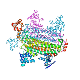
5C90
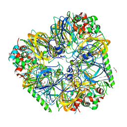
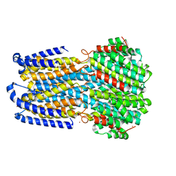
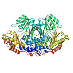
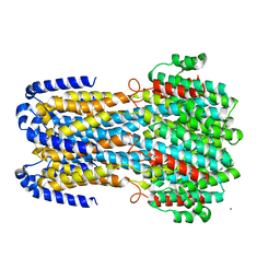
 | | Staphylococcus aureus ClpP mutant - Y63A | | 分子名称: | (4S)-2-METHYL-2,4-PENTANEDIOL, ATP-dependent Clp protease proteolytic subunit | | 著者 | Ye, F, Liu, H, Zhang, J, Gan, J, Yang, C.-G. | | 登録日 | 2015-06-26 | | 公開日 | 2016-05-25 | | 最終更新日 | 2023-11-08 | | 実験手法 | X-RAY DIFFRACTION (1.75 Å) | | 主引用文献 | Characterization of Gain-of-Function Mutant Provides New Insights into ClpP Structure
Acs Chem.Biol., 11, 2016
|
|
6E29
 | |
5M4Y
 | |
1CC0
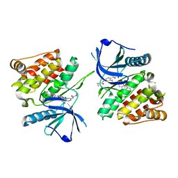
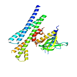
 | | CRYSTAL STRUCTURE OF THE RHOA.GDP-RHOGDI COMPLEX | | 分子名称: | GUANOSINE-5'-DIPHOSPHATE, MAGNESIUM ION, rho GDP dissociation inhibitor alpha, ... | | 著者 | Longenecker, K.L, Read, P, Derewenda, U, Dauter, Z, Garrard, S, Walker, L, Somlyo, A.V, Somlyo, A.P, Nakamoto, R.K, Derewenda, Z.S. | | 登録日 | 1999-03-03 | | 公開日 | 2000-01-07 | | 最終更新日 | 2023-12-27 | | 実験手法 | X-RAY DIFFRACTION (5 Å) | | 主引用文献 | How RhoGDI binds Rho.
Acta Crystallogr.,Sect.D, 55, 1999
|
|
3H7F
 | |
1CHZ
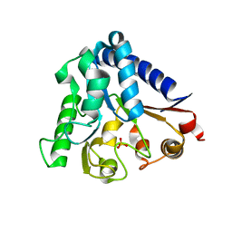
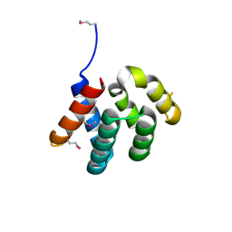
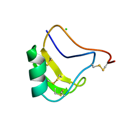 | | A NEW NEUROTOXIN FROM BUTHUS MARTENSII KARSCH | | 分子名称: | CHLORIDE ION, PROTEIN (BMK M2) | | 著者 | He, X.L, Deng, J.P, Li, H.M, Wang, D.C. | | 登録日 | 1999-03-31 | | 公開日 | 2000-03-31 | | 最終更新日 | 2023-08-09 | | 実験手法 | X-RAY DIFFRACTION (1.76 Å) | | 主引用文献 | Structure of a new neurotoxin from the scorpion Buthus martensii Karsch at 1.76 A.
Acta Crystallogr.,Sect.D, 56, 2000
|
|
3IFT
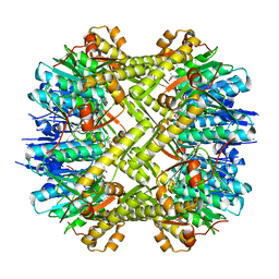
 | | Crystal structure of glycine cleavage system protein H from Mycobacterium tuberculosis, using X-rays from the Compact Light Source. | | 分子名称: | Glycine cleavage system H protein | | 著者 | Edwards, T.E, Abendroth, J, Staker, B, Mayer, C, Phan, I, Kelley, A, Analau, E, Leibly, D, Rifkin, J, Loewen, R, Ruth, R.D, Stewart, L.J, Accelerated Technologies Center for Gene to 3D Structure (ATCG3D) | | 登録日 | 2009-07-25 | | 公開日 | 2009-08-11 | | 最終更新日 | 2023-09-06 | | 実験手法 | X-RAY DIFFRACTION (2 Å) | | 主引用文献 | X-ray structure determination of the glycine cleavage system protein H of Mycobacterium tuberculosis using an inverse Compton synchrotron X-ray source.
J.Struct.Funct.Genom., 11, 2010
|
|
5LG4
 | |
4YKC
 | |
6IV4
 | |
6IV0
 | |
6IVW
 | |
5J8V
 | |
6IV1
 | |
6FKQ
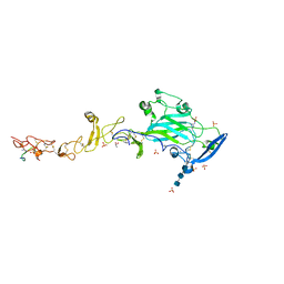 | | THE CRYSTAL STRUCTURE OF A FRAGMENT OF NETRIN-1 IN COMPLEX WITH A FRAGMENT OF DRAXIN | | 分子名称: | 2-acetamido-2-deoxy-beta-D-glucopyranose-(1-4)-2-acetamido-2-deoxy-beta-D-glucopyranose, 2-acetamido-2-deoxy-beta-D-glucopyranose-(1-4)-2-acetamido-2-deoxy-beta-D-glucopyranose-(1-4)-2-acetamido-2-deoxy-beta-D-glucopyranose, CALCIUM ION, ... | | 著者 | Bhowmick, T, Meijers, R. | | 登録日 | 2018-01-24 | | 公開日 | 2018-03-21 | | 最終更新日 | 2024-01-17 | | 実験手法 | X-RAY DIFFRACTION (3.07 Å) | | 主引用文献 | Structural Basis for Draxin-Modulated Axon Guidance and Fasciculation by Netrin-1 through DCC.
Neuron, 97, 2018
|
|
3GTD
 | |
3GVC
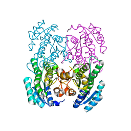 | |
7EW9
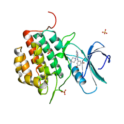
 | | GDP-bound KRAS G12D in complex with TH-Z816 | | 分子名称: | 7-(8-methylnaphthalen-1-yl)-4-[(2~{R})-2-methylpiperazin-1-yl]-2-[[(2~{S})-1-methylpyrrolidin-2-yl]methoxy]-6,8-dihydro-5~{H}-pyrido[3,4-d]pyrimidine, GUANOSINE-5'-DIPHOSPHATE, Isoform 2B of GTPase KRas, ... | | 著者 | Shen, P, Yang, Y, Yang, Y, Zhang, L, Chen, C.-C, Guo, R.-T. | | 登録日 | 2021-05-25 | | 公開日 | 2021-12-29 | | 最終更新日 | 2023-11-29 | | 実験手法 | X-RAY DIFFRACTION (2.13 Å) | | 主引用文献 | KRAS(G12D) can be targeted by potent inhibitors via formation of salt bridge.
Cell Discov, 8, 2022
|
|


