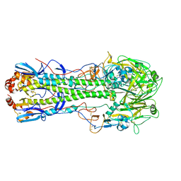
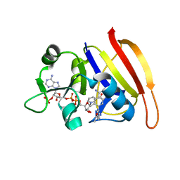
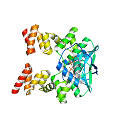
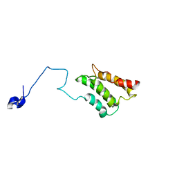
7DTJ
 
 | |
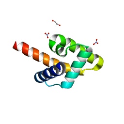
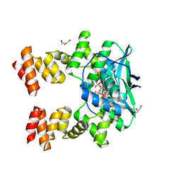
3UA0
 | | N-Terminal Domain of Bombyx mori Fibroin Mediates the Assembly of Silk in Response to pH Decrease | | 分子名称: | Fibroin heavy chain | | 著者 | He, Y.-X, Zhang, N.-N, Chen, B.-Y, Li, W.-F, Chen, Y.-X, Zhou, C.-Z. | | 登録日 | 2011-10-20 | | 公開日 | 2012-03-28 | | 最終更新日 | 2018-01-24 | | 実験手法 | X-RAY DIFFRACTION (3 Å) | | 主引用文献 | N-Terminal Domain of Bombyx mori Fibroin Mediates the Assembly of Silk in Response to pH Decrease.
J.Mol.Biol., 418, 2012
|
|
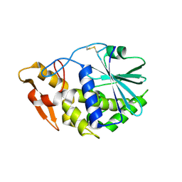
2LQ6
 | | Solution structure of BRD1 PHD2 finger | | 分子名称: | Bromodomain-containing protein 1, ZINC ION | | 著者 | Liu, L, Wu, J. | | 登録日 | 2012-02-25 | | 公開日 | 2012-10-24 | | 最終更新日 | 2024-05-01 | | 実験手法 | SOLUTION NMR | | 主引用文献 | Solution structure of an atypical PHD finger in BRPF2 and its interaction with DNA
J.Struct.Biol., 180, 2012
|
|
5X57
 | | Structure of GAR domain of ACF7 | | 分子名称: | Microtubule-actin cross-linking factor 1, isoforms 1/2/3/5, NICKEL (II) ION | | 著者 | Yang, F, Wang, T, Zhang, Y, Wu, X.Y. | | 登録日 | 2017-02-15 | | 公開日 | 2017-07-05 | | 最終更新日 | 2024-03-27 | | 実験手法 | X-RAY DIFFRACTION (1.45 Å) | | 主引用文献 | ACF7 regulates inflammatory colitis and intestinal wound response by orchestrating tight junction dynamics.
Nat Commun, 8, 2017
|
|
6V49
 | | The crystal structure of hemagglutinin from A/wedge-tailed shearwater/Western Australia/2576/1979 (H15N9) | | 分子名称: | 2-acetamido-2-deoxy-beta-D-glucopyranose, 2-acetamido-2-deoxy-beta-D-glucopyranose-(1-4)-2-acetamido-2-deoxy-beta-D-glucopyranose, Hemagglutinin HA1 chain, ... | | 著者 | Yang, H, Stevens, J. | | 登録日 | 2019-11-27 | | 公開日 | 2020-06-17 | | 最終更新日 | 2023-10-11 | | 実験手法 | X-RAY DIFFRACTION (2.5 Å) | | 主引用文献 | Molecular characterization and three-dimensional structures of avian H8, H11, H14, H15 and swine H4 influenza virus hemagglutinins
Heliyon, 6, 2020
|
|
8J0K
 | | Crystal structure of human TFAP2A in complex with DNA | | 分子名称: | DNA (5'-D(*CP*TP*GP*CP*CP*TP*CP*GP*GP*GP*CP*AP*C)-3'), DNA (5'-D(*GP*TP*GP*CP*CP*CP*GP*AP*GP*GP*CP*AP*G)-3'), GLYCEROL, ... | | 著者 | Liu, K, Xiao, Y.Q, Li, W.F, Min, J.R. | | 登録日 | 2023-04-11 | | 公開日 | 2023-07-05 | | 最終更新日 | 2023-09-06 | | 実験手法 | X-RAY DIFFRACTION (2.1 Å) | | 主引用文献 | Structural basis for specific DNA sequence motif recognition by the TFAP2 transcription factors.
Nucleic Acids Res., 51, 2023
|
|
5XK9
 | | Crystal structure of Isosesquilavandulyl Diphosphate Synthase from Streptomyces sp. strain CNH-189 in complex with GSPP and DMAPP | | 分子名称: | DIMETHYLALLYL DIPHOSPHATE, GERANYL S-THIOLODIPHOSPHATE, MAGNESIUM ION, ... | | 著者 | Ko, T.P, Guo, R.T, Liu, W, Chen, C.C, Gao, J. | | 登録日 | 2017-05-05 | | 公開日 | 2018-01-10 | | 最終更新日 | 2023-11-22 | | 実験手法 | X-RAY DIFFRACTION (2.137 Å) | | 主引用文献 | "Head-to-Middle" and "Head-to-Tail" cis-Prenyl Transferases: Structure of Isosesquilavandulyl Diphosphate Synthase.
Angew. Chem. Int. Ed. Engl., 57, 2018
|
|
5KZA
 | | Crystal structure of the Rous sarcoma virus matrix protein (aa 2-102). Space group I41 | | 分子名称: | 1,2-ETHANEDIOL, NITRATE ION, virus matrix protein | | 著者 | Kingston, R.L, Chan, J, Vogt, V.M. | | 登録日 | 2016-07-24 | | 公開日 | 2017-07-26 | | 最終更新日 | 2024-03-06 | | 実験手法 | X-RAY DIFFRACTION (1.86 Å) | | 主引用文献 | Cholesterol Promotes Protein Binding by Affecting Membrane Electrostatics and Solvation Properties.
Biophys. J., 113, 2017
|
|
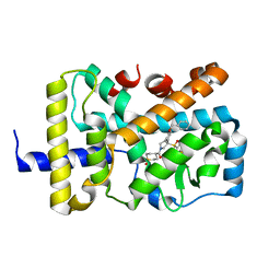
4HW7
 | |
3T10
 | | HSP90 N-terminal domain bound to ACP | | 分子名称: | Heat shock protein HSP 90-alpha, MAGNESIUM ION, PHOSPHOMETHYLPHOSPHONIC ACID ADENYLATE ESTER | | 著者 | Li, J. | | 登録日 | 2011-07-21 | | 公開日 | 2012-01-25 | | 最終更新日 | 2023-11-01 | | 実験手法 | X-RAY DIFFRACTION (1.24 Å) | | 主引用文献 | Structure insights into mechanisms of ATP hydrolysis and the activation of human heat-shock protein 90.
Acta Biochim Biophys Sin (Shanghai), 44, 2012
|
|
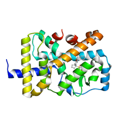
3SRQ
 | |
6W9H
 | |
6W9I
 | |
4PLK
 | |
7BNR
 | |
7BNK
 | |
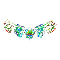
2K3Q
 | |
3GJ9
 | | crystal structure of TIP-1 in complex with c-terminal of Kir2.3 | | 分子名称: | C-terminal peptide from Inward rectifier potassium channel 4, CHLORIDE ION, Tax1-binding protein 3, ... | | 著者 | Shen, Y. | | 登録日 | 2009-03-08 | | 公開日 | 2009-12-15 | | 最終更新日 | 2023-11-01 | | 実験手法 | X-RAY DIFFRACTION (2.8 Å) | | 主引用文献 | Molecular mechanism of inward rectifier potassium channel 2.3 regulation by tax-interacting protein-1
J.Mol.Biol., 392, 2009
|
|
2K3P
 | |
3KTZ
 | | Structure of GAP31 | | 分子名称: | 2-acetamido-2-deoxy-beta-D-glucopyranose, Ribosome-inactivating protein gelonin | | 著者 | Kong, X.-P. | | 登録日 | 2009-11-26 | | 公開日 | 2010-01-26 | | 最終更新日 | 2020-07-29 | | 実験手法 | X-RAY DIFFRACTION (1.6 Å) | | 主引用文献 | A new activity of anti-HIV and anti-tumor protein GAP31: DNA adenosine glycosidase--structural and modeling insight into its functions.
Biochem.Biophys.Res.Commun., 391, 2010
|
|
1MJT
 | | CRYSTAL STRUCTURE OF SANOS, A BACTERIAL NITRIC OXIDE SYNTHASE OXYGENASE PROTEIN, IN COMPLEX WITH NAD+ AND SEITU | | 分子名称: | ETHYLISOTHIOUREA, NICOTINAMIDE-ADENINE-DINUCLEOTIDE, NITRIC-OXIDE SYNTHASE HOMOLOG, ... | | 著者 | Bird, L.E, Ren, J, Stammers, D.K. | | 登録日 | 2002-08-28 | | 公開日 | 2003-01-07 | | 最終更新日 | 2024-02-14 | | 実験手法 | X-RAY DIFFRACTION (2.4 Å) | | 主引用文献 | Crystal Structure of SANOS, a Bacterial Nitric Oxide Synthase Oxygenase Protein from Staphylococcus aureus
Structure, 10, 2002
|
|
3AFP
 | | Crystal structure of the single-stranded DNA binding protein from Mycobacterium leprae (Form I) | | 分子名称: | CADMIUM ION, GLYCEROL, Single-stranded DNA-binding protein | | 著者 | Kaushal, P.S, Singh, P, Sharma, A, Muniyappa, K, Vijayan, M. | | 登録日 | 2010-03-10 | | 公開日 | 2010-10-06 | | 最終更新日 | 2023-11-01 | | 実験手法 | X-RAY DIFFRACTION (2.05 Å) | | 主引用文献 | X-ray and molecular-dynamics studies on Mycobacterium leprae single-stranded DNA-binding protein and comparison with other eubacterial SSB structures
Acta Crystallogr.,Sect.D, 66, 2010
|
|
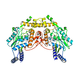
3ND8
 | | Structural characterization for the nucleotide binding ability of subunit A of the A1AO ATP synthase | | 分子名称: | (4R)-2-METHYLPENTANE-2,4-DIOL, (4S)-2-METHYL-2,4-PENTANEDIOL, ACETIC ACID, ... | | 著者 | Kumar, A, Gruber, G. | | 登録日 | 2010-06-07 | | 公開日 | 2011-03-30 | | 最終更新日 | 2023-11-01 | | 実験手法 | X-RAY DIFFRACTION (2.4 Å) | | 主引用文献 | The transition-like state and Pi entrance into the catalytic a subunit of the biological engine A-ATP synthase.
J.Mol.Biol., 408, 2011
|
|
5XK3
 | | Crystal structure of apo form Isosesquilavandulyl Diphosphate Synthase from Streptomyces sp. strain CNH-189 | | 分子名称: | SULFATE ION, Undecaprenyl diphosphate synthase | | 著者 | Ko, T.P, Guo, R.T, Liu, W, Chen, C.C, Gao, J. | | 登録日 | 2017-05-05 | | 公開日 | 2018-01-10 | | 最終更新日 | 2023-11-22 | | 実験手法 | X-RAY DIFFRACTION (1.996 Å) | | 主引用文献 | "Head-to-Middle" and "Head-to-Tail" cis-Prenyl Transferases: Structure of Isosesquilavandulyl Diphosphate Synthase.
Angew. Chem. Int. Ed. Engl., 57, 2018
|
|
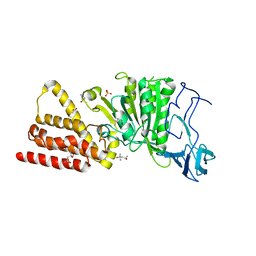
7E12
 | | Crystal structure of PKAc-A11E complex | | 分子名称: | MAGNESIUM ION, PHOSPHOAMINOPHOSPHONIC ACID-ADENYLATE ESTER, THR-ARG-SER-GLU-ILE-ARG-ARG-ALA-SER-THR-ILE-GLU, ... | | 著者 | Qin, J, Lin, L, Yuchi, Z. | | 登録日 | 2021-01-28 | | 公開日 | 2022-04-27 | | 最終更新日 | 2023-11-29 | | 実験手法 | X-RAY DIFFRACTION (2.796 Å) | | 主引用文献 | Structures of PKA-phospholamban complexes reveal a mechanism of familial dilated cardiomyopathy.
Elife, 11, 2022
|
|


