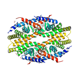
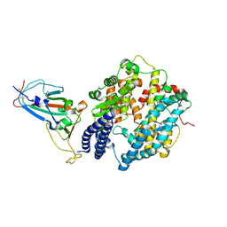
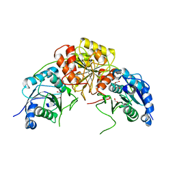
8J67
 
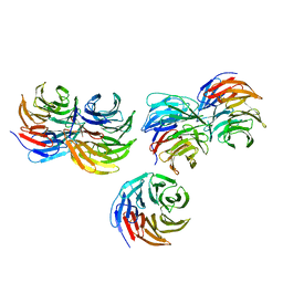
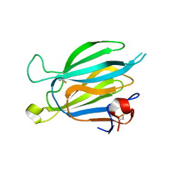 | | Crystal structure of Toxoplasma gondii M2AP | | 分子名称: | MIC2-associated protein | | 著者 | Wang, F.F, Zhang, D.J, Zhang, S, Springer, T.A, Song, G.J. | | 登録日 | 2023-04-24 | | 公開日 | 2023-08-30 | | 最終更新日 | 2023-10-04 | | 実験手法 | X-RAY DIFFRACTION (1.81 Å) | | 主引用文献 | Structural insights into MIC2 recognition by MIC2-associated protein in Toxoplasma gondii.
Commun Biol, 6, 2023
|
|
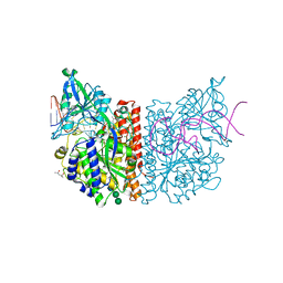
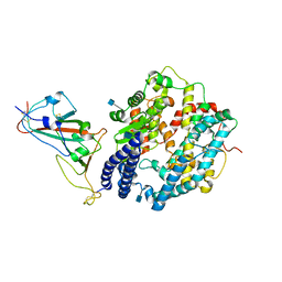
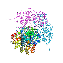
8J64
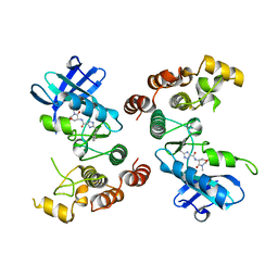
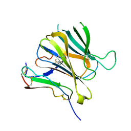 | | Crystal structure of Toxoplasma gondii MIC2-M2AP complex | | 分子名称: | MIC2-associated protein, Micronemal protein MIC2 | | 著者 | Zhang, S, Wang, F.F, Zhang, D.J, Song, G.J, Springer, T.A. | | 登録日 | 2023-04-24 | | 公開日 | 2023-08-30 | | 最終更新日 | 2023-10-04 | | 実験手法 | X-RAY DIFFRACTION (2 Å) | | 主引用文献 | Structural insights into MIC2 recognition by MIC2-associated protein in Toxoplasma gondii.
Commun Biol, 6, 2023
|
|
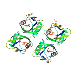
6Z6Y
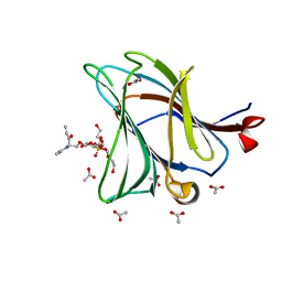 | |
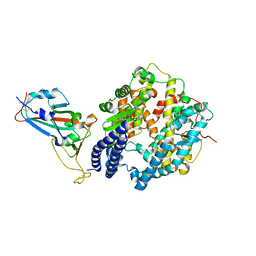
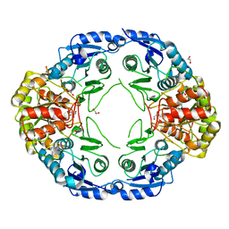
8H0N
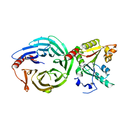 | | Crystal structure of the human METTL1-WDR4 complex | | 分子名称: | tRNA (guanine-N(7)-)-methyltransferase, tRNA (guanine-N(7)-)-methyltransferase non-catalytic subunit WDR4 | | 著者 | Jin, X.H, Guan, Z.Y, Gong, Z, Zhang, D.L. | | 登録日 | 2022-09-30 | | 公開日 | 2023-07-05 | | 最終更新日 | 2023-11-08 | | 実験手法 | X-RAY DIFFRACTION (1.8 Å) | | 主引用文献 | Structural insight into how WDR4 promotes the tRNA N7-methylguanosine methyltransferase activity of METTL1.
Cell Discov, 9, 2023
|
|
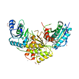
8HID
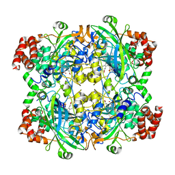 | | HUMAN ERYTHROCYTE CATALSE COMPLEXED WITH BT-Br | | 分子名称: | (~{S})-azanyl-[2-[[3-bromanyl-4-(diethylamino)phenyl]methyl]hydrazinyl]methanethiol, Catalase, PROTOPORPHYRIN IX CONTAINING FE | | 著者 | Lin, H.-Y, Yang, G.-F. | | 登録日 | 2022-11-19 | | 公開日 | 2023-10-04 | | 実験手法 | X-RAY DIFFRACTION (2.2 Å) | | 主引用文献 | A catalase inhibitor: Targeting the NADPH-binding site for castration-resistant prostate cancer therapy.
Redox Biol, 63, 2023
|
|
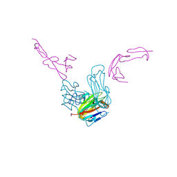
6PTO
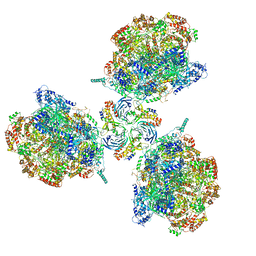 | | Structure of Ctf4 trimer in complex with three CMG helicases | | 分子名称: | ADENOSINE-5'-TRIPHOSPHATE, Cell division control protein 45, DNA polymerase alpha-binding protein, ... | | 著者 | Yuan, Z, Georgescu, R, Bai, L, Santos, R, Donnell, M, Li, H. | | 登録日 | 2019-07-16 | | 公開日 | 2019-11-20 | | 最終更新日 | 2019-12-18 | | 実験手法 | ELECTRON MICROSCOPY (7 Å) | | 主引用文献 | Ctf4 organizes sister replisomes and Pol alpha into a replication factory.
Elife, 8, 2019
|
|
6PTJ
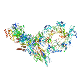 | | Structure of Ctf4 trimer in complex with one CMG helicase | | 分子名称: | Cell division control protein 45, DNA polymerase alpha-binding protein, DNA replication complex GINS protein PSF1, ... | | 著者 | Yuan, Z, Georgescu, R, Bai, L, Santos, R, Donnell, M, Li, H. | | 登録日 | 2019-07-15 | | 公開日 | 2019-11-20 | | 最終更新日 | 2019-12-18 | | 実験手法 | ELECTRON MICROSCOPY (3.8 Å) | | 主引用文献 | Ctf4 organizes sister replisomes and Pol alpha into a replication factory.
Elife, 8, 2019
|
|
6PTN
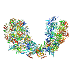 | | Structure of Ctf4 trimer in complex with two CMG helicases | | 分子名称: | ADENOSINE-5'-TRIPHOSPHATE, Cell division control protein 45, DNA polymerase alpha-binding protein, ... | | 著者 | Yuan, Z, Georgescu, R, Bai, L, Santos, R, Donnell, M, Li, H. | | 登録日 | 2019-07-16 | | 公開日 | 2019-11-20 | | 最終更新日 | 2019-12-18 | | 実験手法 | ELECTRON MICROSCOPY (5.8 Å) | | 主引用文献 | Ctf4 organizes sister replisomes and Pol alpha into a replication factory.
Elife, 8, 2019
|
|
5TBP
 | | Crystal Structure of RXR-alpha ligand binding domain complexed with synthetic modulator K8003 | | 分子名称: | ACETATE ION, DIMETHYL SULFOXIDE, GLYCEROL, ... | | 著者 | Aleshin, A.E, Liddington, R.C, Su, Y, Zhang, X. | | 登録日 | 2016-09-12 | | 公開日 | 2017-08-09 | | 最終更新日 | 2023-10-04 | | 実験手法 | X-RAY DIFFRACTION (2.6 Å) | | 主引用文献 | Modulation of nongenomic activation of PI3K signalling by tetramerization of N-terminally-cleaved RXR alpha.
Nat Commun, 8, 2017
|
|
6RTI
 | | X-ray structure of human glutamate carboxypeptidase II (GCPII) in complex with aptamer A9g | | 分子名称: | (2S)-2-(PHOSPHONOMETHYL)PENTANEDIOIC ACID, (4S)-2-METHYL-2,4-PENTANEDIOL, 2-acetamido-2-deoxy-beta-D-glucopyranose-(1-4)-2-acetamido-2-deoxy-beta-D-glucopyranose, ... | | 著者 | Motlova, L, Kolenko, P, Barinka, C. | | 登録日 | 2019-05-24 | | 公開日 | 2020-06-10 | | 最終更新日 | 2024-01-24 | | 実験手法 | X-RAY DIFFRACTION (2.2 Å) | | 主引用文献 | Structural basis of prostate-specific membrane antigen recognition by the A9g RNA aptamer.
Nucleic Acids Res., 48, 2020
|
|
5GP0
 | | Crystal structure of geraniol-NUDX1 complex | | 分子名称: | GERANYL DIPHOSPHATE, GLYCEROL, Nudix hydrolase 1 | | 著者 | Liu, J, Guan, Z, Zou, T, Yin, P. | | 登録日 | 2016-07-30 | | 公開日 | 2017-10-04 | | 最終更新日 | 2023-11-08 | | 実験手法 | X-RAY DIFFRACTION (1.702 Å) | | 主引用文献 | Structural Insights into the Substrate Recognition Mechanism of Arabidopsis GPP-Bound NUDX1 for Noncanonical Monoterpene Biosynthesis.
Mol Plant, 11, 2018
|
|
7WBQ
 | |
7WBP
 | |
7WBL
 | |
7LAW
 | | crystal structure of GITR complex with GITR-L | | 分子名称: | 2-acetamido-2-deoxy-beta-D-glucopyranose, Tumor necrosis factor ligand superfamily member 18, Tumor necrosis factor receptor superfamily member 18 | | 著者 | Longenecker, K.L, Rogers, B, Bigelow, L, Judge, R.A, Alvarez, H. | | 登録日 | 2021-01-07 | | 公開日 | 2022-03-09 | | 最終更新日 | 2023-10-18 | | 実験手法 | X-RAY DIFFRACTION (2.752 Å) | | 主引用文献 | An anti-PD-1-GITR-L bispecific agonist induces GITR clustering-mediated T cell activation for cancer immunotherapy.
Nat Cancer, 3, 2022
|
|
8ECE
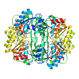 | | E. coli L-asparaginase II mutant (V27T) in complex with L-Glu | | 分子名称: | 1,2-ETHANEDIOL, GLUTAMIC ACID, L-asparaginase 2 | | 著者 | Strzelczyk, P, Wlodawer, A, Lubkowski, J. | | 登録日 | 2022-09-01 | | 公開日 | 2022-11-16 | | 最終更新日 | 2023-10-25 | | 実験手法 | X-RAY DIFFRACTION (1.86 Å) | | 主引用文献 | The E. coli L-asparaginase V27T mutant: structural and functional characterization and comparison with theoretical predictions.
Febs Lett., 596, 2022
|
|
8ECD
 | | E. coli L-asparaginase II mutant (V27T) in complex with L-Asp | | 分子名称: | 1,2-ETHANEDIOL, ASPARTIC ACID, CITRIC ACID, ... | | 著者 | Strzelczyk, P, Wlodawer, A, Lubkowski, J. | | 登録日 | 2022-09-01 | | 公開日 | 2022-11-16 | | 最終更新日 | 2023-10-25 | | 実験手法 | X-RAY DIFFRACTION (1.62 Å) | | 主引用文献 | The E. coli L-asparaginase V27T mutant: structural and functional characterization and comparison with theoretical predictions.
Febs Lett., 596, 2022
|
|
7R6A
 | |
7R69
 | |
7R6B
 | | Crystal structure of mutant R43D/L124D/R125A/C273S of L-Asparaginase I from Yersinia pestis | | 分子名称: | 1,2-ETHANEDIOL, CALCIUM ION, L-asparaginase I | | 著者 | Strzelczyk, P, Wlodawer, A, Lubkowski, J. | | 登録日 | 2021-06-22 | | 公開日 | 2022-07-06 | | 最終更新日 | 2023-10-25 | | 実験手法 | X-RAY DIFFRACTION (2.03 Å) | | 主引用文献 | The dimeric form of bacterial l-asparaginase YpAI is fully active.
Febs J., 290, 2023
|
|
5YZV
 | | Biophysical and structural characterization of the thermostable WD40 domain of a prokaryotic protein, Thermomonospora curvata PkwA | | 分子名称: | Probable serine/threonine-protein kinase PkwA | | 著者 | Li, D.Y, Shen, C, Du, Y, Qiao, F.F, Kong, T, Yuan, L.R, Zhang, D.L, Wu, X.H, Wu, Y.D. | | 登録日 | 2017-12-15 | | 公開日 | 2018-10-03 | | 最終更新日 | 2024-03-27 | | 実験手法 | X-RAY DIFFRACTION (2.6 Å) | | 主引用文献 | Biophysical and structural characterization of the thermostable WD40 domain of a prokaryotic protein, Thermomonospora curvata PkwA
Sci Rep, 8, 2018
|
|
5TX5
 | | Rip1 Kinase ( flag 1-294, C34A, C127A, C233A, C240A) with GSK772 | | 分子名称: | 3-benzyl-N-[(3S)-5-methyl-4-oxo-2,3,4,5-tetrahydro-1,5-benzoxazepin-3-yl]-1H-1,2,4-triazole-5-carboxamide, Receptor-interacting serine/threonine-protein kinase 1 | | 著者 | Campobasso, N, Ward, P, Thrope, J. | | 登録日 | 2016-11-15 | | 公開日 | 2017-07-05 | | 最終更新日 | 2024-03-06 | | 実験手法 | X-RAY DIFFRACTION (2.56 Å) | | 主引用文献 | Discovery of a First-in-Class Receptor Interacting Protein 1 (RIP1) Kinase Specific Clinical Candidate (GSK2982772) for the Treatment of Inflammatory Diseases.
J. Med. Chem., 60, 2017
|
|
6TZS
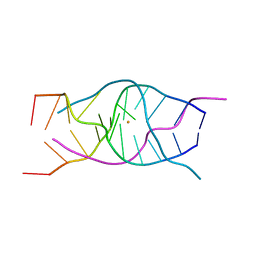
 | | A DNA i-motif/duplex hybrid | | 分子名称: | DNA (5'-D(*CP*CP*AP*GP*GP*CP*TP*GP*(CBR)P*AP*A)-3') | | 著者 | Chu, B, Paukstelis, P.J. | | 登録日 | 2019-08-13 | | 公開日 | 2019-10-16 | | 最終更新日 | 2024-03-13 | | 実験手法 | X-RAY DIFFRACTION (2.6 Å) | | 主引用文献 | A DNA G-quadruplex/i-motif hybrid.
Nucleic Acids Res., 47, 2019
|
|
6TZQ
 | |
6TZR
 | |


