Loading
PDBj
メニューPDBj@FacebookPDBj@TwitterPDBj@YouTubewwPDB FoundationwwPDB
RCSB PDBPDBeBMRBAdv. SearchSearch help
Search by PDB author
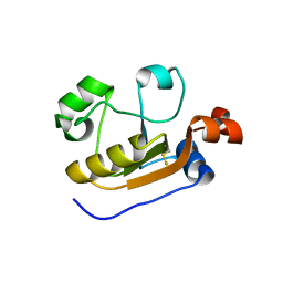
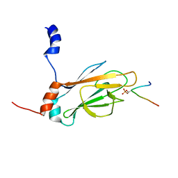
1J4P
DownloadVisualize
BU of 1j4p by Molmil
NMR STRUCTURE OF THE FHA1 DOMAIN OF RAD53 IN COMPLEX WITH A RAD9-DERIVED PHOSPHOTHREONINE (AT T155) PEPTIDE
分子名称: DNA REPAIR PROTEIN RAD9, PROTEIN KINASE SPK1
著者Yuan, C, Yongkiettrakul, S, Byeon, I.-J.L, Zhou, S, Tsai, M.-D.
登録日2001-10-22
公開日2001-12-05
最終更新日2023-12-27
実験手法SOLUTION NMR
主引用文献Solution structures of two FHA1-phosphothreonine peptide complexes provide insight into the structural basis of the ligand specificity of FHA1 from yeast Rad53.
J.Mol.Biol., 314, 2001
6LI6
DownloadVisualize
BU of 6li6 by Molmil
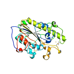
Crystal structure of MCR-1-S treated by Au(PEt3)Cl
分子名称: GOLD ION, Probable phosphatidylethanolamine transferase Mcr-1, TRIETHYLPHOSPHANE
著者Zhang, Q, Wang, M, Sun, H.
登録日2019-12-10
公開日2020-09-16
最終更新日2020-10-28
実験手法X-RAY DIFFRACTION (1.68 Å)
主引用文献Resensitizing carbapenem- and colistin-resistant bacteria to antibiotics using auranofin.
Nat Commun, 11, 2020
2L16
DownloadVisualize
BU of 2l16 by Molmil
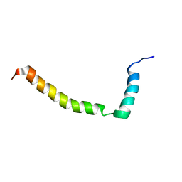
Solution structure of Bacillus subtilits TatAd protein in DPC micelles
分子名称: Sec-independent protein translocase protein tatAd
著者Hu, Y, Jin, C.
登録日2010-07-23
公開日2010-09-08
最終更新日2024-05-01
実験手法SOLUTION NMR
主引用文献Solution NMR structure of the TatA component of the twin-arginine protein transport system from gram-positive bacterium Bacillus subtilis
J.Am.Chem.Soc., 132, 2010
2L54
DownloadVisualize
BU of 2l54 by Molmil
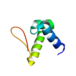
Solution structure of the Zalpha domain mutant of ADAR1 (N43A,Y47A)
分子名称: Double-stranded RNA-specific adenosine deaminase
著者Zhao, J, Pervushin, K, Feng, S, Droge, P.
登録日2010-10-24
公開日2011-01-12
最終更新日2024-05-01
実験手法SOLUTION NMR
主引用文献Alternate rRNA secondary structures as regulators of translation
Nat.Struct.Mol.Biol., 18, 2011
6KQP
DownloadVisualize
BU of 6kqp by Molmil
NSD1 SET domain in complex with SAM
分子名称: Histone-lysine N-methyltransferase, H3 lysine-36 and H4 lysine-20 specific, S-ADENOSYLMETHIONINE, ...
著者Cho, H.J, Cierpicki, T.
登録日2019-08-18
公開日2020-09-02
最終更新日2023-11-22
実験手法X-RAY DIFFRACTION (2.4 Å)
主引用文献Covalent inhibition of NSD1 histone methyltransferase.
Nat.Chem.Biol., 16, 2020
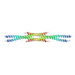
3CVF
DownloadVisualize
BU of 3cvf by Molmil
Crystal Structure of the carboxy terminus of Homer3
分子名称: Homer protein homolog 3
著者Hayashi, M.K, Stearns, M.H, Giannini, V, Xu, R.-M, Sala, C, Hayashi, Y.
登録日2008-04-18
公開日2009-03-31
最終更新日2023-11-15
実験手法X-RAY DIFFRACTION (2.9 Å)
主引用文献The postsynaptic density proteins Homer and Shank form a polymeric network structure.
Cell(Cambridge,Mass.), 137, 2009
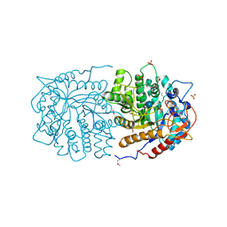
3QOM
DownloadVisualize
BU of 3qom by Molmil
Crystal structure of 6-phospho-beta-glucosidase from Lactobacillus plantarum
分子名称: 6-phospho-beta-glucosidase, ACETATE ION, PHOSPHATE ION, ...
著者Michalska, K, Hatzos-Skintges, C, Bearden, J, Kohler, M, Joachimiak, A, Midwest Center for Structural Genomics (MCSG)
登録日2011-02-10
公開日2011-03-09
最終更新日2020-07-29
実験手法X-RAY DIFFRACTION (1.498 Å)
主引用文献GH1-family 6-P-beta-glucosidases from human microbiome lactic acid bacteria.
Acta Crystallogr.,Sect.D, 69, 2013
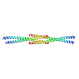
3CVE
DownloadVisualize
BU of 3cve by Molmil
Crystal Structure of the carboxy terminus of Homer1
分子名称: Homer protein homolog 1
著者Hayashi, M.K, Stearns, M.H, Giannini, V, Xu, R.-M, Sala, C, Hayashi, Y.
登録日2008-04-18
公開日2009-03-31
最終更新日2021-10-20
実験手法X-RAY DIFFRACTION (1.75 Å)
主引用文献The postsynaptic density proteins Homer and Shank form a polymeric network structure.
Cell(Cambridge,Mass.), 137, 2009
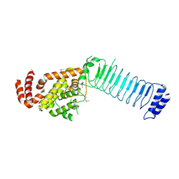
3CVR
DownloadVisualize
BU of 3cvr by Molmil
Crystal structure of the full length IpaH3
分子名称: Invasion plasmid antigen
著者Zhu, Y, Shao, F.
登録日2008-04-19
公開日2008-11-11
最終更新日2011-07-13
実験手法X-RAY DIFFRACTION (2.8 Å)
主引用文献Structure of a Shigella effector reveals a new class of ubiquitin ligases
Nat.Struct.Mol.Biol., 15, 2008
5X5P
DownloadVisualize
BU of 5x5p by Molmil
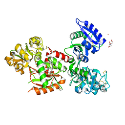
Human serum transferrin bound to ruthenium NTA
分子名称: FE (III) ION, MALONATE ION, NITRILOTRIACETIC ACID, ...
著者Sun, H, Wang, M.
登録日2017-02-17
公開日2018-02-21
最終更新日2023-11-22
実験手法X-RAY DIFFRACTION (2.7 Å)
主引用文献Binding of ruthenium and osmium at non‐iron sites of transferrin accounts for their iron-independent cellular uptake.
J.Inorg.Biochem., 234, 2022
7E17
DownloadVisualize
BU of 7e17 by Molmil
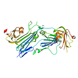
Structure of dimeric uPAR
分子名称: 2-acetamido-2-deoxy-beta-D-glucopyranose, Urokinase plasminogen activator surface receptor
著者Cai, Y, Huang, M.
登録日2021-02-01
公開日2021-12-22
最終更新日2023-11-29
実験手法X-RAY DIFFRACTION (2.96 Å)
主引用文献Crystal structure and cellular functions of uPAR dimer
Nat Commun, 13, 2022
7EOK
DownloadVisualize
BU of 7eok by Molmil
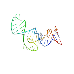
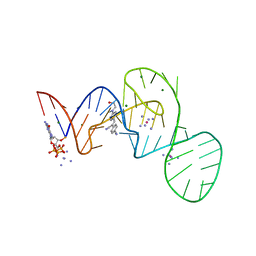
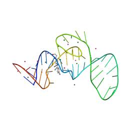
Crystal structure of the Pepper aptamer in complex with HBC485
分子名称: 4-[(~{Z})-1-cyano-2-[5-[2-(dimethylamino)ethyl-methyl-amino]pyrazin-2-yl]ethenyl]benzenecarbonitrile, GUANOSINE-5'-TRIPHOSPHATE, MAGNESIUM ION, ...
著者Huang, K.Y, Ren, A.M.
登録日2021-04-22
公開日2021-11-24
最終更新日2023-11-29
実験手法X-RAY DIFFRACTION (2.7 Å)
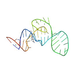
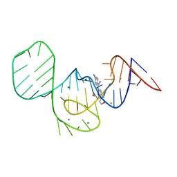
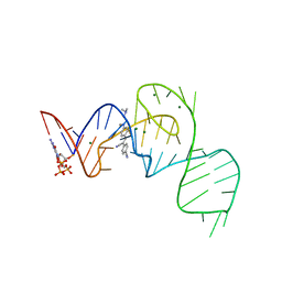
主引用文献Structure-based investigation of fluorogenic Pepper aptamer.
Nat.Chem.Biol., 17, 2021
7EOO
DownloadVisualize
BU of 7eoo by Molmil
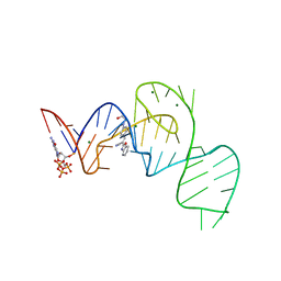
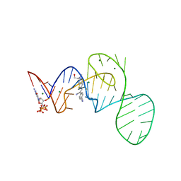
Crystal structure of the Pepper aptamer in complex with HBC525
分子名称: (~{E})-2-(1,3-benzoxazol-2-yl)-3-[4-[2-hydroxyethyl(methyl)amino]phenyl]prop-2-enenitrile, GUANOSINE-5'-TRIPHOSPHATE, MAGNESIUM ION, ...
著者Huang, K.Y, Ren, A.M.
登録日2021-04-22
公開日2021-11-24
最終更新日2023-11-29
実験手法X-RAY DIFFRACTION (2.23 Å)
主引用文献Structure-based investigation of fluorogenic Pepper aptamer.
Nat.Chem.Biol., 17, 2021
7EOG
DownloadVisualize
BU of 7eog by Molmil
Crystal structure of the Pepper aptamer in complex with HBC, iridium hexammine soak
分子名称: 4-[(~{Z})-1-cyano-2-[4-[2-hydroxyethyl(methyl)amino]phenyl]ethenyl]benzenecarbonitrile, GUANOSINE-5'-TRIPHOSPHATE, IRIDIUM HEXAMMINE ION, ...
著者Huang, K.Y, Ren, A.M.
登録日2021-04-22
公開日2021-11-24
最終更新日2024-05-29
実験手法X-RAY DIFFRACTION (1.5 Å)
主引用文献Structure-based investigation of fluorogenic Pepper aptamer.
Nat.Chem.Biol., 17, 2021
7EOP
DownloadVisualize
BU of 7eop by Molmil
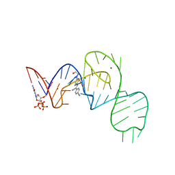
Crystal structure of the Pepper aptamer in complex with HBC620
分子名称: 4-[(~{Z})-1-cyano-2-[5-[2-hydroxyethyl(methyl)amino]thieno[3,2-b]thiophen-2-yl]ethenyl]benzenecarbonitrile, GUANOSINE-5'-TRIPHOSPHATE, MAGNESIUM ION, ...
著者Huang, K.Y, Ren, A.M.
登録日2021-04-22
公開日2021-11-24
最終更新日2023-11-29
実験手法X-RAY DIFFRACTION (1.8 Å)
主引用文献Structure-based investigation of fluorogenic Pepper aptamer.
Nat.Chem.Biol., 17, 2021
7EOM
DownloadVisualize
BU of 7eom by Molmil
Crystal structure of the Pepper aptamer in complex with HBC508
分子名称: 4-[(~{Z})-1-cyano-2-[6-[2-hydroxyethyl(methyl)amino]pyridin-3-yl]ethenyl]benzenecarbonitrile, GUANOSINE-5'-TRIPHOSPHATE, MAGNESIUM ION, ...
著者Huang, K.Y, Ren, A.M.
登録日2021-04-22
公開日2021-11-24
最終更新日2023-11-29
実験手法X-RAY DIFFRACTION (2.703 Å)
主引用文献Structure-based investigation of fluorogenic Pepper aptamer.
Nat.Chem.Biol., 17, 2021
7EOL
DownloadVisualize
BU of 7eol by Molmil
Crystal structure of the Pepper aptamer in complex with HBC497
分子名称: 4-[(~{Z})-1-cyano-2-[5-[2-hydroxyethyl(methyl)amino]pyrazin-2-yl]ethenyl]benzenecarbonitrile, GUANOSINE-5'-TRIPHOSPHATE, MAGNESIUM ION, ...
著者Huang, K.Y, Ren, A.M.
登録日2021-04-22
公開日2021-11-24
最終更新日2023-11-29
実験手法X-RAY DIFFRACTION (2.309 Å)
主引用文献Structure-based investigation of fluorogenic Pepper aptamer.
Nat.Chem.Biol., 17, 2021
7EOJ
DownloadVisualize
BU of 7eoj by Molmil
Crystal structure of the Pepper aptamer in complex with HBC, cesium soak
分子名称: 4-[(~{Z})-1-cyano-2-[4-[2-hydroxyethyl(methyl)amino]phenyl]ethenyl]benzenecarbonitrile, CESIUM ION, MAGNESIUM ION, ...
著者Huang, K.Y, Ren, A.M.
登録日2021-04-22
公開日2021-11-24
最終更新日2023-11-29
実験手法X-RAY DIFFRACTION (1.77 Å)
主引用文献Structure-based investigation of fluorogenic Pepper aptamer.
Nat.Chem.Biol., 17, 2021
7EOH
DownloadVisualize
BU of 7eoh by Molmil
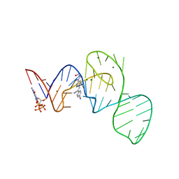
Crystal structure of the Pepper aptamer in complex with HBC
分子名称: 4-[(~{Z})-1-cyano-2-[4-[2-hydroxyethyl(methyl)amino]phenyl]ethenyl]benzenecarbonitrile, MAGNESIUM ION, Pepper (49-MER)
著者Huang, K.Y, Ren, A.M.
登録日2021-04-22
公開日2021-11-24
最終更新日2023-11-29
実験手法X-RAY DIFFRACTION (1.637 Å)
主引用文献Structure-based investigation of fluorogenic Pepper aptamer.
Nat.Chem.Biol., 17, 2021
7EOI
DownloadVisualize
BU of 7eoi by Molmil
Crystal structure of the Pepper aptamer in complex with HBC, manganese soak
分子名称: 4-[(~{Z})-1-cyano-2-[4-[2-hydroxyethyl(methyl)amino]phenyl]ethenyl]benzenecarbonitrile, MAGNESIUM ION, MANGANESE (II) ION, ...
著者Huang, K.Y, Ren, A.M.
登録日2021-04-22
公開日2021-11-24
最終更新日2023-11-29
実験手法X-RAY DIFFRACTION (1.92 Å)
主引用文献Structure-based investigation of fluorogenic Pepper aptamer.
Nat.Chem.Biol., 17, 2021
7EON
DownloadVisualize
BU of 7eon by Molmil
Crystal structure of the Pepper aptamer in complex with HBC514
分子名称: 4-[(~{Z})-1-cyano-2-[4-[2-(dimethylamino)ethyl-methyl-amino]phenyl]ethenyl]benzenecarbonitrile, GUANOSINE-5'-TRIPHOSPHATE, MAGNESIUM ION, ...
著者Huang, K.Y, Ren, A.M.
登録日2021-04-22
公開日2021-11-24
最終更新日2023-11-29
実験手法X-RAY DIFFRACTION (2.3 Å)
主引用文献Structure-based investigation of fluorogenic Pepper aptamer.
Nat.Chem.Biol., 17, 2021
6K6R
DownloadVisualize
BU of 6k6r by Molmil
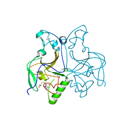
Crystal structure of Saccharomyces cerevisiae single domain sulfurtranferase RDL2
分子名称: Thiosulfate sulfurtransferase RDL2, mitochondrial
著者Li, H.J, Wang, Q.D.
登録日2019-06-04
公開日2019-11-06
最終更新日2023-11-22
実験手法X-RAY DIFFRACTION (2.471 Å)
主引用文献Saccharomyces cerevisiaeRhodanese RDL2 Uses the ArgResidue of the Active-Site Loop for Thiosulfate Decomposition
Antioxidants, 10, 2021
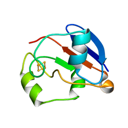
1YJJ
DownloadVisualize
BU of 1yjj by Molmil
RDC-refined Solution NMR structure of oxidized putidaredoxin
分子名称: FE2/S2 (INORGANIC) CLUSTER, Putidaredoxin
著者Jain, N.U, Tjioe, E, Savidor, A, Boulie, J.
登録日2005-01-14
公開日2005-06-28
最終更新日2024-05-22
実験手法SOLUTION NMR
主引用文献Redox-dependent structural differences in putidaredoxin derived from homologous structure refinement via residual dipolar couplings.
Biochemistry, 44, 2005
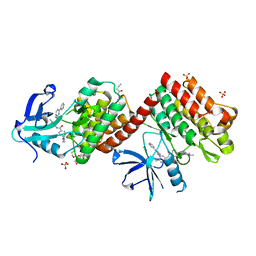
6NPE
DownloadVisualize
BU of 6npe by Molmil
C-abl Kinase domain with the activator(cmpd6), 2-cyano-N-(4-(3,4-dichlorophenyl)thiazol-2-yl)acetamide
分子名称: 2-cyano-~{N}-[4-(3,4-dichlorophenyl)-1,3-thiazol-2-yl]ethanamide, 4-(4-METHYL-PIPERAZIN-1-YLMETHYL)-N-[4-METHYL-3-(4-PYRIDIN-3-YL-PYRIMIDIN-2-YLAMINO)-PHENYL]-BENZAMIDE, NONAETHYLENE GLYCOL, ...
著者campobasso, N.
登録日2019-01-17
公開日2019-03-13
最終更新日2024-03-13
実験手法X-RAY DIFFRACTION (2.15 Å)
主引用文献Identification and Optimization of Novel Small c-Abl Kinase Activators Using Fragment and HTS Methodologies.
J. Med. Chem., 62, 2019
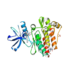
6NPV
DownloadVisualize
BU of 6npv by Molmil
C-abl Kinase domain with the activator(cmpd51), N-(1-(3,4-dichlorophenyl)-4-(2-hydroxyethyl)-4,5-dihydro-1H-pyrazol-3-yl)isonicotinamide
分子名称: 4-(4-METHYL-PIPERAZIN-1-YLMETHYL)-N-[4-METHYL-3-(4-PYRIDIN-3-YL-PYRIMIDIN-2-YLAMINO)-PHENYL]-BENZAMIDE, GLYCEROL, NONAETHYLENE GLYCOL, ...
著者campobasso, N.
登録日2019-01-18
公開日2019-03-13
最終更新日2024-03-13
実験手法X-RAY DIFFRACTION (1.86 Å)
主引用文献Identification and Optimization of Novel Small c-Abl Kinase Activators Using Fragment and HTS Methodologies.
J. Med. Chem., 62, 2019

222624

件を2024-07-17に公開中

PDB statisticsPDBj update infoContact PDBjnumon