3A8J
 
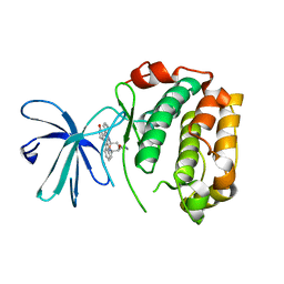
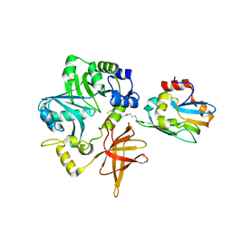 | | Crystal Structure of ET-EHred complex | | 分子名称: | Aminomethyltransferase, Glycine cleavage system H protein | | 著者 | Okamura-Ikeda, K, Hosaka, H. | | 登録日 | 2009-10-06 | | 公開日 | 2010-04-07 | | 最終更新日 | 2023-11-01 | | 実験手法 | X-RAY DIFFRACTION (1.98 Å) | | 主引用文献 | Crystal structure of aminomethyltransferase in complex with dihydrolipoyl-H-protein of the glycine cleavage system: implications for recognition of lipoyl protein substrate, disease-related mutations, and reaction mechanism
J.Biol.Chem., 285, 2010
|
|
1J1E
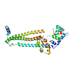 | | Crystal structure of the 52kDa domain of human cardiac troponin in the Ca2+ saturated form | | 分子名称: | CALCIUM ION, Troponin C, Troponin I, ... | | 著者 | Takeda, S, Yamashita, A, Maeda, K, Maeda, Y. | | 登録日 | 2002-12-03 | | 公開日 | 2003-07-15 | | 最終更新日 | 2023-10-25 | | 実験手法 | X-RAY DIFFRACTION (3.3 Å) | | 主引用文献 | Structure of the core domain of human cardiac troponin in the Ca2+-saturated form
Nature, 424, 2003
|
|
3AB9
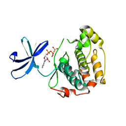
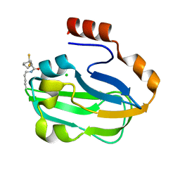 | | Crystal Structure of lipoylated E. coli H-protein (reduced form) | | 分子名称: | CALCIUM ION, CHLORIDE ION, Glycine cleavage system H protein | | 著者 | Okamura-Ikeda, K, Maita, N. | | 登録日 | 2009-12-04 | | 公開日 | 2010-04-07 | | 最終更新日 | 2023-11-01 | | 実験手法 | X-RAY DIFFRACTION (1.65 Å) | | 主引用文献 | Crystal structure of aminomethyltransferase in complex with dihydrolipoyl-H-protein of the glycine cleavage system: implications for recognition of lipoyl protein substrate, disease-related mutations, and reaction mechanism
J.Biol.Chem., 285, 2010
|
|
1J1D
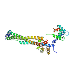 | | Crystal structure of the 46kDa domain of human cardiac troponin in the Ca2+ saturated form | | 分子名称: | CALCIUM ION, Troponin C, Troponin I, ... | | 著者 | Takeda, S, Yamashita, A, Maeda, K, Maeda, Y. | | 登録日 | 2002-12-03 | | 公開日 | 2003-07-15 | | 最終更新日 | 2023-12-27 | | 実験手法 | X-RAY DIFFRACTION (2.61 Å) | | 主引用文献 | Structure of the core domain of human cardiac troponin in the Ca2+-saturated form
Nature, 424, 2003
|
|
1A2X
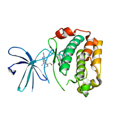
 | | COMPLEX OF TROPONIN C WITH A 47 RESIDUE (1-47) FRAGMENT OF TROPONIN I | | 分子名称: | CALCIUM ION, TROPONIN C, TROPONIN I | | 著者 | Vassylyev, D.G, Takeda, S, Wakatsuki, S, Maeda, K, Maeda, Y. | | 登録日 | 1998-01-13 | | 公開日 | 1998-07-15 | | 最終更新日 | 2024-02-07 | | 実験手法 | X-RAY DIFFRACTION (2.3 Å) | | 主引用文献 | Crystal structure of troponin C in complex with troponin I fragment at 2.3-A resolution.
Proc.Natl.Acad.Sci.USA, 95, 1998
|
|
2DEK
 | |
2DQX
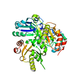
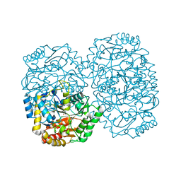
 | | mutant beta-amylase (W55R) from soy bean | | 分子名称: | Beta-amylase | | 著者 | Ishikawa, K. | | 登録日 | 2006-06-01 | | 公開日 | 2007-05-08 | | 最終更新日 | 2023-10-25 | | 実験手法 | X-RAY DIFFRACTION (2.2 Å) | | 主引用文献 | Kinetic and structural analysis of enzyme sliding on a substrate: multiple attack in beta-amylase
Biochemistry, 46, 2007
|
|
2Z7Q
 | |
5DGQ
 | |
5DGR
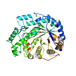
 | | Crystal structure of GH9 exo-beta-D-glucosaminidase PBPRA0520, glucosamine complex | | 分子名称: | 2-amino-2-deoxy-beta-D-glucopyranose, Putative endoglucanase-related protein, SODIUM ION | | 著者 | Suzuki, K, Honda, Y, Fushinobu, S. | | 登録日 | 2015-08-28 | | 公開日 | 2015-12-09 | | 最終更新日 | 2023-11-08 | | 実験手法 | X-RAY DIFFRACTION (1.9 Å) | | 主引用文献 | The crystal structure of an inverting glycoside hydrolase family 9 exo-beta-D-glucosaminidase and the design of glycosynthase.
Biochem.J., 473, 2016
|
|
2Z7S
 | |
2Z7R
 | |
3A8U
 | |


