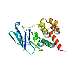
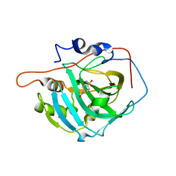
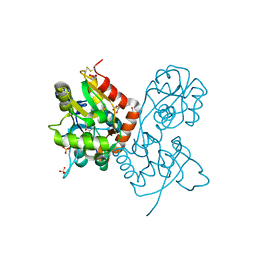
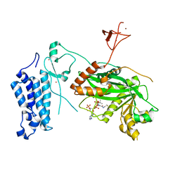
8CBA
 
 | |
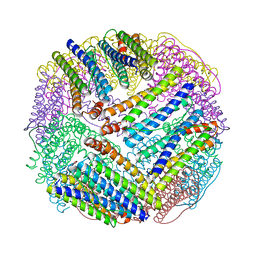
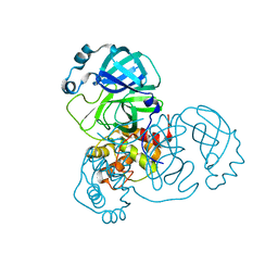
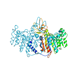
1EP7
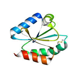 | | CRYSTAL STRUCTURE OF WT THIOREDOXIN H FROM CHLAMYDOMONAS REINHARDTII | | 分子名称: | THIOREDOXIN CH1, H-TYPE | | 著者 | Menchise, V, Corbier, C, Didierjean, C, Saviano, M, Benedetti, E, Jacquot, J.P, Aubry, A. | | 登録日 | 2000-03-28 | | 公開日 | 2001-12-12 | | 最終更新日 | 2011-07-13 | | 実験手法 | X-RAY DIFFRACTION (2.1 Å) | | 主引用文献 | Crystal structure of the wild-type and D30A mutant thioredoxin h of Chlamydomonas reinhardtii and implications for the catalytic mechanism.
Biochem.J., 359, 2001
|
|
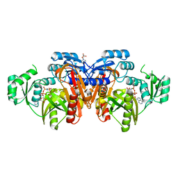
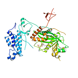
8CCT
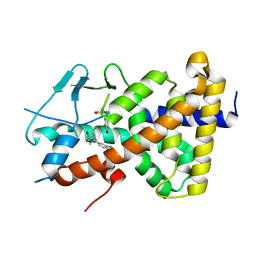 | | Crystal structure of the human PXR ligand-binding domain in complex with 2,2'-dichloro bisphenol A | | 分子名称: | 2-chloranyl-4-[2-(3-chloranyl-4-oxidanyl-phenyl)propan-2-yl]phenol, Nuclear receptor subfamily 1 group I member 2 | | 著者 | Derosa, Q, Grimaldi, M, Carivenc, C, Boulahtouf, A, Bourguet, W, Balaguer, P. | | 登録日 | 2023-01-27 | | 公開日 | 2024-02-07 | | 実験手法 | X-RAY DIFFRACTION (2.9 Å) | | 主引用文献 | Crystal structure of the hPXR-LBD in complex with 2,2'-dichloro bisphenol A
To Be Published
|
|
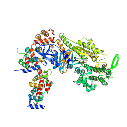
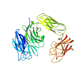
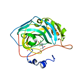
2LU3
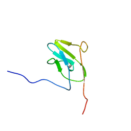 | |
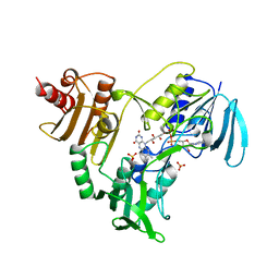
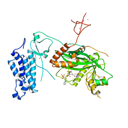
8CDX
 | |
8C8A
 | | The NMR structure of the MAX28 effector from Magnaporthe oryzae | | 分子名称: | But2 domain-containing protein | | 著者 | Lahfa, M, Padilla, A, de Guillen, K, Kroj, T, Roumestand, C, Barthe, P. | | 登録日 | 2023-01-19 | | 公開日 | 2024-02-14 | | 実験手法 | SOLUTION NMR | | 主引用文献 | 1H, 15N Backbone and side-chain NMR assignments for a MAX effector from Magnaporthe oryzae
To Be Published
|
|
1EUM
 | | CRYSTAL STRUCTURE OF THE E.COLI FERRITIN ECFTNA | | 分子名称: | FERRITIN 1 | | 著者 | Stillman, T.J, Hempstead, P.D, Artymiuk, P.J, Andrews, S.C, Hudson, A.J, Treffry, A, Guest, J.R, Harrison, P.M. | | 登録日 | 2000-04-17 | | 公開日 | 2001-03-28 | | 最終更新日 | 2024-02-07 | | 実験手法 | X-RAY DIFFRACTION (2.05 Å) | | 主引用文献 | The high-resolution X-ray crystallographic structure of the ferritin (EcFtnA) of Escherichia coli; comparison with human H ferritin (HuHF) and the structures of the Fe(3+) and Zn(2+) derivatives.
J.Mol.Biol., 307, 2001
|
|
8CDM
 | | Plasmodium falciparum Myosin A full-length, post-rigor state complexed to the inhibitor KNX-002 | | 分子名称: | 1,2-ETHANEDIOL, 1-(4-methoxyphenyl)-~{N}-[(3-thiophen-2-yl-1~{H}-pyrazol-4-yl)methyl]cyclopropan-1-amine, Myosin A tail domain interacting protein, ... | | 著者 | Moussaoui, D, Robblee, J.P, Robert-Paganin, J, Auguin, D, Fisher, F, Fagnant, P.M, MacFarlane, J.E, Mueller-Dieckmann, C, Baum, J, Trybus, K.M, Houdusse, A. | | 登録日 | 2023-01-31 | | 公開日 | 2024-02-21 | | 実験手法 | X-RAY DIFFRACTION (2.35 Å) | | 主引用文献 | Plasmodium falciparum Myosin A full-length, post-rigor state complexed to the inhibitor KNX-002
To Be Published
|
|
1NHP
 | | CRYSTALLOGRAPHIC ANALYSES OF NADH PEROXIDASE CYS42ALA AND CYS42SER MUTANTS: ACTIVE SITE STRUCTURE, MECHANISTIC IMPLICATIONS, AND AN UNUSUAL ENVIRONMENT OF ARG303 | | 分子名称: | FLAVIN-ADENINE DINUCLEOTIDE, NADH PEROXIDASE, SULFATE ION | | 著者 | Mande, S.S, Claiborne, A, Hol, W.G.J. | | 登録日 | 1994-12-09 | | 公開日 | 1995-02-14 | | 最終更新日 | 2024-02-14 | | 実験手法 | X-RAY DIFFRACTION (2 Å) | | 主引用文献 | Crystallographic analyses of NADH peroxidase Cys42Ala and Cys42Ser mutants: active site structures, mechanistic implications, and an unusual environment of Arg 303.
Biochemistry, 34, 1995
|
|
1WVJ
 | | Exploring the GluR2 ligand-binding core in complex with the bicyclic AMPA analogue (S)-4-AHCP | | 分子名称: | 3-(3-HYDROXY-7,8-DIHYDRO-6H-CYCLOHEPTA[D]ISOXAZOL-4-YL)-L-ALANINE, GLYCEROL, SULFATE ION, ... | | 著者 | Nielsen, B.B, Pickering, D.S, Greenwood, J.R, Brehm, L, Gajhede, M, Schousboe, A, Kastrup, J.S. | | 登録日 | 2004-12-15 | | 公開日 | 2005-04-26 | | 最終更新日 | 2023-10-25 | | 実験手法 | X-RAY DIFFRACTION (1.75 Å) | | 主引用文献 | Exploring the GluR2 ligand-binding core in complex with the bicyclical AMPA analogue (S)-4-AHCP
FEBS J., 272, 2005
|
|
8CDC
 | | Native 3CLpro from SARS-CoV-2 at 1.54 A | | 分子名称: | 3C-like proteinase | | 著者 | Mazzei, L, Jovanovic, A, Acton, T.B, Ciurli, S, Montelione, G.T. | | 登録日 | 2023-01-30 | | 公開日 | 2024-02-21 | | 実験手法 | X-RAY DIFFRACTION (1.54 Å) | | 主引用文献 | Expression, purification, and characterization of SARS-CoV2 3CLpro in a mature form
To Be Published
|
|
1EYZ
 | | STRUCTURE OF ESCHERICHIA COLI PURT-ENCODED GLYCINAMIDE RIBONUCLEOTIDE TRANSFORMYLASE COMPLEXED WITH MG AND AMPPNP | | 分子名称: | 3[N-MORPHOLINO]PROPANE SULFONIC ACID, CHLORIDE ION, MAGNESIUM ION, ... | | 著者 | Thoden, J.B, Firestine, S, Nixon, A, Benkovic, S.J, Holden, H.M. | | 登録日 | 2000-05-09 | | 公開日 | 2000-08-02 | | 最終更新日 | 2024-02-07 | | 実験手法 | X-RAY DIFFRACTION (1.75 Å) | | 主引用文献 | Molecular structure of Escherichia coli PurT-encoded glycinamide ribonucleotide transformylase.
Biochemistry, 39, 2000
|
|
1EUT
 | | SIALIDASE, LARGE 68KD FORM, COMPLEXED WITH GALACTOSE | | 分子名称: | SIALIDASE, SODIUM ION | | 著者 | Gaskell, A, Crennell, S.J, Taylor, G.L. | | 登録日 | 1996-06-21 | | 公開日 | 1997-01-11 | | 最終更新日 | 2011-07-13 | | 実験手法 | X-RAY DIFFRACTION (2.5 Å) | | 主引用文献 | The three domains of a bacterial sialidase: a beta-propeller, an immunoglobulin module and a galactose-binding jelly-roll.
Structure, 3, 1995
|
|
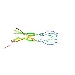
8CKM
 | | Semaphorin-5A TSR 3-4 domains | | 分子名称: | Semaphorin-5A | | 著者 | Nagy, G.N, Duman, R, Harlos, K, El Omari, K, Wagner, A, Jones, E.Y. | | 登録日 | 2023-02-15 | | 公開日 | 2024-02-28 | | 実験手法 | X-RAY DIFFRACTION (2.72 Å) | | 主引用文献 | Semaphorin-5A TSR 3-4 domains
To Be Published
|
|
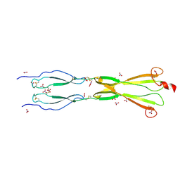
8CKG
 | | Semaphorin-5A TSR 3-4 domains in complex with sulfate | | 分子名称: | SULFATE ION, Semaphorin-5A, alpha-D-mannopyranose | | 著者 | Nagy, G.N, Duman, R, Harlos, K, El Omari, K, Wagner, A, Jones, E.Y. | | 登録日 | 2023-02-15 | | 公開日 | 2024-02-28 | | 実験手法 | X-RAY DIFFRACTION (1.714 Å) | | 主引用文献 | Semaphorin-5A TSR 3-4 domains in complex with sulfate
To Be Published
|
|
8CKK
 | | Semaphorin-5A TSR 3-4 domains in complex with nitrate | | 分子名称: | NITRATE ION, Semaphorin-5A, alpha-D-mannopyranose | | 著者 | Nagy, G.N, Duman, R, Harlos, K, El Omari, K, Wagner, A, Jones, E.Y. | | 登録日 | 2023-02-15 | | 公開日 | 2024-02-28 | | 実験手法 | X-RAY DIFFRACTION (1.56 Å) | | 主引用文献 | Semaphorin-5A TSR 3-4 domains in complex with nitrate
To Be Published
|
|
8CKL
 | | Semaphorin-5A TSR 3-4 domains in complex with sucrose octasulfate (SOS) | | 分子名称: | 2,3,4,6-tetra-O-sulfonato-alpha-D-glucopyranose-(1-2)-1,3,4,6-tetra-O-sulfo-beta-D-fructofuranose, Semaphorin-5A, alpha-D-mannopyranose | | 著者 | Nagy, G.N, Duman, R, Harlos, K, El Omari, K, Wagner, A, Jones, E.Y. | | 登録日 | 2023-02-15 | | 公開日 | 2024-02-28 | | 実験手法 | X-RAY DIFFRACTION (2.56 Å) | | 主引用文献 | Semaphorin-5A TSR 3-4 domains in complex with sucrose octasulfate (SOS)
To Be Published
|
|
1E9K
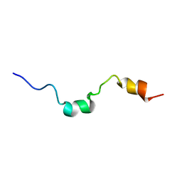 | | The structure of the RACK1 interaction sites located within the unique N-terminal region of the cAMP-specific phosphodiesterase, PDE4D5. | | 分子名称: | cAMP-specific 3',5'-cyclic phosphodiesterase 4D | | 著者 | Bolger, G.B, Smith, K.J, McCahill, A, Hyde, E.I, Steele, M.R, Houslay, M.D. | | 登録日 | 2000-10-20 | | 公開日 | 2001-10-18 | | 最終更新日 | 2024-05-15 | | 実験手法 | SOLUTION NMR | | 主引用文献 | 1H NMR structural and functional characterisation of a cAMP-specific phosphodiesterase-4D5 (PDE4D5) N-terminal region peptide that disrupts PDE4D5 interaction with the signalling scaffold proteins, beta-arrestin and RACK1.
Cell. Signal., 19, 2007
|
|
8CMZ
 | | Crystal structure of CREBBP-R1446C histone acetyltransferase domain in complex with Coenzyme A | | 分子名称: | COENZYME A, ZINC ION, histone acetyltransferase | | 著者 | Mechaly, A.E, Zhang, W, Haouz, A, Green, M, Rodrigues-Lima, F. | | 登録日 | 2023-02-21 | | 公開日 | 2024-03-06 | | 実験手法 | X-RAY DIFFRACTION (2.252 Å) | | 主引用文献 | Crystal structure of CREBBP-R1446C histone acetyltransferase domain in complex with Coenzyme A
To Be Published
|
|
8CN0
 | | Crystal structure of CREBBP-Y1482N histone acetyltransferase domain in complex with Coenzyme A | | 分子名称: | COENZYME A, ZINC ION, histone acetyltransferase | | 著者 | Mechaly, A.E, Zhang, W, Haouz, A, Green, M, Rodrigues-Lima, F. | | 登録日 | 2023-02-21 | | 公開日 | 2024-03-06 | | 実験手法 | X-RAY DIFFRACTION (2.44 Å) | | 主引用文献 | Crystal structure of CREBBP-Y1482N histone acetyltransferase domain in complex with Coenzyme A
To Be Published
|
|
1EW2
 | | CRYSTAL STRUCTURE OF A HUMAN PHOSPHATASE | | 分子名称: | 2-acetamido-2-deoxy-beta-D-glucopyranose, MAGNESIUM ION, PHOSPHATASE, ... | | 著者 | Le Du, M.H, Stigbrand, T, Taussig, M.J, Menez, A, Stura, E.A. | | 登録日 | 2000-04-21 | | 公開日 | 2001-04-04 | | 最終更新日 | 2020-07-29 | | 実験手法 | X-RAY DIFFRACTION (1.82 Å) | | 主引用文献 | Crystal structure of alkaline phosphatase from human placenta at 1.8 A resolution. Implication for a substrate specificity.
J.Biol.Chem., 276, 2001
|
|
3EFT
 | | Crystal structure of the complex between Carbonic Anhydrase II and a spin-labeled sulfonamide incorporating TEMPO moiety | | 分子名称: | 3-chloro-4-{[(1-hydroxy-2,2,6,6-tetramethylpiperidin-4-yl)carbamothioyl]amino}benzenesulfonamide, Carbonic anhydrase 2, MERCURY (II) ION, ... | | 著者 | Temperini, C, Cecchi, A, Scozzafava, A, Supuran, C.T. | | 登録日 | 2008-09-10 | | 公開日 | 2009-09-08 | | 最終更新日 | 2023-11-01 | | 実験手法 | X-RAY DIFFRACTION (1.85 Å) | | 主引用文献 | Dissecting the Inhibition Mechanism of Cytosolic versus Transmembrane Carbonic Anhydrases by ESR
J.Phys.Chem.B, 113, 2009
|
|
8CNA
 | | Crystal structure of CREBBP-R1446C histone acetyltransferase domain in complex with a bisubstrate inhibitor, Lys-CoA | | 分子名称: | ZINC ION, [(2R,3S,4R,5R)-5-(6-amino-9H-purin-9-yl)-4-hydroxy-3-(phosphonooxy)tetrahydrofuran-2-yl]methyl (3R,20R)-20-carbamoyl-3-hydroxy-2,2-dimethyl-4,8,14,22-tetraoxo-12-thia-5,9,15,21-tetraazatricos-1-yl dihydrogen diphosphate, histone acetyltransferase | | 著者 | Mechaly, A.E, Zhang, W, Haouz, A, Green, M, Rodrigues-Lima, F. | | 登録日 | 2023-02-22 | | 公開日 | 2024-03-06 | | 実験手法 | X-RAY DIFFRACTION (2.463 Å) | | 主引用文献 | Crystal structure of CREBBP-R1446C histone acetyltransferase domain in complex with a bisubstrate inhibitor, Lys-CoA
To Be Published
|
|
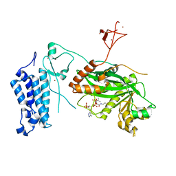
3EFZ
 | | Crystal Structure of a 14-3-3 protein from cryptosporidium parvum (cgd1_2980) | | 分子名称: | 1,2-ETHANEDIOL, 14-3-3 protein | | 著者 | Wernimont, A.K, Dong, A, Qiu, W, Lew, J, Wasney, G.A, Vedadi, M, Kozieradzki, I, Zhao, Y, Ren, H, Alam, Z, Lin, Y.H, Sundstrom, M, Weigelt, J, Arrowsmith, C.H, Edwards, A.M, Bochkarev, A, Hui, R, Brokx, S, Structural Genomics Consortium (SGC) | | 登録日 | 2008-09-10 | | 公開日 | 2008-09-23 | | 最終更新日 | 2012-02-22 | | 実験手法 | X-RAY DIFFRACTION (2.08 Å) | | 主引用文献 | Characterization of 14-3-3 proteins from Cryptosporidium parvum.
Plos One, 6, 2011
|
|
8CND
 | | Crystal structure of CREBBP-Y1482N histone acetyltransferase domain in complex with a bisubstrate inhibitor, Lys-CoA | | 分子名称: | ZINC ION, [(2R,3S,4R,5R)-5-(6-amino-9H-purin-9-yl)-4-hydroxy-3-(phosphonooxy)tetrahydrofuran-2-yl]methyl (3R,20R)-20-carbamoyl-3-hydroxy-2,2-dimethyl-4,8,14,22-tetraoxo-12-thia-5,9,15,21-tetraazatricos-1-yl dihydrogen diphosphate, histone acetyltransferase | | 著者 | Mechaly, A.E, Zhang, W, Haouz, A, Green, M, Rodrigues-Lima, F. | | 登録日 | 2023-02-22 | | 公開日 | 2024-03-06 | | 実験手法 | X-RAY DIFFRACTION (2.972 Å) | | 主引用文献 | Crystal structure of CREBBP-Y1482N histone acetyltransferase domain in complex with a bisubstrate inhibitor, Lys-CoA
To Be Published
|
|


