Loading
PDBj
メニューPDBj@FacebookPDBj@TwitterPDBj@YouTubewwPDB FoundationwwPDB
RCSB PDBPDBeBMRBAdv. SearchSearch help
Search by PDB author
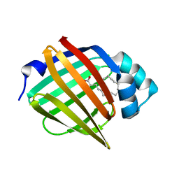
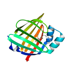
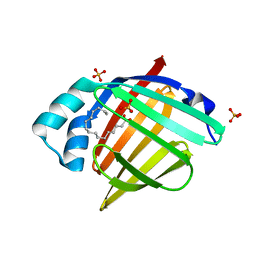
6XVR
DownloadVisualize
BU of 6xvr by Molmil
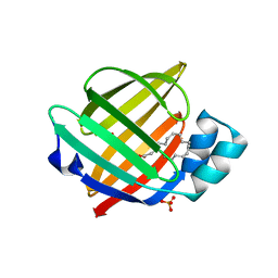
Human myelin protein P2 mutant L35S
分子名称: Myelin P2 protein, PALMITIC ACID, SULFATE ION
著者Ruskamo, S, Lehtimaki, M, Kursula, P.
登録日2020-01-22
公開日2020-04-08
最終更新日2024-01-24
実験手法X-RAY DIFFRACTION (2 Å)
主引用文献Cryo-EM, X-ray diffraction, and atomistic simulations reveal determinants for the formation of a supramolecular myelin-like proteolipid lattice.
J.Biol.Chem., 295, 2020
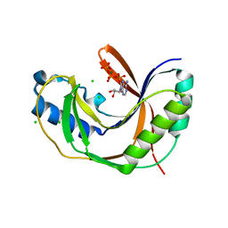
6ZMM
DownloadVisualize
BU of 6zmm by Molmil
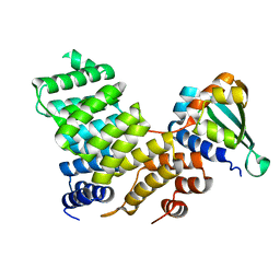
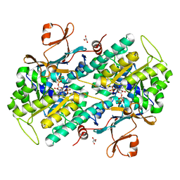
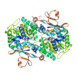
Crystal structure of human NDRG1
分子名称: CHLORIDE ION, Protein NDRG1
著者Mustonen, V, Kursula, P, Ruskamo, S.
登録日2020-07-03
公開日2020-12-23
最終更新日2024-01-31
実験手法X-RAY DIFFRACTION (2.96 Å)
主引用文献Crystal and solution structure of NDRG1, a membrane-binding protein linked to myelination and tumour suppression.
Febs J., 288, 2021
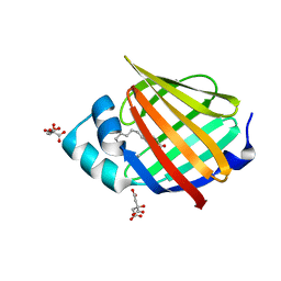
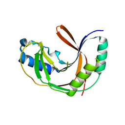
6XU5
DownloadVisualize
BU of 6xu5 by Molmil
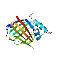
Human myelin protein P2 mutant N2D
分子名称: CITRIC ACID, Myelin P2 protein, PALMITIC ACID
著者Ruskamo, S, Lehtimaki, M, Kursula, P.
登録日2020-01-17
公開日2020-04-08
最終更新日2024-01-24
実験手法X-RAY DIFFRACTION (1.65 Å)
主引用文献Cryo-EM, X-ray diffraction, and atomistic simulations reveal determinants for the formation of a supramolecular myelin-like proteolipid lattice.
J.Biol.Chem., 295, 2020
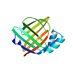
6XVS
DownloadVisualize
BU of 6xvs by Molmil
Human myelin protein P2 mutant P38G, unliganded
分子名称: GLYCEROL, Myelin P2 protein
著者Ruskamo, S, Lehtimaki, M, Kursula, P.
登録日2020-01-22
公開日2020-04-08
最終更新日2024-01-24
実験手法X-RAY DIFFRACTION (1.8 Å)
主引用文献Cryo-EM, X-ray diffraction, and atomistic simulations reveal determinants for the formation of a supramolecular myelin-like proteolipid lattice.
J.Biol.Chem., 295, 2020
6XUW
DownloadVisualize
BU of 6xuw by Molmil
Human myelin protein P2 mutant L27D
分子名称: CHLORIDE ION, Myelin P2 protein, PALMITIC ACID
著者Ruskamo, S, Lehtimaki, M, Kursula, P.
登録日2020-01-21
公開日2020-04-08
最終更新日2024-01-24
実験手法X-RAY DIFFRACTION (2.31 Å)
主引用文献Cryo-EM, X-ray diffraction, and atomistic simulations reveal determinants for the formation of a supramolecular myelin-like proteolipid lattice.
J.Biol.Chem., 295, 2020
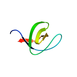
6HYF
DownloadVisualize
BU of 6hyf by Molmil
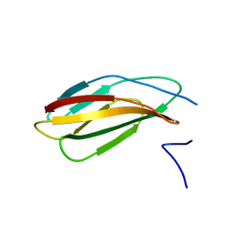
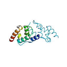
Crystal structure of the third FNIII domain from rat beta4 integrin, a binding site for periaxin
分子名称: Integrin beta-4
著者Raasakka, A, Kursula, P.
登録日2018-10-20
公開日2019-05-08
最終更新日2024-01-24
実験手法X-RAY DIFFRACTION (1.6 Å)
主引用文献Direct Binding of the Flexible C-Terminal Segment of Periaxin to beta 4 Integrin Suggests a Molecular Basis for CMT4F.
Front Mol Neurosci, 12, 2019
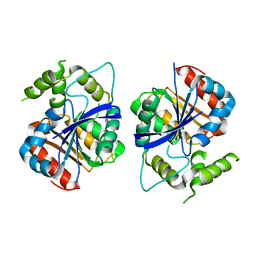
5LDQ
DownloadVisualize
BU of 5ldq by Molmil
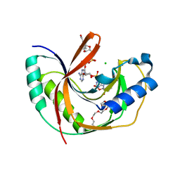
Crystal structure of E.coli LigT complexed with NADP+
分子名称: CHLORIDE ION, NADP NICOTINAMIDE-ADENINE-DINUCLEOTIDE PHOSPHATE, PENTAETHYLENE GLYCOL, ...
著者Myllykoski, M, Kursula, P.
登録日2016-06-27
公開日2017-01-18
最終更新日2024-01-10
実験手法X-RAY DIFFRACTION (1.7 Å)
主引用文献Structural aspects of nucleotide ligand binding by a bacterial 2H phosphoesterase.
PLoS ONE, 12, 2017
5LXX
DownloadVisualize
BU of 5lxx by Molmil
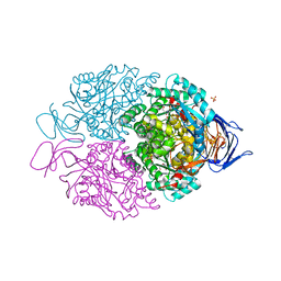
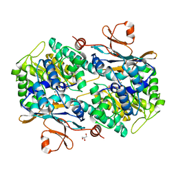
High-resolution structure of human collapsin response mediator protein 2
分子名称: 2-[BIS-(2-HYDROXY-ETHYL)-AMINO]-2-HYDROXYMETHYL-PROPANE-1,3-DIOL, Dihydropyrimidinase-related protein 2, SULFATE ION
著者Myllykoski, M, Hensley, K, Kursula, P.
登録日2016-09-23
公開日2017-01-11
最終更新日2024-01-17
実験手法X-RAY DIFFRACTION (1.25 Å)
主引用文献Collapsin response mediator protein 2: high-resolution crystal structure sheds light on small-molecule binding, post-translational modifications, and conformational flexibility.
Amino Acids, 49, 2017
5LDJ
DownloadVisualize
BU of 5ldj by Molmil
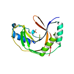
Crystal structure of E.coli LigT complexed with phosphate
分子名称: PHOSPHATE ION, RNA 2',3'-cyclic phosphodiesterase
著者Myllykoski, M, Kursula, P.
登録日2016-06-27
公開日2017-01-18
最終更新日2024-01-10
実験手法X-RAY DIFFRACTION (2.802 Å)
主引用文献Structural aspects of nucleotide ligand binding by a bacterial 2H phosphoesterase.
PLoS ONE, 12, 2017
5LDK
DownloadVisualize
BU of 5ldk by Molmil
Crystal structure of E.coli LigT complexed with ATP
分子名称: ADENOSINE-5'-TRIPHOSPHATE, RNA 2',3'-cyclic phosphodiesterase
著者Myllykoski, M, Kursula, P.
登録日2016-06-27
公開日2017-01-18
最終更新日2024-01-10
実験手法X-RAY DIFFRACTION (2.099 Å)
主引用文献Structural aspects of nucleotide ligand binding by a bacterial 2H phosphoesterase.
PLoS ONE, 12, 2017
5LDP
DownloadVisualize
BU of 5ldp by Molmil
Crystal structure of E.coli LigT complexed with ATP
分子名称: ADENOSINE-5'-TRIPHOSPHATE, CHLORIDE ION, MAGNESIUM ION, ...
著者Myllykoski, M, Kursula, P.
登録日2016-06-27
公開日2017-01-18
最終更新日2024-01-10
実験手法X-RAY DIFFRACTION (1.8 Å)
主引用文献Structural aspects of nucleotide ligand binding by a bacterial 2H phosphoesterase.
PLoS ONE, 12, 2017
5O99
DownloadVisualize
BU of 5o99 by Molmil
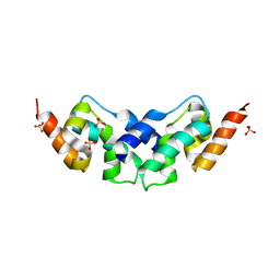
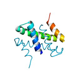
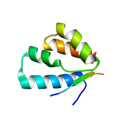
Unconventional SH3 domain from the postsynaptic density scaffold protein Shank3
分子名称: 2-[BIS-(2-HYDROXY-ETHYL)-AMINO]-2-HYDROXYMETHYL-PROPANE-1,3-DIOL, SH3 and multiple ankyrin repeat domains protein 3
著者Ponna, S.K, Myllykoski, M, Boeckers, T.M, Kursula, P.
登録日2017-06-16
公開日2017-07-05
最終更新日2024-05-08
実験手法X-RAY DIFFRACTION (0.871 Å)
主引用文献Structure of an unconventional SH3 domain from the postsynaptic density protein Shank3 at ultrahigh resolution.
Biochem. Biophys. Res. Commun., 490, 2017
6S2M
DownloadVisualize
BU of 6s2m by Molmil
Perdeuterated human myelin protein P2 at 0.72-A resolution
分子名称: Myelin P2 protein, PALMITIC ACID, VACCENIC ACID
著者Laulumaa, S, Kursula, P.
登録日2019-06-21
公開日2019-08-28
最終更新日2024-01-24
実験手法X-RAY DIFFRACTION (0.72 Å)
主引用文献Sub-Atomic Resolution Crystal Structures Reveal Conserved Geometric Outliers at Functional Sites.
Molecules, 24, 2019
2C0L
DownloadVisualize
BU of 2c0l by Molmil
TPR DOMAIN OF HUMAN PEX5P IN COMPLEX WITH HUMAN MSCP2
分子名称: NONSPECIFIC LIPID-TRANSFER PROTEIN, PEROXISOMAL TARGETING SIGNAL 1 RECEPTOR
著者Stanley, W.A, Kursula, P, Wilmanns, M.
登録日2005-09-05
公開日2006-11-15
最終更新日2024-05-08
実験手法X-RAY DIFFRACTION (2.3 Å)
主引用文献Recognition of a Functional Peroxisome Type 1 Target by the Dynamic Import Receptor Pex5P.
Mol.Cell, 24, 2006
6S2S
DownloadVisualize
BU of 6s2s by Molmil
Hydrogenated human myelin protein P2 at 0.86-A resolution
分子名称: CITRIC ACID, Myelin P2 protein, PALMITIC ACID, ...
著者Laulumaa, S, Kursula, P.
登録日2019-06-21
公開日2019-08-28
最終更新日2024-01-24
実験手法X-RAY DIFFRACTION (0.86 Å)
主引用文献Sub-Atomic Resolution Crystal Structures Reveal Conserved Geometric Outliers at Functional Sites.
Molecules, 24, 2019
6SID
DownloadVisualize
BU of 6sid by Molmil
Crystal structure of the C-lobe of drosophila Arc 1 at atomic resolution
分子名称: Activity-regulated cytoskeleton associated protein 1
著者Hallin, E.I, Kursula, P.
登録日2019-08-09
公開日2019-08-28
最終更新日2024-06-19
実験手法X-RAY DIFFRACTION (1.05 Å)
主引用文献Crystal and solution structures reveal oligomerization of individual capsid homology domains of Drosophila Arc.
Plos One, 16, 2021
6STS
DownloadVisualize
BU of 6sts by Molmil
Human myelin protein P2 mutant R30Q
分子名称: Myelin P2 protein, PALMITIC ACID, SULFATE ION
著者Ruskamo, S, Lehtimaki, M, Kursula, P.
登録日2019-09-11
公開日2020-04-08
最終更新日2024-01-24
実験手法X-RAY DIFFRACTION (3 Å)
主引用文献Cryo-EM, X-ray diffraction, and atomistic simulations reveal determinants for the formation of a supramolecular myelin-like proteolipid lattice.
J.Biol.Chem., 295, 2020
6TA0
DownloadVisualize
BU of 6ta0 by Molmil
Human NAMPT in complex with nicotinic acid and phosphoribosyl pyrophosphate
分子名称: 1-O-pyrophosphono-5-O-phosphono-alpha-D-ribofuranose, GLYCEROL, NICOTINIC ACID, ...
著者Houry, D, Raasakka, A, Kursula, P, Ziegler, M.
登録日2019-10-29
公開日2020-11-18
最終更新日2024-01-24
実験手法X-RAY DIFFRACTION (1.58 Å)
主引用文献Identification of structural determinants of NAMPT activity and substrate selectivity
To Be Published
6TA2
DownloadVisualize
BU of 6ta2 by Molmil
Human NAMPT in complex with nicotinic acid mononucleotide and phosphate
分子名称: CHLORIDE ION, GLYCEROL, NICOTINATE MONONUCLEOTIDE, ...
著者Houry, D, Raasakka, A, Kursula, P, Ziegler, M.
登録日2019-10-29
公開日2020-11-18
最終更新日2024-01-24
実験手法X-RAY DIFFRACTION (1.68 Å)
主引用文献Identification of structural determinants of NAMPT activity and substrate selectivity
To Be Published
6SIE
DownloadVisualize
BU of 6sie by Molmil
Crystal structure of the C-lobe of drosophila Arc 2
分子名称: Activity-regulated cytoskeleton associated protein 2, SULFATE ION
著者Hallin, E.I, Kursula, P.
登録日2019-08-09
公開日2019-08-28
最終更新日2024-10-16
実験手法X-RAY DIFFRACTION (2.8 Å)
主引用文献Crystal and solution structures reveal oligomerization of individual capsid homology domains of Drosophila Arc.
Plos One, 16, 2021
6SIB
DownloadVisualize
BU of 6sib by Molmil
Crystal structure of the domain-swapped N-lobe dimer of drosophila Arc 2
分子名称: Activity-regulated cytoskeleton associated protein 2
著者Hallin, E.I, Kursula, P.
登録日2019-08-09
公開日2019-08-28
最終更新日2024-06-19
実験手法X-RAY DIFFRACTION (1.9 Å)
主引用文献Crystal and solution structures reveal oligomerization of individual capsid homology domains of Drosophila Arc.
Plos One, 16, 2021
6TAC
DownloadVisualize
BU of 6tac by Molmil
Human NAMPT deletion mutant in complex with nicotinamide mononucleotide, pyrophosphate, and Mg2+
分子名称: BETA-NICOTINAMIDE RIBOSE MONOPHOSPHATE, GLYCEROL, MAGNESIUM ION, ...
著者Houry, D, Raasakka, A, Kursula, P, Ziegler, M.
登録日2019-10-29
公開日2020-11-18
最終更新日2024-01-24
実験手法X-RAY DIFFRACTION (1.6 Å)
主引用文献Identification of structural determinants of NAMPT activity and substrate selectivity
To Be Published
2J9Q
DownloadVisualize
BU of 2j9q by Molmil
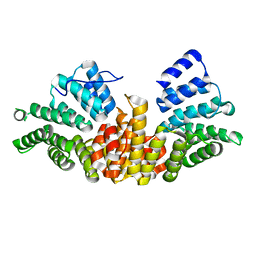
A novel conformation for the TPR domain of pex5p
分子名称: PEROXISOMAL TARGETING SIGNAL 1 RECEPTOR, STRONTIUM ION
著者Stanley, W.A, Wilmanns, M, Kursula, P.
登録日2006-11-15
公開日2007-04-17
最終更新日2023-12-13
実験手法X-RAY DIFFRACTION (2.65 Å)
主引用文献A Previously Unobserved Conformation for the Human Pex5P Receptor Suggests Roles for Intrinsic Flexibility and Rigid Domain Motions in Ligand Binding
Bmc Struct.Biol., 7, 2007
6TQ0
DownloadVisualize
BU of 6tq0 by Molmil
Crystal structure of the human Arc N-lobe bound to repeat peptide 5 from GKAP
分子名称: Activity-regulated cytoskeleton-associated protein, repeat peptide 5 from GKAP
著者Hallin, E.I, Bramham, C.R, Kursula, P.
登録日2019-12-15
公開日2021-01-13
最終更新日2024-01-24
実験手法X-RAY DIFFRACTION (1.95 Å)
主引用文献Structural properties and peptide ligand binding of the capsid homology domains of human Arc.
Biochem Biophys Rep, 26, 2021
2C0M
DownloadVisualize
BU of 2c0m by Molmil
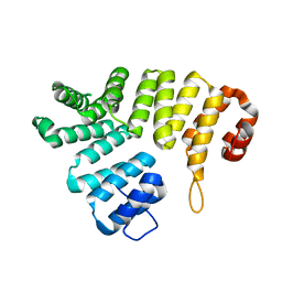
apo form of the TPR domain of the pex5p receptor
分子名称: PEROXISOMAL TARGETING SIGNAL 1 RECEPTOR
著者Stanley, W.A, Kursula, P, Wilmanns, M.
登録日2005-09-05
公開日2006-11-15
最終更新日2024-05-08
実験手法X-RAY DIFFRACTION (2.5 Å)
主引用文献Recognition of a Functional Peroxisome Type 1 Target by the Dynamic Import Receptor Pex5P.
Mol.Cell, 24, 2006

226707

件を2024-10-30に公開中

PDB statisticsPDBj update infoContact PDBjnumon