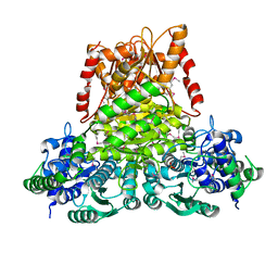
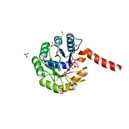
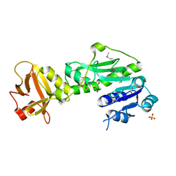
3V4Z
 
 | | D-alanine--D-alanine ligase from Yersinia pestis | | 分子名称: | D-alanine--D-alanine ligase, DI(HYDROXYETHYL)ETHER, TRIETHYLENE GLYCOL | | 著者 | Osipiuk, J, Nocek, B, Mulligan, R, Papazisi, L, Anderson, W.F, Joachimiak, A, Center for Structural Genomics of Infectious Diseases (CSGID) | | 登録日 | 2011-12-15 | | 公開日 | 2011-12-28 | | 最終更新日 | 2023-09-13 | | 実験手法 | X-RAY DIFFRACTION (2.69 Å) | | 主引用文献 | D-alanine--D-alanine ligase from Yersinia pestis.
To be Published
|
|
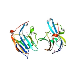
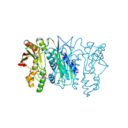
3KOM
 | | Crystal structure of apo transketolase from Francisella tularensis | | 分子名称: | Transketolase | | 著者 | Anderson, S.M, Wawrzak, Z, Skarina, T, Gordon, E, Kwon, K, Savchenko, A, Anderson, W.F, Center for Structural Genomics of Infectious Diseases (CSGID) | | 登録日 | 2009-11-13 | | 公開日 | 2009-12-01 | | 最終更新日 | 2021-10-13 | | 実験手法 | X-RAY DIFFRACTION (1.6 Å) | | 主引用文献 | Crystal structure of apo transketolase from Francisella tularensis
TO BE PUBLISHED
|
|
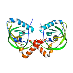
3QN3
 | | Phosphopyruvate hydratase from Campylobacter jejuni. | | 分子名称: | (4S)-2-METHYL-2,4-PENTANEDIOL, Enolase, GLYCEROL, ... | | 著者 | Osipiuk, J, Gu, M, Kwon, K, Anderson, W.F, Joachimiak, A, Center for Structural Genomics of Infectious Diseases (CSGID) | | 登録日 | 2011-02-07 | | 公開日 | 2011-02-23 | | 最終更新日 | 2023-09-13 | | 実験手法 | X-RAY DIFFRACTION (2.13 Å) | | 主引用文献 | Phosphopyruvate hydratase from Campylobacter jejuni.
To be Published
|
|
3QFG
 | | Structure of a putative lipoprotein from Staphylococcus aureus subsp. aureus NCTC 8325 | | 分子名称: | Uncharacterized protein | | 著者 | Filippova, E.V, Halavaty, A, Shuvalova, L, Minasov, G, Dubrovska, I, Winsor, J, Kiryukhina, O, Papazisi, L, Bagnoli, F, Falugi, F, Bottomley, M, Grandi, G, Anderson, W.F, Center for Structural Genomics of Infectious Diseases (CSGID) | | 登録日 | 2011-01-21 | | 公開日 | 2011-02-02 | | 最終更新日 | 2017-11-08 | | 実験手法 | X-RAY DIFFRACTION (3.08 Å) | | 主引用文献 | Structure of a putative lipoprotein from Staphylococcus aureus subsp. aureus NCTC 8325
To be Published
|
|
3LG3
 | | 1.4A Crystal Structure of Isocitrate Lyase from Yersinia pestis CO92 | | 分子名称: | Isocitrate lyase | | 著者 | Sharma, S.S, Brunzelle, J.S, Skarina, T, Savchenko, A, Anderson, W.F, Center for Structural Genomics of Infectious Diseases (CSGID) | | 登録日 | 2010-01-19 | | 公開日 | 2010-04-14 | | 最終更新日 | 2011-12-14 | | 実験手法 | X-RAY DIFFRACTION (1.4 Å) | | 主引用文献 | 1.4A Crystal Structure of Isocitrate Lyase from Yersinia pestis CO92
To be Published
|
|
3L92
 | | Phosphopantetheine adenylyltransferase from Yersinia pestis complexed with coenzyme A. | | 分子名称: | COENZYME A, Phosphopantetheine adenylyltransferase | | 著者 | Osipiuk, J, Maltseva, N, Makowska-grzyska, M, Kwon, K, Anderson, W.F, Joachimiak, A, Center for Structural Genomics of Infectious Diseases (CSGID) | | 登録日 | 2010-01-04 | | 公開日 | 2010-01-19 | | 最終更新日 | 2023-09-06 | | 実験手法 | X-RAY DIFFRACTION (1.89 Å) | | 主引用文献 | X-ray crystal structure of phosphopantetheine adenylyltransferase from Yersinia pestis.
To be Published
|
|
3QJG
 | | Epidermin biosynthesis protein EpiD from Staphylococcus aureus | | 分子名称: | CHLORIDE ION, Epidermin biosynthesis protein EpiD, FLAVIN MONONUCLEOTIDE | | 著者 | Osipiuk, J, Makowska-Grzyska, M, Kwon, K, Anderson, W.F, Joachimiak, A, Center for Structural Genomics of Infectious Diseases (CSGID) | | 登録日 | 2011-01-28 | | 公開日 | 2011-02-09 | | 最終更新日 | 2023-09-13 | | 実験手法 | X-RAY DIFFRACTION (2.04 Å) | | 主引用文献 | Epidermin biosynthesis protein EpiD from Staphylococcus aureus.
To be Published
|
|
3NX3
 | | Crystal structure of acetylornithine aminotransferase (argD) from Campylobacter jejuni | | 分子名称: | Acetylornithine aminotransferase, MAGNESIUM ION | | 著者 | Anderson, S.M, Wawrzak, Z, Onopriyenko, O, Skarina, T, Anderson, W.F, Savchenko, A, Center for Structural Genomics of Infectious Diseases (CSGID) | | 登録日 | 2010-07-12 | | 公開日 | 2010-08-25 | | 最終更新日 | 2011-07-13 | | 実験手法 | X-RAY DIFFRACTION (1.8 Å) | | 主引用文献 |
|
|
3MZZ
 | | Crystal Structure of Rhodanese-like Domain Protein from Staphylococcus aureus | | 分子名称: | Rhodanese-like domain protein | | 著者 | Kim, Y, Chruszcz, M, Minor, W, Edwards, A, Anderson, W.F, Joachimiak, A, Center for Structural Genomics of Infectious Diseases (CSGID) | | 登録日 | 2010-05-13 | | 公開日 | 2010-06-30 | | 最終更新日 | 2022-04-13 | | 実験手法 | X-RAY DIFFRACTION (2 Å) | | 主引用文献 | Crystal Structure of Rhodanese-like Domain Protein from Staphylococcus aureus
To be Published
|
|
3R2T
 | | 2.2 Angstrom Resolution Crystal Structure of Superantigen-like Protein from Staphylococcus aureus subsp. aureus NCTC 8325. | | 分子名称: | Putative uncharacterized protein, SULFATE ION | | 著者 | Minasov, G, Halavaty, A, Shuvalova, L, Dubrovska, I, Winsor, J, Kiryukhina, O, Falugi, F, Bottomley, M, Bagnoli, F, Grandi, G, Anderson, W.F, Center for Structural Genomics of Infectious Diseases (CSGID) | | 登録日 | 2011-03-14 | | 公開日 | 2011-03-23 | | 最終更新日 | 2023-09-13 | | 実験手法 | X-RAY DIFFRACTION (2.21 Å) | | 主引用文献 | 2.2 Angstrom Resolution Crystal Structure of Superantigen-like Protein from Staphylococcus aureus subsp. aureus NCTC 8325.
TO BE PUBLISHED
|
|
3OET
 | | D-Erythronate-4-Phosphate Dehydrogenase complexed with NAD | | 分子名称: | Erythronate-4-phosphate dehydrogenase, NICOTINAMIDE-ADENINE-DINUCLEOTIDE | | 著者 | Filippova, E.V, Wawrzak, Z, Onopriyenko, O, Savchenko, A, Edwards, A, Anderson, W.F, Center for Structural Genomics of Infectious Diseases (CSGID) | | 登録日 | 2010-08-13 | | 公開日 | 2010-08-25 | | 最終更新日 | 2023-12-06 | | 実験手法 | X-RAY DIFFRACTION (2.36 Å) | | 主引用文献 | D-Erythronate-4-Phosphate Dehydrogenase complexed with NAD
To be Published
|
|
3IV8
 | | N-acetylglucosamine-6-phosphate deacetylase from Vibrio cholerae complexed with fructose 6-phosphate | | 分子名称: | 6-O-phosphono-beta-D-fructofuranose, N-acetylglucosamine-6-phosphate deacetylase, NICKEL (II) ION, ... | | 著者 | Osipiuk, J, Maltseva, N, Stam, J, Anderson, W.F, Joachimiak, A, Center for Structural Genomics of Infectious Diseases (CSGID) | | 登録日 | 2009-08-31 | | 公開日 | 2009-09-08 | | 最終更新日 | 2023-09-06 | | 実験手法 | X-RAY DIFFRACTION (2.53 Å) | | 主引用文献 | X-ray crystal structure of N-acetylglucosamine-6-phosphate deacetylase from Vibrio cholerae complexed with fructose 6-phosphate.
To be Published
|
|
3TL2
 | | Crystal structure of Bacillus anthracis str. Ames malate dehydrogenase in closed conformation. | | 分子名称: | 1,2-ETHANEDIOL, Malate dehydrogenase, THIOCYANATE ION | | 著者 | Blus, B.J, Chruszcz, M, Tkaczuk, K.L, Osinski, T, Cymborowski, M, Kudritska, M, Grimshaw, S, Savchenko, A, Anderson, W.F, Minor, W, Center for Structural Genomics of Infectious Diseases (CSGID) | | 登録日 | 2011-08-29 | | 公開日 | 2011-09-21 | | 最終更新日 | 2023-09-13 | | 実験手法 | X-RAY DIFFRACTION (1.7 Å) | | 主引用文献 | Crystal structure of Bacillus anthracis str. Ames malate dehydrogenase in closed conformation.
TO BE PUBLISHED
|
|
3QU1
 | | Peptide deformylase from Vibrio cholerae | | 分子名称: | CHLORIDE ION, Peptide deformylase 2, SULFATE ION, ... | | 著者 | Osipiuk, J, Mulligan, R, Papazisi, L, Anderson, W.F, Joachimiak, A, Center for Structural Genomics of Infectious Diseases (CSGID) | | 登録日 | 2011-02-23 | | 公開日 | 2011-03-09 | | 最終更新日 | 2023-09-13 | | 実験手法 | X-RAY DIFFRACTION (1.8 Å) | | 主引用文献 | Peptide deformylase from Vibrio cholerae.
To be Published
|
|
3O04
 | | Crystal structure of the beta-keto-acyl carrier protein synthase II (lmo2201) from Listeria monocytogenes | | 分子名称: | beta-keto-acyl carrier protein synthase II | | 著者 | Anderson, S.M, Wawrzak, Z, Onopriyenko, O, Anderson, W.F, Savchenko, A, Center for Structural Genomics of Infectious Diseases (CSGID) | | 登録日 | 2010-07-19 | | 公開日 | 2010-08-04 | | 最終更新日 | 2021-10-06 | | 実験手法 | X-RAY DIFFRACTION (1.85 Å) | | 主引用文献 | Crystal structure of the beta-keto-acyl carrier protein synthase II (lmo2201) from Listeria monocytogenes
To be Published
|
|
3IGS
 | | Structure of the Salmonella enterica N-acetylmannosamine-6-phosphate 2-epimerase | | 分子名称: | 2-acetamido-2-deoxy-6-O-phosphono-alpha-D-glucopyranose, CHLORIDE ION, N-acetylmannosamine-6-phosphate 2-epimerase 2, ... | | 著者 | Anderson, S.M, Wawrzak, Z, Gordon, E, Skarina, T, Papazisi, L, Anderson, W.F, Savchenko, A, Center for Structural Genomics of Infectious Diseases (CSGID) | | 登録日 | 2009-07-28 | | 公開日 | 2009-08-04 | | 最終更新日 | 2020-07-29 | | 実験手法 | X-RAY DIFFRACTION (1.5 Å) | | 主引用文献 |
|
|
3O6V
 | | Crystal structure of Uridine Phosphorylase from Vibrio cholerae O1 biovar El Tor | | 分子名称: | FORMIC ACID, GLYCEROL, Uridine phosphorylase | | 著者 | Maltseva, N, Kim, Y, Hasseman, J, Anderson, W.F, Joachimiak, A, Center for Structural Genomics of Infectious Diseases (CSGID) | | 登録日 | 2010-07-29 | | 公開日 | 2010-10-13 | | 最終更新日 | 2023-09-06 | | 実験手法 | X-RAY DIFFRACTION (1.695 Å) | | 主引用文献 | Crystal structure of Uridine Phosphorylase from Vibrio cholerae O1 biovar El Tor
To be Published
|
|
3O1K
 | | Crystal structure of putative dihydroneopterin aldolase (FolB) from Vibrio cholerae O1 biovar El Tor str. N16961 | | 分子名称: | 1,2-ETHANEDIOL, Dihydroneopterin aldolase FolB, putative | | 著者 | Nocek, B, Zhou, M, Papazisi, L, Anderson, W, Joachimiak, A, Center for Structural Genomics of Infectious Diseases (CSGID) | | 登録日 | 2010-07-21 | | 公開日 | 2010-09-08 | | 最終更新日 | 2024-02-21 | | 実験手法 | X-RAY DIFFRACTION (1.95 Å) | | 主引用文献 | Crystal structure of putative dihydroneopterin aldolase (FolB) from Vibrio cholerae O1 biovar El Tor str. N16961
To be Published
|
|
3P4E
 | | Phosphoribosylformylglycinamidine cyclo-ligase from Vibrio cholerae | | 分子名称: | 1,2-ETHANEDIOL, ADENOSINE MONOPHOSPHATE, CITRIC ACID, ... | | 著者 | Osipiuk, J, Zhou, M, Papazisi, L, Anderson, W.F, Joachimiak, A, Center for Structural Genomics of Infectious Diseases (CSGID) | | 登録日 | 2010-10-06 | | 公開日 | 2010-10-20 | | 最終更新日 | 2023-09-06 | | 実験手法 | X-RAY DIFFRACTION (1.77 Å) | | 主引用文献 | Phosphoribosylformylglycinamidine cyclo-ligase from Vibrio cholerae.
To be Published
|
|
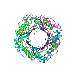
3LAY
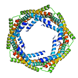 | | Alpha-Helical barrel formed by the decamer of the zinc resistance-associated protein (STM4172) from Salmonella enterica subsp. enterica serovar Typhimurium str. LT2 | | 分子名称: | Zinc resistance-associated protein | | 著者 | Filippova, E.V, Minasov, G, Shuvalova, L, Winsor, J, Dubrovska, I, Anderson, W.F, Center for Structural Genomics of Infectious Diseases (CSGID) | | 登録日 | 2010-01-07 | | 公開日 | 2010-01-19 | | 最終更新日 | 2024-02-21 | | 実験手法 | X-RAY DIFFRACTION (2.7 Å) | | 主引用文献 | Alpha-Helical barrel formed by the decamer of the zinc resistance-associated protein (STM4172) from Salmonella enterica subsp. enterica serovar Typhimurium str. LT2
To be Published
|
|
3Q12
 | | Pantoate-beta-alanine ligase from Yersinia pestis in complex with pantoate. | | 分子名称: | CHLORIDE ION, PANTOATE, Pantoate--beta-alanine ligase | | 著者 | Osipiuk, J, Maltseva, N, Kwon, K, Anderson, W.F, Joachimiak, A, Center for Structural Genomics of Infectious Diseases (CSGID) | | 登録日 | 2010-12-16 | | 公開日 | 2011-02-02 | | 最終更新日 | 2023-09-13 | | 実験手法 | X-RAY DIFFRACTION (1.58 Å) | | 主引用文献 | Pantoate-beta-alanine ligase from Yersinia pestis.
To be Published
|
|
3Q7I
 | | Glucose-6-phosphate isomerase from Francisella tularensis complexed with 6-phosphogluconic acid. | | 分子名称: | 2-(N-MORPHOLINO)-ETHANESULFONIC ACID, 6-PHOSPHOGLUCONIC ACID, CALCIUM ION, ... | | 著者 | Osipiuk, J, Maltseva, N, Hasseman, J, Anderson, W.F, Joachimiak, A, Center for Structural Genomics of Infectious Diseases (CSGID) | | 登録日 | 2011-01-04 | | 公開日 | 2011-02-02 | | 最終更新日 | 2023-09-13 | | 実験手法 | X-RAY DIFFRACTION (1.54 Å) | | 主引用文献 | Glucose-6-phosphate isomerase from Francisella tularensis.
To be Published
|
|
3Q0I
 | | Methionyl-tRNA formyltransferase from Vibrio cholerae | | 分子名称: | GLYCEROL, Methionyl-tRNA formyltransferase, SULFATE ION | | 著者 | Osipiuk, J, Gu, M, Papazisi, L, Anderson, W.F, Joachimiak, A, Center for Structural Genomics of Infectious Diseases (CSGID) | | 登録日 | 2010-12-15 | | 公開日 | 2010-12-29 | | 最終更新日 | 2017-11-08 | | 実験手法 | X-RAY DIFFRACTION (1.89 Å) | | 主引用文献 | Methionyl-tRNA formyltransferase from Vibrio cholerae.
To be Published
|
|
3N4J
 | | Putative RNA methyltransferase from Yersinia pestis | | 分子名称: | RNA methyltransferase, SULFATE ION | | 著者 | Osipiuk, J, Maltseva, N, Peterson, S, Anderson, W.F, Joachimiak, A, Center for Structural Genomics of Infectious Diseases (CSGID) | | 登録日 | 2010-05-21 | | 公開日 | 2010-06-23 | | 最終更新日 | 2023-09-06 | | 実験手法 | X-RAY DIFFRACTION (1.47 Å) | | 主引用文献 | X-ray crystal structure of putative RNA methyltransferase from Yersinia pestis.
To be Published
|
|
3R9U
 | | Thioredoxin-disulfide reductase from Campylobacter jejuni. | | 分子名称: | 1,2-ETHANEDIOL, FLAVIN-ADENINE DINUCLEOTIDE, Thioredoxin reductase | | 著者 | Osipiuk, J, Zhou, M, Kwon, K, Anderson, K.F, Joachimiak, A, Center for Structural Genomics of Infectious Diseases (CSGID) | | 登録日 | 2011-03-25 | | 公開日 | 2011-04-06 | | 最終更新日 | 2017-11-08 | | 実験手法 | X-RAY DIFFRACTION (2.36 Å) | | 主引用文献 | Thioredoxin-disulfide reductase from Campylobacter jejuni.
To be Published
|
|


