Loading
PDBj
メニューPDBj@FacebookPDBj@TwitterPDBj@YouTubewwPDB FoundationwwPDB
RCSB PDBPDBeBMRBAdv. SearchSearch help
Search by PDB author
1Q87
DownloadVisualize
BU of 1q87 by Molmil
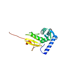
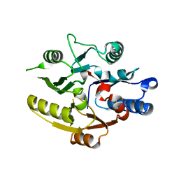
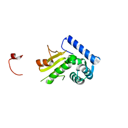
Crystal structure of the C-domain of the T.vaginalis Inr binding protein, IBP39 (tetragonal form)
分子名称: 39 kDa initiator binding protein
著者Schumacher, M.A, Johnson, P.J.
登録日2003-08-20
公開日2003-11-18
最終更新日2023-08-16
実験手法X-RAY DIFFRACTION (2.32 Å)
主引用文献Structural Basis of Core Promoter Recognition in a Primitive Eukaryote
Cell(Cambridge,Mass.), 115, 2003
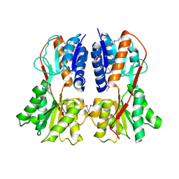
1RZR
DownloadVisualize
BU of 1rzr by Molmil
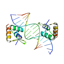
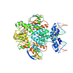
crystal structure of transcriptional regulator-phosphoprotein-DNA complex
分子名称: 5'-D(*CP*TP*GP*AP*AP*AP*GP*CP*GP*CP*TP*AP*AP*CP*AP*G)-3', 5'-D(*CP*TP*GP*TP*TP*AP*GP*CP*GP*CP*TP*TP*TP*CP*AP*G)-3', Glucose-resistance amylase regulator, ...
著者Schumacher, M.A, Allen, G.S, Brennan, R.G.
登録日2003-12-27
公開日2004-10-12
最終更新日2011-07-13
実験手法X-RAY DIFFRACTION (2.8 Å)
主引用文献Structural basis for allosteric control of the transcription regulator CcpA by the phosphoprotein HPr-Ser46-P.
Cell(Cambridge,Mass.), 118, 2004
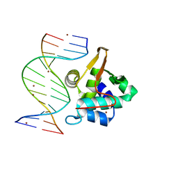
1PP8
DownloadVisualize
BU of 1pp8 by Molmil
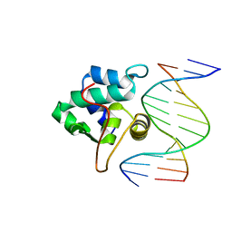
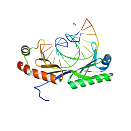
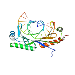
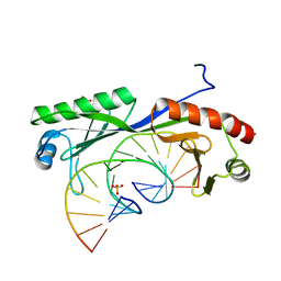
crystal structure of the T. vaginalis IBP39 Initiator binding domain (IBD) bound to the alpha-SCS Inr element
分子名称: 39 kDa initiator binding protein, ALPHA-SCS INR, SULFATE ION
著者Schumacher, M.A, Lau, A.O.T, Johnson, P.J.
登録日2003-06-16
公開日2003-11-18
最終更新日2024-02-14
実験手法X-RAY DIFFRACTION (3.05 Å)
主引用文献Structural Basis of Core Promoter Recognition in a Primitive Eukaryote
Cell(Cambridge,Mass.), 115, 2003
1SDL
DownloadVisualize
BU of 1sdl by Molmil
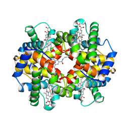
CROSS-LINKED, CARBONMONOXY HEMOGLOBIN A
分子名称: 1,3,5-BENZENETRICARBOXYLIC ACID, CARBON MONOXIDE, HEMOGLOBIN A, ...
著者Schumacher, M.A, Dixon, M.M, Kluger, R, Jones, R.T, Brennan, R.G.
登録日1996-02-26
公開日1996-08-01
最終更新日2024-06-05
実験手法X-RAY DIFFRACTION (1.8 Å)
主引用文献Allosteric transition intermediates modelled by crosslinked haemoglobins.
Nature, 375, 1995
1SDK
DownloadVisualize
BU of 1sdk by Molmil
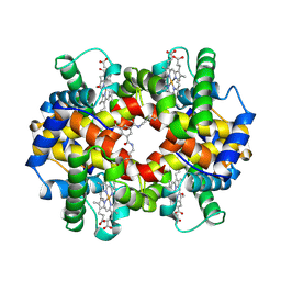
CROSS-LINKED, CARBONMONOXY HEMOGLOBIN A
分子名称: 1,3,5-BENZENETRICARBOXYLIC ACID, CARBON MONOXIDE, HEMOGLOBIN A, ...
著者Schumacher, M.A, Dixon, M.M, Kluger, R, Jones, R.T, Brennan, R.G.
登録日1996-02-26
公開日1996-08-01
最終更新日2024-06-05
実験手法X-RAY DIFFRACTION (1.8 Å)
主引用文献Allosteric transition intermediates modelled by crosslinked haemoglobins.
Nature, 375, 1995
1Q89
DownloadVisualize
BU of 1q89 by Molmil
Crystal structure of the C-domain of the T.vaginalis Inr binding protein, IBP39 (cubic crystal form)
分子名称: 39 kDa initiator binding protein
著者Schumacher, M.A, Johnson, P.J.
登録日2003-08-20
公開日2003-11-18
最終更新日2023-08-16
実験手法X-RAY DIFFRACTION (2.75 Å)
主引用文献Structural Basis of Core Promoter Recognition in a Primitive Eukaryote
Cell(Cambridge,Mass.), 115, 2003
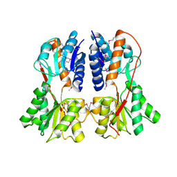
1SXH
DownloadVisualize
BU of 1sxh by Molmil
apo structure of B. megaterium transcription regulator
分子名称: Glucose-resistance amylase regulator
著者Schumacher, M.A, Allen, G.S, Diel, M, Seidel, G, Hillen, W, Brennan, R.G.
登録日2004-03-30
公開日2004-10-19
最終更新日2024-04-03
実験手法X-RAY DIFFRACTION (2.75 Å)
主引用文献Structural studies on the apo transcription factor form B. megaterium
Cell(Cambridge,Mass.), 118, 2004
1PP7
DownloadVisualize
BU of 1pp7 by Molmil
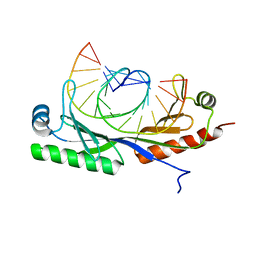
Crystal structure of the T. vaginalis Initiator binding protein bound to the ferredoxin Inr
分子名称: 39 kDa initiator binding protein, FERREDOXIN INR, ZINC ION
著者Schumacher, M.A, Lau, A.O.T, Johnson, P.J.
登録日2003-06-16
公開日2003-11-18
最終更新日2024-04-03
実験手法X-RAY DIFFRACTION (2.45 Å)
主引用文献Structural Basis of Core Promoter Recognition in a Primitive Eukaryote
Cell(Cambridge,Mass.), 115, 2003
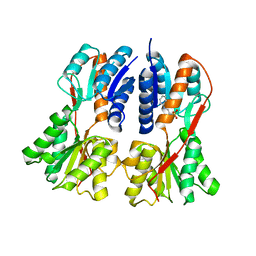
1SXI
DownloadVisualize
BU of 1sxi by Molmil
Structure of apo transcription regulator B. megaterium
分子名称: Glucose-resistance amylase regulator, MAGNESIUM ION
著者Schumacher, M.A, Allen, G.S, Diel, M, Seidel, G, Hillen, W, Brennan, R.G.
登録日2004-03-30
公開日2004-10-19
最終更新日2023-11-15
実験手法X-RAY DIFFRACTION (3 Å)
主引用文献Structural studies on the apo transcription factor form B. megaterium
Cell(Cambridge,Mass.), 118, 2004
1SXG
DownloadVisualize
BU of 1sxg by Molmil
Structural studies on the apo transcription factor form B. megaterium
分子名称: 2-PHENYLAMINO-ETHANESULFONIC ACID, Glucose-resistance amylase regulator
著者Schumacher, M.A, Allen, G.S, Diel, M, Seidel, G, Hillen, W, Brennan, R.G.
登録日2004-03-30
公開日2004-10-19
最終更新日2024-04-03
実験手法X-RAY DIFFRACTION (2.75 Å)
主引用文献Structural studies on the apo transcription factor form B. megaterium
Cell(Cambridge,Mass.), 118, 2004
7RMW
DownloadVisualize
BU of 7rmw by Molmil
Crystal structure of B. subtilis PurR bound to ppGpp
分子名称: GUANOSINE-5',3'-TETRAPHOSPHATE, Pur operon repressor
著者Schumacher, M.A.
登録日2021-07-28
公開日2021-12-22
最終更新日2023-10-18
実験手法X-RAY DIFFRACTION (2.45 Å)
主引用文献The nucleotide messenger (p)ppGpp is an anti-inducer of the purine synthesis transcription regulator PurR in Bacillus.
Nucleic Acids Res., 50, 2022
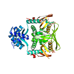
6E4N
DownloadVisualize
BU of 6e4n by Molmil
Structure of the T. brucei TbRGG2 RRM domain: apo R3 crystal form
分子名称: RNA-binding protein, putative
著者Schumacher, M.A.
登録日2018-07-18
公開日2018-12-12
最終更新日2023-10-11
実験手法X-RAY DIFFRACTION (1.801 Å)
主引用文献The RRM of the kRNA-editing protein TbRGG2 uses multiple surfaces to bind and remodel RNA.
Nucleic Acids Res., 47, 2019
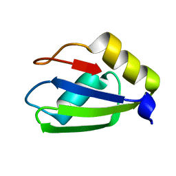
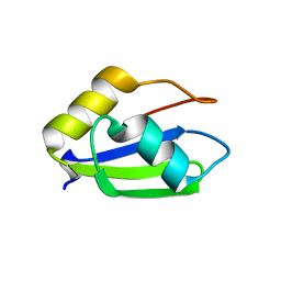
6E4O
DownloadVisualize
BU of 6e4o by Molmil
Structure of apo T. brucei RRM: P4(1)2(1)2 form
分子名称: RNA-binding protein, putative
著者Schumacher, M.A.
登録日2018-07-18
公開日2018-12-12
最終更新日2023-10-11
実験手法X-RAY DIFFRACTION (1.8 Å)
主引用文献The RRM of the kRNA-editing protein TbRGG2 uses multiple surfaces to bind and remodel RNA.
Nucleic Acids Res., 47, 2019
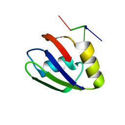
6E4P
DownloadVisualize
BU of 6e4p by Molmil
Structure of the T. brucei RRM domain in complex with RNA
分子名称: RNA (5'-R(P*UP*UP*UP*U)-3'), RNA-binding protein, putative
著者Schumacher, M.A.
登録日2018-07-18
公開日2018-12-12
最終更新日2023-10-11
実験手法X-RAY DIFFRACTION (1.949 Å)
主引用文献The RRM of the kRNA-editing protein TbRGG2 uses multiple surfaces to bind and remodel RNA.
Nucleic Acids Res., 47, 2019
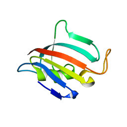
5HT1
DownloadVisualize
BU of 5ht1 by Molmil
Structure of apo C. glabrata FKBP12
分子名称: FK506-binding protein 1
著者Schumacher, M.A.
登録日2016-01-26
公開日2016-09-14
最終更新日2024-03-06
実験手法X-RAY DIFFRACTION (2.651 Å)
主引用文献Structures of Pathogenic Fungal FKBP12s Reveal Possible Self-Catalysis Function.
Mbio, 7, 2016
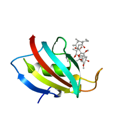
5HUA
DownloadVisualize
BU of 5hua by Molmil
Structure of C. glabrata FKBP12-FK506 complex
分子名称: 8-DEETHYL-8-[BUT-3-ENYL]-ASCOMYCIN, FK506-binding protein 1
著者Schumacher, M.A.
登録日2016-01-27
公開日2016-09-14
最終更新日2024-03-06
実験手法X-RAY DIFFRACTION (1.3 Å)
主引用文献Structures of Pathogenic Fungal FKBP12s Reveal Possible Self-Catalysis Function.
Mbio, 7, 2016
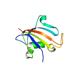
5HTG
DownloadVisualize
BU of 5htg by Molmil
Structure of apo P1 form of Candida albicans FKBP12
分子名称: FK506-binding protein 1
著者Schumacher, M.A.
登録日2016-01-26
公開日2016-09-14
最終更新日2024-03-06
実験手法X-RAY DIFFRACTION (2.4 Å)
主引用文献Structures of Pathogenic Fungal FKBP12s Reveal Possible Self-Catalysis Function.
Mbio, 7, 2016
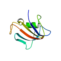
5I98
DownloadVisualize
BU of 5i98 by Molmil
Structure of apo FKBP12(P104G) from C. albicans
分子名称: FK506-binding protein 1
著者Schumacher, M.A.
登録日2016-02-19
公開日2016-09-14
最終更新日2024-03-06
実験手法X-RAY DIFFRACTION (1.999 Å)
主引用文献Structures of Pathogenic Fungal FKBP12s Reveal Possible Self-Catalysis Function.
Mbio, 7, 2016
8DPK
DownloadVisualize
BU of 8dpk by Molmil
structure of T. brucei RESC5
分子名称: RESC5
著者Schumacher, M.A, Salinas, R, Cannistraci, E.
登録日2022-07-15
公開日2023-02-22
最終更新日2024-05-22
実験手法X-RAY DIFFRACTION (1.94 Å)
主引用文献Structure of the T. brucei kinetoplastid RNA editing substrate-binding complex core component, RESC5.
Plos One, 18, 2023
8CSH
DownloadVisualize
BU of 8csh by Molmil
Structure of the DNA binding domain of pSK1 Par partition protein bound to centromere DNA
分子名称: DNA (5'-D(*TP*AP*GP*TP*TP*AP*GP*GP*TP*AP*CP*CP*TP*AP*AP*CP*A)-3'), DNA (5'-D(P*TP*GP*TP*TP*AP*GP*GP*TP*AP*CP*CP*TP*AP*AP*C)-3'), Par
著者Schumacher, M.A.
登録日2022-05-12
公開日2022-08-10
最終更新日2024-02-14
実験手法X-RAY DIFFRACTION (2.25 Å)
主引用文献Molecular Analysis of pSK1 par: A Novel Plasmid Partitioning System Encoded by Staphylococcal Multiresistance Plasmids.
J.Mol.Biol., 434, 2022
4YJ1
DownloadVisualize
BU of 4yj1 by Molmil
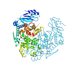
Crystal structure of T. brucei MRB1590-ADP bound to poly-U RNA
分子名称: ADENOSINE-5'-DIPHOSPHATE, MAGNESIUM ION, Uncharacterized protein
著者Schumacher, M.A.
登録日2015-03-03
公開日2015-08-12
最終更新日2024-02-28
実験手法X-RAY DIFFRACTION (2.05 Å)
主引用文献Structures of the T. brucei kRNA editing factor MRB1590 reveal unique RNA-binding pore motif contained within an ABC-ATPase fold.
Nucleic Acids Res., 43, 2015
6UEP
DownloadVisualize
BU of 6uep by Molmil
Structure of A. thaliana TBP bound to a DNA site with a C-C mismatch
分子名称: DNA (5'-D(*GP*CP*TP*AP*TP*AP*AP*AP*CP*GP*GP*GP*CP*A)-3'), DNA (5'-D(*TP*GP*CP*CP*CP*CP*TP*TP*TP*AP*TP*AP*GP*C)-3'), FORMIC ACID, ...
著者Schumacher, M.A, Al-hashimi, H.
登録日2019-09-22
公開日2020-09-02
最終更新日2024-03-13
実験手法X-RAY DIFFRACTION (2.05 Å)
主引用文献DNA mismatches reveal conformational penalties in protein-DNA recognition.
Nature, 587, 2020
6UEO
DownloadVisualize
BU of 6ueo by Molmil
Structure of A. thaliana TBP-AC mismatch DNA site
分子名称: DNA (5'-D(*GP*CP*TP*AP*TP*AP*AP*AP*AP*GP*GP*GP*CP*A)-3'), DNA (5'-D(*TP*GP*CP*CP*CP*CP*TP*TP*TP*AP*TP*AP*GP*C)-3'), TATA-box-binding protein 1
著者Schumacher, M.A.
登録日2019-09-22
公開日2020-09-02
最終更新日2023-10-11
実験手法X-RAY DIFFRACTION (2 Å)
主引用文献DNA mismatches reveal conformational penalties in protein-DNA recognition.
Nature, 587, 2020
6UER
DownloadVisualize
BU of 6uer by Molmil
Crystal form 2: Structure of TBP bound to C-C mismatch at pH 7
分子名称: DNA (5'-D(*GP*CP*TP*AP*TP*AP*AP*AP*CP*GP*GP*GP*CP*A)-3'), DNA (5'-D(*TP*GP*CP*CP*CP*CP*TP*TP*TP*AP*TP*AP*GP*C)-3'), TATA-box-binding protein 1
著者Schumacher, M.A, Al-Hashimi, H.
登録日2019-09-22
公開日2020-09-02
最終更新日2023-10-11
実験手法X-RAY DIFFRACTION (2.5 Å)
主引用文献DNA mismatches reveal conformational penalties in protein-DNA recognition.
Nature, 587, 2020
6UEQ
DownloadVisualize
BU of 6ueq by Molmil
Structure of TBP bound to C-C mismatch containing TATA site
分子名称: DNA (5'-D(*GP*CP*TP*AP*TP*AP*AP*AP*AP*CP*GP*GP*CP*A)-3'), DNA (5'-D(*TP*GP*CP*CP*CP*TP*TP*TP*TP*AP*TP*AP*GP*C)-3'), SULFATE ION, ...
著者Schumacher, M.A, Al-Hashimi, H.
登録日2019-09-22
公開日2020-09-02
最終更新日2023-10-11
実験手法X-RAY DIFFRACTION (2.4 Å)
主引用文献DNA mismatches reveal conformational penalties in protein-DNA recognition.
Nature, 587, 2020

223166

件を2024-07-31に公開中

PDB statisticsPDBj update infoContact PDBjnumon