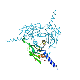
8WQH
 
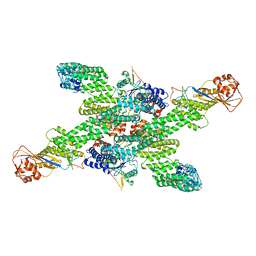 | | cryo-EM structure of neddylated CUL2-RBX1-ELOB-ELOC-FEM1B bound with the C-degron of CCDC89 (conformation 2) | | 分子名称: | Coiled-coil domain-containing protein 89, Cullin-2, E3 ubiquitin-protein ligase RBX1, ... | | 著者 | Chen, X, Zhang, K, Xu, C. | | 登録日 | 2023-10-11 | | 公開日 | 2024-04-03 | | 最終更新日 | 2024-05-08 | | 実験手法 | ELECTRON MICROSCOPY (3.44 Å) | | 主引用文献 | Mechanism of Psi-Pro/C-degron recognition by the CRL2 FEM1B ubiquitin ligase.
Nat Commun, 15, 2024
|
|
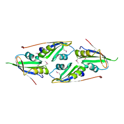
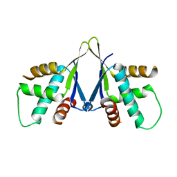
6L0X
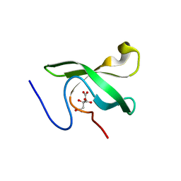 | | The First Tudor Domain of PHF20L1 | | 分子名称: | CITRIC ACID, GLYCEROL, PHD finger protein 20-like protein 1 | | 著者 | Lv, M.Q, Gao, J. | | 登録日 | 2019-09-27 | | 公開日 | 2020-09-23 | | 最終更新日 | 2023-11-22 | | 実験手法 | X-RAY DIFFRACTION (1.3 Å) | | 主引用文献 | Conformational Selection in Ligand Recognition by the First Tudor Domain of PHF20L1.
J Phys Chem Lett, 11, 2020
|
|
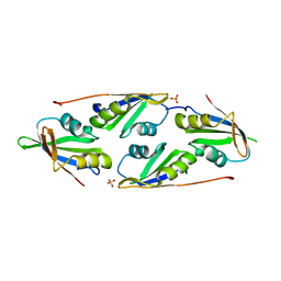
6L1P
 | | Crystal structure of PHF20L1 in complex with Hit 1 | | 分子名称: | 4-(1-methyl-3,6-dihydro-2H-pyridin-4-yl)phenol, GLYCEROL, PHD finger protein 20-like protein 1, ... | | 著者 | Lv, M.Q, Gao, J. | | 登録日 | 2019-09-29 | | 公開日 | 2020-09-23 | | 最終更新日 | 2023-11-22 | | 実験手法 | X-RAY DIFFRACTION (1.231 Å) | | 主引用文献 | Conformational Selection in Ligand Recognition by the First Tudor Domain of PHF20L1.
J Phys Chem Lett, 11, 2020
|
|
6L1C
 | |
6L1I
 | |
6L10
 | | PHF20L1 Tudor1 - MES | | 分子名称: | 2-(N-MORPHOLINO)-ETHANESULFONIC ACID, PHD finger protein 20-like protein 1, SULFATE ION | | 著者 | Lv, M.Q, Gao, J. | | 登録日 | 2019-09-27 | | 公開日 | 2020-09-23 | | 最終更新日 | 2023-11-22 | | 実験手法 | X-RAY DIFFRACTION (1.6 Å) | | 主引用文献 | Conformational Selection in Ligand Recognition by the First Tudor Domain of PHF20L1.
J Phys Chem Lett, 11, 2020
|
|
6L1F
 | |
6H02
 | |
8WLG
 | | Crystal structure of Cypovirus Polyhedra mutant fused with c-Myc fragment | | 分子名称: | Polyhedrin,Myc proto-oncogene protein | | 著者 | Kojima, M, Ueno, T, Abe, S, Hirata, K. | | 登録日 | 2023-09-29 | | 公開日 | 2024-06-05 | | 最終更新日 | 2024-07-03 | | 実験手法 | X-RAY DIFFRACTION (2.55 Å) | | 主引用文献 | High-throughput structure determination of an intrinsically disordered protein using cell-free protein crystallization.
Proc.Natl.Acad.Sci.USA, 121, 2024
|
|
8X8S
 | | Crystal structure of Cypovirus Polyhedra mutant fused with c-Myc fragment | | 分子名称: | Polyhedrin,Myc proto-oncogene protein | | 著者 | Kojima, M, Ueno, T, Abe, S, Hirata, K. | | 登録日 | 2023-11-28 | | 公開日 | 2024-06-05 | | 最終更新日 | 2024-07-03 | | 実験手法 | X-RAY DIFFRACTION (2.04 Å) | | 主引用文献 | High-throughput structure determination of an intrinsically disordered protein using cell-free protein crystallization.
Proc.Natl.Acad.Sci.USA, 121, 2024
|
|
8X8V
 | | Crystal structure of Cypovirus Polyhedra mutant fused with c-Myc fragment | | 分子名称: | Polyhedrin,Myc proto-oncogene protein | | 著者 | Kojima, M, Ueno, T, Abe, S, Hirata, K. | | 登録日 | 2023-11-29 | | 公開日 | 2024-06-05 | | 最終更新日 | 2024-07-03 | | 実験手法 | X-RAY DIFFRACTION (2 Å) | | 主引用文献 | High-throughput structure determination of an intrinsically disordered protein using cell-free protein crystallization.
Proc.Natl.Acad.Sci.USA, 121, 2024
|
|
7E3O
 | |
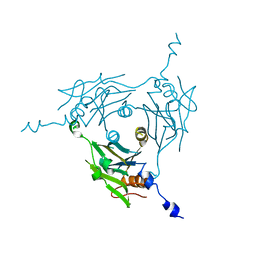
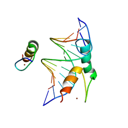
7D1L
 | | complex structure of two RRM domains | | 分子名称: | Embryonic developmental protein tofu-6, Uncharacterized protein | | 著者 | Wang, X, Liao, S, Xu, C. | | 登録日 | 2020-09-14 | | 公開日 | 2021-08-25 | | 最終更新日 | 2024-10-09 | | 実験手法 | X-RAY DIFFRACTION (1.95 Å) | | 主引用文献 | Molecular basis for PICS-mediated piRNA biogenesis and cell division.
Nat Commun, 12, 2021
|
|
7D2Y
 | | complex of two RRM domains | | 分子名称: | Embryonic developmental protein tofu-6, RRM2, SULFATE ION | | 著者 | Wang, X, Liao, S, Xu, C. | | 登録日 | 2020-09-17 | | 公開日 | 2021-08-25 | | 最終更新日 | 2023-11-29 | | 実験手法 | X-RAY DIFFRACTION (2.68 Å) | | 主引用文献 | Molecular basis for PICS-mediated piRNA biogenesis and cell division.
Nat Commun, 12, 2021
|
|
7EJO
 | | Structure of ERH-2 bound to TOST-1 | | 分子名称: | Enhancer of rudimentary homolog 2, Enhancer of rudimentary homolog 2,Protein tost-1 | | 著者 | Wang, X, Xu, C. | | 登録日 | 2021-04-02 | | 公開日 | 2021-08-25 | | 最終更新日 | 2023-11-29 | | 実験手法 | X-RAY DIFFRACTION (2.191 Å) | | 主引用文献 | Molecular basis for PICS-mediated piRNA biogenesis and cell division.
Nat Commun, 12, 2021
|
|
7EJS
 | | Structure of ERH-2 bound to PICS-1 | | 分子名称: | Enhancer of rudimentary homolog 2,Protein pid-3 | | 著者 | Wang, X, Xu, C. | | 登録日 | 2021-04-02 | | 公開日 | 2021-08-25 | | 最終更新日 | 2023-11-29 | | 実験手法 | X-RAY DIFFRACTION (2.387 Å) | | 主引用文献 | Molecular basis for PICS-mediated piRNA biogenesis and cell division.
Nat Commun, 12, 2021
|
|
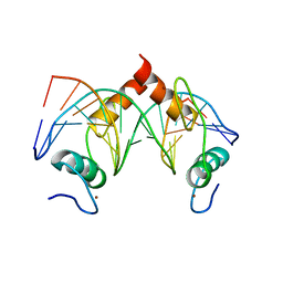
7CNG
 | | Structure of CDK5R1 bound FEM1B | | 分子名称: | Protein fem-1 homolog B,Peptide from Cyclin-dependent kinase 5 activator 1, SULFATE ION | | 著者 | Chen, X, Liao, S, Xu, C. | | 登録日 | 2020-07-31 | | 公開日 | 2020-10-21 | | 最終更新日 | 2023-11-29 | | 実験手法 | X-RAY DIFFRACTION (3.49 Å) | | 主引用文献 | Molecular basis for arginine C-terminal degron recognition by Cul2 FEM1 E3 ligase.
Nat.Chem.Biol., 17, 2021
|
|
7Y3K
 | | Structure of SALL4 ZFC4 bound with 16 bp AT-rich dsDNA | | 分子名称: | DNA (16-mer), Sal-like protein 4, ZINC ION | | 著者 | Ru, W, Xu, C. | | 登録日 | 2022-06-11 | | 公開日 | 2022-10-26 | | 最終更新日 | 2023-11-29 | | 実験手法 | X-RAY DIFFRACTION (2.501 Å) | | 主引用文献 | Structural studies of SALL family protein zinc finger cluster domains in complex with DNA reveal preferential binding to an AATA tetranucleotide motif.
J.Biol.Chem., 298, 2022
|
|
7Y3I
 | | Structure of DNA bound SALL4 | | 分子名称: | DNA (12-mer), Sal-like protein 4, ZINC ION | | 著者 | Ru, W, Xu, C. | | 登録日 | 2022-06-10 | | 公開日 | 2022-10-26 | | 最終更新日 | 2024-05-29 | | 実験手法 | X-RAY DIFFRACTION (2.45 Å) | | 主引用文献 | Structural studies of SALL family protein zinc finger cluster domains in complex with DNA reveal preferential binding to an AATA tetranucleotide motif.
J.Biol.Chem., 298, 2022
|
|
7Y3L
 | | Structure of SALL3 ZFC4 bound with 12 bp AT-rich dsDNA | | 分子名称: | DNA (12-mer), Sal-like protein 3, ZINC ION | | 著者 | Ru, W, Xu, C. | | 登録日 | 2022-06-11 | | 公開日 | 2022-10-26 | | 最終更新日 | 2023-11-29 | | 実験手法 | X-RAY DIFFRACTION (2.5 Å) | | 主引用文献 | Structural studies of SALL family protein zinc finger cluster domains in complex with DNA reveal preferential binding to an AATA tetranucleotide motif.
J.Biol.Chem., 298, 2022
|
|
7Y3M
 | | Structure of SALL4 ZFC1 bound with 16 bp AT-rich dsDNA | | 分子名称: | DNA (16-mer), Sal-like protein 4, ZINC ION | | 著者 | Ru, W, Xu, C. | | 登録日 | 2022-06-11 | | 公開日 | 2022-10-26 | | 最終更新日 | 2023-11-29 | | 実験手法 | X-RAY DIFFRACTION (2.723 Å) | | 主引用文献 | Structural studies of SALL family protein zinc finger cluster domains in complex with DNA reveal preferential binding to an AATA tetranucleotide motif.
J.Biol.Chem., 298, 2022
|
|
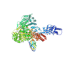
8JAR
 | | Structure of CRL2APPBP2 bound with RxxGPAA degron (dimer) | | 分子名称: | Amyloid protein-binding protein 2, Cullin-2, Elongin-B, ... | | 著者 | Zhao, S, Zhang, K, Xu, C. | | 登録日 | 2023-05-07 | | 公開日 | 2023-10-18 | | 最終更新日 | 2023-10-25 | | 実験手法 | ELECTRON MICROSCOPY (3.3 Å) | | 主引用文献 | Molecular basis for C-degron recognition by CRL2 APPBP2 ubiquitin ligase.
Proc.Natl.Acad.Sci.USA, 120, 2023
|
|
8JAL
 | | Structure of CRL2APPBP2 bound with RxxGP degron (dimer) | | 分子名称: | Amyloid protein-binding protein 2, Cullin-2, Elongin-B, ... | | 著者 | Zhao, S, Zhang, K, Xu, C. | | 登録日 | 2023-05-06 | | 公開日 | 2023-10-18 | | 最終更新日 | 2023-10-25 | | 実験手法 | ELECTRON MICROSCOPY (3.3 Å) | | 主引用文献 | Molecular basis for C-degron recognition by CRL2 APPBP2 ubiquitin ligase.
Proc.Natl.Acad.Sci.USA, 120, 2023
|
|
8JAS
 | | Structure of CRL2APPBP2 bound with RxxGPAA degron (tetramer) | | 分子名称: | Amyloid protein-binding protein 2, Cullin-2, E3 ubiquitin-protein ligase RBX1, ... | | 著者 | Zhao, S, Zhang, K, Xu, C. | | 登録日 | 2023-05-07 | | 公開日 | 2023-10-18 | | 最終更新日 | 2023-10-25 | | 実験手法 | ELECTRON MICROSCOPY (3.54 Å) | | 主引用文献 | Molecular basis for C-degron recognition by CRL2 APPBP2 ubiquitin ligase.
Proc.Natl.Acad.Sci.USA, 120, 2023
|
|
8JAQ
 | | Structure of CRL2APPBP2 bound with RxxGP degron (tetramer) | | 分子名称: | Amyloid protein-binding protein 2, Cullin-2, E3 ubiquitin-protein ligase RBX1, ... | | 著者 | Zhao, S, Zhang, K, Xu, C. | | 登録日 | 2023-05-06 | | 公開日 | 2023-10-18 | | 最終更新日 | 2023-10-25 | | 実験手法 | ELECTRON MICROSCOPY (3.26 Å) | | 主引用文献 | Molecular basis for C-degron recognition by CRL2 APPBP2 ubiquitin ligase.
Proc.Natl.Acad.Sci.USA, 120, 2023
|
|


