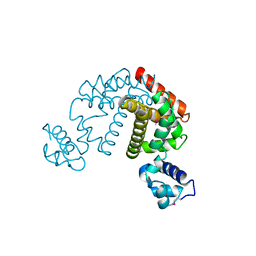
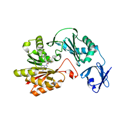
3W9Z
 
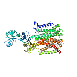
 | | Crystal structure of DusC | | 分子名称: | FLAVIN MONONUCLEOTIDE, tRNA-dihydrouridine synthase C | | 著者 | Chen, M, Yu, J, Tanaka, Y, Tanaka, I, Yao, M. | | 登録日 | 2013-04-19 | | 公開日 | 2013-07-31 | | 最終更新日 | 2024-03-20 | | 実験手法 | X-RAY DIFFRACTION (2.1 Å) | | 主引用文献 | Structure of dihydrouridine synthase C (DusC) from Escherichia coli
Acta Crystallogr.,Sect.F, 69, 2013
|
|
2D6Y
 | | Crystal Structure of transcriptional factor SCO4008 from Streptomyces coelicolor A3(2) | | 分子名称: | L(+)-TARTARIC ACID, putative tetR family regulatory protein | | 著者 | Hayashi, T, Tanaka, Y, Sakai, N, Yao, M, Tamura, T, Tanaka, I. | | 登録日 | 2005-11-15 | | 公開日 | 2006-10-31 | | 最終更新日 | 2013-09-04 | | 実験手法 | X-RAY DIFFRACTION (2.3 Å) | | 主引用文献 | SCO4008, a Putative TetR Transcriptional Repressor from Streptomyces coelicolor A3(2), Regulates Transcription of sco4007 by Multidrug Recognition.
J.Mol.Biol., 425, 2013
|
|
2DG7
 | | Crystal structure of the putative transcriptional regulator SCO0337 from Streptomyces coelicolor A3(2) | | 分子名称: | putative transcriptional regulator | | 著者 | Hayashi, T, Tanaka, Y, Sakai, N, Yao, M, Tamura, T, Tanaka, I. | | 登録日 | 2006-03-08 | | 公開日 | 2007-03-13 | | 最終更新日 | 2011-07-13 | | 実験手法 | X-RAY DIFFRACTION (2.3 Å) | | 主引用文献 | Crystal structure of the putative transcriptional regulator SCO0337 from Streptomyces coelicolor A3(2)
To be Published
|
|
2DG6
 | | Crystal structure of the putative transcriptional regulator SCO5550 from Streptomyces coelicolor A3(2) | | 分子名称: | putative transcriptional regulator | | 著者 | Hayashi, T, Tanaka, Y, Sakai, N, Yao, M, Tamura, T, Tanaka, I. | | 登録日 | 2006-03-08 | | 公開日 | 2007-03-13 | | 最終更新日 | 2018-03-28 | | 実験手法 | X-RAY DIFFRACTION (2.2 Å) | | 主引用文献 | Structural and genomic DNA analysis of a putative transcription factor SCO5550 from Streptomyces coelicolor A3(2): regulating the expression of gene sco5551 as a transcriptional activator with a novel dimer shape
Biochem. Biophys. Res. Commun., 435, 2013
|
|
2E7D
 | | Crystal structure of a NEAT domain from Staphylococcus aureus | | 分子名称: | ACETATE ION, GLYCEROL, Hypothetical protein IsdH, ... | | 著者 | Suenaga, A, Tanaka, Y, Yao, M, Kumagai, I, Tanaka, I, Tsumoto, K. | | 登録日 | 2007-01-09 | | 公開日 | 2008-01-22 | | 最終更新日 | 2024-03-13 | | 実験手法 | X-RAY DIFFRACTION (2.2 Å) | | 主引用文献 | Structural basis for multimeric heme complexation through a specific protein-heme interaction: the case of the third neat domain of IsdH from Staphylococcus aureus
J.Biol.Chem., 283, 2008
|
|
2ZKL
 | | Crystal Structure of capsular polysaccharide assembling protein CapF from staphylococcus aureus | | 分子名称: | Capsular polysaccharide synthesis enzyme Cap5F, GLYCEROL, ZINC ION | | 著者 | Miyafusa, T, Tanaka, Y, Yao, M, Tanaka, I, Tsumoto, K. | | 登録日 | 2008-03-25 | | 公開日 | 2009-03-31 | | 最終更新日 | 2024-03-13 | | 実験手法 | X-RAY DIFFRACTION (2.61 Å) | | 主引用文献 | Crystal structure of capsular polysaccharide assembling protein from Staphylococcus aureus
to be published
|
|
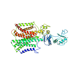
3B0U
 | | tRNA-dihydrouridine synthase from Thermus thermophilus in complex with tRNA fragment | | 分子名称: | FLAVIN MONONUCLEOTIDE, RNA (5'-R(*GP*GP*(H2U)P*A)-3'), tRNA-dihydrouridine synthase | | 著者 | Yu, F, Tanaka, Y, Yamashita, K, Nakamura, A, Yao, M, Tanaka, I. | | 登録日 | 2011-06-14 | | 公開日 | 2011-12-14 | | 最終更新日 | 2023-11-01 | | 実験手法 | X-RAY DIFFRACTION (1.948 Å) | | 主引用文献 | Molecular basis of dihydrouridine formation on tRNA
Proc.Natl.Acad.Sci.USA, 108, 2011
|
|
3B0P
 | | tRNA-dihydrouridine synthase from Thermus thermophilus | | 分子名称: | FLAVIN MONONUCLEOTIDE, tRNA-dihydrouridine synthase | | 著者 | Yu, F, Tanaka, Y, Yamashita, K, Nakamura, A, Yao, M, Tanaka, I. | | 登録日 | 2011-06-12 | | 公開日 | 2011-12-14 | | 最終更新日 | 2024-03-13 | | 実験手法 | X-RAY DIFFRACTION (1.7 Å) | | 主引用文献 | Molecular basis of dihydrouridine formation on tRNA
Proc.Natl.Acad.Sci.USA, 108, 2011
|
|
3AZV
 | | Crystal structure of the receptor binding domain | | 分子名称: | D/C mosaic neurotoxin, SULFATE ION | | 著者 | Nuemket, N, Tanaka, Y, Tsukamoto, K, Tsuji, T, Nakamura, K, Kozaki, S, Yao, M, Tanaka, I. | | 登録日 | 2011-06-02 | | 公開日 | 2011-12-28 | | 最終更新日 | 2017-10-11 | | 実験手法 | X-RAY DIFFRACTION (3.1 Å) | | 主引用文献 | Structural and mutational analyses of the receptor binding domain of botulinum D/C mosaic neurotoxin: insight into the ganglioside binding mechanism
Biochem.Biophys.Res.Commun., 411, 2011
|
|
3AZW
 | | Crystal structure of the receptor binding domain | | 分子名称: | D/C mosaic neurotoxin, SULFATE ION | | 著者 | Nuemket, N, Tanaka, Y, Tsukamoto, K, Tsuji, T, Nakamura, K, Kozaki, S, Yao, M, Tanaka, I. | | 登録日 | 2011-06-02 | | 公開日 | 2011-12-28 | | 最終更新日 | 2024-03-13 | | 実験手法 | X-RAY DIFFRACTION (2.99 Å) | | 主引用文献 | Structural and mutational analyses of the receptor binding domain of botulinum D/C mosaic neurotoxin: insight into the ganglioside binding mechanism
Biochem.Biophys.Res.Commun., 411, 2011
|
|
3B0V
 | | tRNA-dihydrouridine synthase from Thermus thermophilus in complex with tRNA | | 分子名称: | FLAVIN MONONUCLEOTIDE, tRNA, tRNA-dihydrouridine synthase | | 著者 | Yu, F, Tanaka, Y, Yamashita, K, Nakamura, A, Yao, M, Tanaka, I. | | 登録日 | 2011-06-14 | | 公開日 | 2011-12-14 | | 最終更新日 | 2017-10-11 | | 実験手法 | X-RAY DIFFRACTION (3.51 Å) | | 主引用文献 | Molecular basis of dihydrouridine formation on tRNA
Proc.Natl.Acad.Sci.USA, 108, 2011
|
|
8J4C
 | | YeeE(TsuA)-YeeD(TsuB) complex for thiosulfate uptake | | 分子名称: | (2R)-2,3-dihydroxypropyl (9Z)-octadec-9-enoate, Spirochaeta thermophila YeeE(TsuA)-YeeD(TsuB),UPF0033 domain-containing protein, SirA-like domain-containing protein (chimera), ... | | 著者 | Ikei, M, Miyazaki, R, Monden, K, Naito, Y, Takeuchi, A, Takahashi, Y.S, Tanaka, Y, Ichikawa, M, Tsukazaki, T. | | 登録日 | 2023-04-19 | | 公開日 | 2024-03-27 | | 最終更新日 | 2024-10-09 | | 実験手法 | X-RAY DIFFRACTION (3.34 Å) | | 主引用文献 | YeeD is an essential partner for YeeE-mediated thiosulfate uptake in bacteria and regulates thiosulfate ion decomposition.
Plos Biol., 22, 2024
|
|
8K1R
 | | YeeE(TsuA)-YeeD(TsuB) complex for thiosulfate uptake | | 分子名称: | (2R)-2,3-dihydroxypropyl (9Z)-octadec-9-enoate, Spirochaeta thermophila YeeE(TsuA)-YeeD(TsuB),UPF0033 domain-containing protein, SirA-like domain-containing protein (chimera) | | 著者 | Ikei, M, Miyazaki, R, Monden, K, Naito, Y, Takeuchi, A, Takahashi, Y.S, Tanaka, Y, Ichikawa, M, Tsukazaki, T. | | 登録日 | 2023-07-11 | | 公開日 | 2024-03-27 | | 最終更新日 | 2024-10-09 | | 実験手法 | X-RAY DIFFRACTION (2.6 Å) | | 主引用文献 | YeeD is an essential partner for YeeE-mediated thiosulfate uptake in bacteria and regulates thiosulfate ion decomposition.
Plos Biol., 22, 2024
|
|
5XAM
 | |
5XAP
 | | Crystal structure of SecDF in I form (C2 space group) | | 分子名称: | (2R)-2,3-dihydroxypropyl (9Z)-octadec-9-enoate, DI(HYDROXYETHYL)ETHER, Protein translocase subunit SecD | | 著者 | Tsukazaki, T, Tanaka, Y, Furukwa, A. | | 登録日 | 2017-03-14 | | 公開日 | 2017-05-17 | | 最終更新日 | 2023-11-22 | | 実験手法 | X-RAY DIFFRACTION (2.605 Å) | | 主引用文献 | Tunnel Formation Inferred from the I-Form Structures of the Proton-Driven Protein Secretion Motor SecDF
Cell Rep, 19, 2017
|
|
5XAN
 | | Crystal structure of SecDF in I form (P212121 space group) | | 分子名称: | (2R)-2,3-dihydroxypropyl (9Z)-octadec-9-enoate, POLYETHYLENE GLYCOL (N=34), Protein translocase subunit SecD | | 著者 | Tsukazaki, T, Tanaka, Y, Furukwa, A. | | 登録日 | 2017-03-14 | | 公開日 | 2017-05-17 | | 最終更新日 | 2023-11-22 | | 実験手法 | X-RAY DIFFRACTION (2.75 Å) | | 主引用文献 | Tunnel Formation Inferred from the I-Form Structures of the Proton-Driven Protein Secretion Motor SecDF
Cell Rep, 19, 2017
|
|
3VZR
 | |
3VZQ
 | |
3VSE
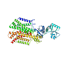
 | | Crystal structure of methyltransferase | | 分子名称: | Putative uncharacterized protein, S-ADENOSYL-L-HOMOCYSTEINE | | 著者 | Kita, S, Tanaka, Y, Yao, M, Tanaka, I. | | 登録日 | 2012-04-25 | | 公開日 | 2013-04-10 | | 最終更新日 | 2024-03-20 | | 実験手法 | X-RAY DIFFRACTION (2.099 Å) | | 主引用文献 | Crystal structure of a putative methyltransferase SAV1081 from Staphylococcus aureus
Protein Pept.Lett., 20, 2012
|
|
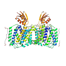
3VZP
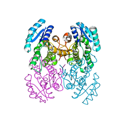
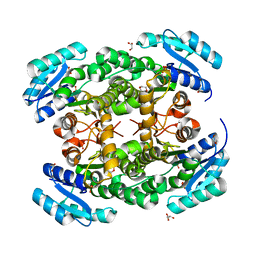 | | Crystal structure of PhaB from Ralstonia eutropha | | 分子名称: | 1,4-DIETHYLENE DIOXIDE, Acetoacetyl-CoA reductase, GLYCEROL, ... | | 著者 | Ikeda, K, Tanaka, Y, Tanaka, I, Yao, M. | | 登録日 | 2012-10-15 | | 公開日 | 2013-08-28 | | 最終更新日 | 2023-11-08 | | 実験手法 | X-RAY DIFFRACTION (1.792 Å) | | 主引用文献 | Directed evolution and structural analysis of NADPH-dependent Acetoacetyl Coenzyme A (Acetoacetyl-CoA) reductase from Ralstonia eutropha reveals two mutations responsible for enhanced kinetics
Appl.Environ.Microbiol., 79, 2013
|
|
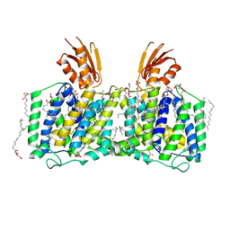
3VZS
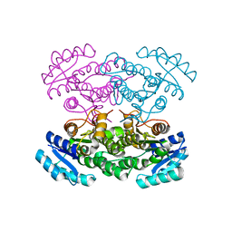
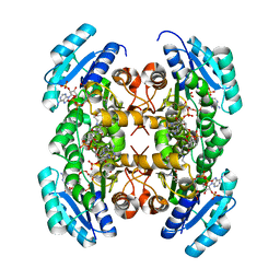 | | Crystal structure of PhaB from Ralstonia eutropha in complex with Acetoacetyl-CoA and NADP | | 分子名称: | ACETOACETYL-COENZYME A, Acetoacetyl-CoA reductase, NADP NICOTINAMIDE-ADENINE-DINUCLEOTIDE PHOSPHATE, ... | | 著者 | Ikeda, K, Tanaka, Y, Tanaka, I, Yao, M. | | 登録日 | 2012-10-15 | | 公開日 | 2013-08-28 | | 最終更新日 | 2023-11-08 | | 実験手法 | X-RAY DIFFRACTION (2.14 Å) | | 主引用文献 | Directed evolution and structural analysis of NADPH-dependent Acetoacetyl Coenzyme A (Acetoacetyl-CoA) reductase from Ralstonia eutropha reveals two mutations responsible for enhanced kinetics
Appl.Environ.Microbiol., 79, 2013
|
|
3W9W
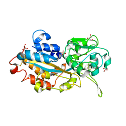 | | Crystal structure of DING protein | | 分子名称: | DING protein, GLYCEROL, PHOSPHATE ION | | 著者 | Gai, Z.Q, Nakamura, A, Tanaka, Y, Hirano, N, Tanaka, I, Yao, M. | | 登録日 | 2013-04-17 | | 公開日 | 2013-10-30 | | 最終更新日 | 2024-10-09 | | 実験手法 | X-RAY DIFFRACTION (1.35 Å) | | 主引用文献 | Crystal structure analysis, overexpression and refolding behaviour of a DING protein with single mutation.
J.SYNCHROTRON RADIAT., 20, 2013
|
|
8IFC
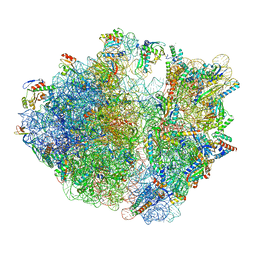 | | Arbekacin-bound E.coli 70S ribosome in the PURE system | | 分子名称: | 16S ribosomal RNA, 23S ribosomal RNA, 30S ribosomal protein S10, ... | | 著者 | Tomono, J, Asano, K, Chiashi, T, Tanaka, Y, Yokoyama, T. | | 登録日 | 2023-02-17 | | 公開日 | 2024-02-14 | | 最終更新日 | 2024-06-19 | | 実験手法 | ELECTRON MICROSCOPY (2.9 Å) | | 主引用文献 | Direct visualization of ribosomes in the cell-free system revealed the functional evolution of aminoglycoside.
J.Biochem., 175, 2024
|
|
8IFD
 | | Dibekacin-added human 80S ribosome | | 分子名称: | 18S ribosomal RNA, 28S ribosomal RNA, 40S ribosomal protein S10, ... | | 著者 | Tomono, J, Asano, K, Chiashi, T, Tanaka, Y, Yokoyama, T. | | 登録日 | 2023-02-17 | | 公開日 | 2024-02-14 | | 最終更新日 | 2024-06-19 | | 実験手法 | ELECTRON MICROSCOPY (2.59 Å) | | 主引用文献 | Direct visualization of ribosomes in the cell-free system revealed the functional evolution of aminoglycoside.
J.Biochem., 175, 2024
|
|
8IFE
 | | Arbekacin-added human 80S ribosome | | 分子名称: | 18S ribosomal RNA, 28S ribosomal RNA, 40S ribosomal protein S10, ... | | 著者 | Tomono, J, Asano, K, Chiashi, T, Tanaka, Y, Yokoyama, T. | | 登録日 | 2023-02-17 | | 公開日 | 2024-02-14 | | 最終更新日 | 2024-06-19 | | 実験手法 | ELECTRON MICROSCOPY (2.57 Å) | | 主引用文献 | Direct visualization of ribosomes in the cell-free system revealed the functional evolution of aminoglycoside.
J.Biochem., 175, 2024
|
|


