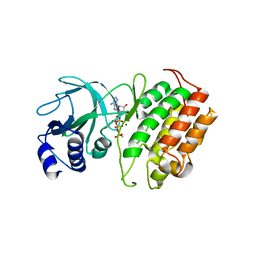
8YHH
 
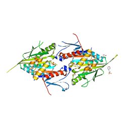 | | The Crystal Structure of Mitotic Kinesin Eg5 from Biortus | | 分子名称: | 1,2-ETHANEDIOL, 2-(N-MORPHOLINO)-ETHANESULFONIC ACID, ADENOSINE-5'-DIPHOSPHATE, ... | | 著者 | Wang, F, Cheng, W, Lv, Z, Qi, J, Wu, B. | | 登録日 | 2024-02-28 | | 公開日 | 2024-03-13 | | 実験手法 | X-RAY DIFFRACTION (1.95 Å) | | 主引用文献 | The Crystal Structure of Mitotic Kinesin Eg5 from Biortus
To Be Published
|
|
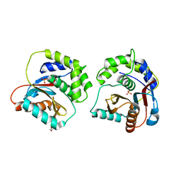
8YGX
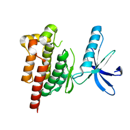 | |
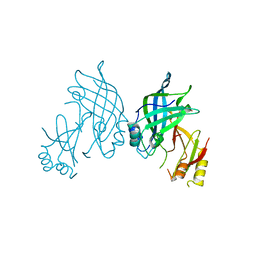
8YHS
 | | The Crystal Structure of BRDT from Biortus. | | 分子名称: | 1,2-ETHANEDIOL, 2,4-dimethyl-6-[6-(oxan-4-yl)-1-[(1~{S})-1-phenylethyl]imidazo[4,5-c]pyridin-2-yl]pyridazin-3-one, Bromodomain testis-specific protein | | 著者 | Wang, F, Cheng, W, Lv, Z, Meng, Q, Lu, Y. | | 登録日 | 2024-02-28 | | 公開日 | 2024-03-13 | | 実験手法 | X-RAY DIFFRACTION (1.5 Å) | | 主引用文献 | The Crystal Structure of BRDT from Biortus.
To Be Published
|
|
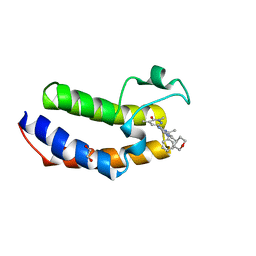
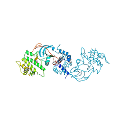
8YHL
 | | The Crystal Structure of Tgf-Beta Type I Receptor (Alk5) from Biortus | | 分子名称: | 1,2-ETHANEDIOL, ACETIC ACID, TGF-beta receptor type-1, ... | | 著者 | Wang, F, Cheng, W, Lv, Z, Meng, Q, Xu, Y. | | 登録日 | 2024-02-28 | | 公開日 | 2024-03-13 | | 最終更新日 | 2024-11-20 | | 実験手法 | X-RAY DIFFRACTION (1.47 Å) | | 主引用文献 | The Crystal Structure of Tgf-Beta Type I Receptor (Alk5) from Biortus
To Be Published
|
|
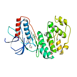
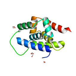
8YGZ
 | | The Crystal Structure of TGF beta R2 kinase domain from Biortus. | | 分子名称: | 1,2-ETHANEDIOL, DI(HYDROXYETHYL)ETHER, TGF-beta receptor type-2 | | 著者 | Wang, F, Cheng, W, Lv, Z, Ju, C, Wang, J. | | 登録日 | 2024-02-27 | | 公開日 | 2024-03-13 | | 最終更新日 | 2024-10-23 | | 実験手法 | X-RAY DIFFRACTION (2.1 Å) | | 主引用文献 | The Crystal Structure of TGF beta R2 kinase domain from Biortus.
To Be Published
|
|
8YGW
 | | The Crystal Structure of MAPK11 from Biortus | | 分子名称: | 1-(5-TERT-BUTYL-2-P-TOLYL-2H-PYRAZOL-3-YL)-3-[4-(2-MORPHOLIN-4-YL-ETHOXY)-NAPHTHALEN-1-YL]-UREA, Mitogen-activated protein kinase 11 | | 著者 | Wang, F, Cheng, W, Yuan, Z, Lin, D, Guo, S. | | 登録日 | 2024-02-27 | | 公開日 | 2024-03-13 | | 実験手法 | X-RAY DIFFRACTION (3.301 Å) | | 主引用文献 | The Crystal Structure of MAPK11 from Biortus.
To Be Published
|
|
8YHW
 | | The Crystal Structure of NF-kB-inducing Kinase (NIK) from Biortus | | 分子名称: | 1,2-ETHANEDIOL, 3,6,9,12,15,18,21-HEPTAOXATRICOSANE-1,23-DIOL, MAGNESIUM ION, ... | | 著者 | Wang, F, Cheng, W, Lv, Z, Meng, Q, Xu, Y. | | 登録日 | 2024-02-28 | | 公開日 | 2024-03-13 | | 実験手法 | X-RAY DIFFRACTION (2.9 Å) | | 主引用文献 | The Crystal Structure of NF-kB-inducing Kinase (NIK) from Biortus
To Be Published
|
|
8YHP
 | | Structure of the PGK1 from Biortus. | | 分子名称: | 1,2-ETHANEDIOL, Phosphoglycerate kinase 1 | | 著者 | Wang, F, Cheng, W, Lv, Z, Ju, C, Wang, J. | | 登録日 | 2024-02-28 | | 公開日 | 2024-03-13 | | 実験手法 | X-RAY DIFFRACTION (1.95 Å) | | 主引用文献 | Structure of the PGK1 from Biortus.
To Be Published
|
|
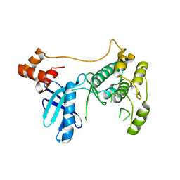
8YHV
 | |
7D4A
 | | The Crystal Structure of human JMJD2A Tudor domain from Biortus | | 分子名称: | Lysine-specific demethylase 4A, SULFATE ION | | 著者 | Wang, F, Lv, Z, Cheng, W, Lin, D, Ju, C, Bao, X, Zhu, B. | | 登録日 | 2020-09-23 | | 公開日 | 2020-10-07 | | 最終更新日 | 2023-11-29 | | 実験手法 | X-RAY DIFFRACTION (2.201 Å) | | 主引用文献 | The Crystal Structure of human JMJD2A from Biortus.
To Be Published
|
|
7DSF
 | | The Crystal Structure of human SPR from Biortus. | | 分子名称: | ACETATE ION, NADP NICOTINAMIDE-ADENINE-DINUCLEOTIDE PHOSPHATE, Sepiapterin reductase, ... | | 著者 | Wang, F, Lv, Z, Cheng, W, Lin, D, Meng, Q, Zhang, B, Huang, Y. | | 登録日 | 2020-12-31 | | 公開日 | 2021-01-13 | | 最終更新日 | 2023-11-29 | | 実験手法 | X-RAY DIFFRACTION (1.8 Å) | | 主引用文献 | The Crystal Structure of human SPR from Biortus.
To Be Published
|
|
7DS7
 | | The Crystal Structure of Leaf-branch compost cutinase from Biortus. | | 分子名称: | CITRIC ACID, GLYCEROL, IMIDAZOLE, ... | | 著者 | Wang, F, Lv, Z, Cheng, W, Lin, D, Chu, F, Xu, X, Tan, J. | | 登録日 | 2020-12-30 | | 公開日 | 2021-01-13 | | 最終更新日 | 2024-10-23 | | 実験手法 | X-RAY DIFFRACTION (2.15 Å) | | 主引用文献 | The Crystal Structure of Leaf-branch compost cutinase from Biortus.
To Be Published
|
|
8ZH5
 | |
8XLD
 | | Structure of the GFP:GFP-nanobody complex from Biortus. | | 分子名称: | 1,2-ETHANEDIOL, Nanobody(Staygold-S2G10)-Nanobody(Staygold-S4F1), ZINC ION, ... | | 著者 | Wang, F, Cheng, W, Yuan, Z, Lin, D, Bao, C. | | 登録日 | 2023-12-25 | | 公開日 | 2024-03-06 | | 最終更新日 | 2024-11-20 | | 実験手法 | X-RAY DIFFRACTION (2.1 Å) | | 主引用文献 | Structure of the GFP:GFP-nanobody complex from Biortus.
To Be Published
|
|
8XOV
 | | The Crystal Structure of N-terminal kinase domain of human RSK-1 from Biortus. | | 分子名称: | 1,2-ETHANEDIOL, PHOSPHOAMINOPHOSPHONIC ACID-ADENYLATE ESTER, Ribosomal protein S6 kinase alpha-1 | | 著者 | Wang, F, Cheng, W, Lv, Z, Meng, Q, Zhang, B. | | 登録日 | 2024-01-02 | | 公開日 | 2024-03-06 | | 実験手法 | X-RAY DIFFRACTION (2.55 Å) | | 主引用文献 | The Crystal Structure of N-terminal kinase domain of human RSK-1 from Biortus.
To Be Published
|
|
8XQU
 | | The Crystal Structure of ClpC1-NTD from Biortus. | | 分子名称: | 1,2-ETHANEDIOL, ATP-dependent Clp protease ATP-binding subunit ClpC1, DI(HYDROXYETHYL)ETHER, ... | | 著者 | Wang, F, Cheng, W, Lv, Z, Ju, C, Ni, C. | | 登録日 | 2024-01-05 | | 公開日 | 2024-03-06 | | 実験手法 | X-RAY DIFFRACTION (1.85 Å) | | 主引用文献 | The Crystal Structure of ClpC1-NTD from Biortus.
To Be Published
|
|
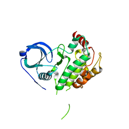
8XQD
 | |
8XPG
 | | The Crystal Structure of polo box domain of Plk4 from Biortus. | | 分子名称: | SULFATE ION, Serine/threonine-protein kinase PLK4 | | 著者 | Wang, F, Cheng, W, Lv, Z, Meng, Q, Zhang, B. | | 登録日 | 2024-01-03 | | 公開日 | 2024-03-06 | | 実験手法 | X-RAY DIFFRACTION (2.7 Å) | | 主引用文献 | The Crystal Structure of polo box domain of Plk4 from Biortus.
To Be Published
|
|
8XPX
 | | The Crystal Structure of PARP12 from Biortus. | | 分子名称: | 1,2-ETHANEDIOL, ACETATE ION, DI(HYDROXYETHYL)ETHER, ... | | 著者 | Wang, F, Cheng, W, Lv, Z, Qi, J, Shen, Z. | | 登録日 | 2024-01-04 | | 公開日 | 2024-03-06 | | 実験手法 | X-RAY DIFFRACTION (1.75 Å) | | 主引用文献 | The Crystal Structure of PARP12 from Biortus.
To Be Published
|
|
8XI8
 | |
8XPN
 | | The Crystal Structure of USP8 from Biortus. | | 分子名称: | 1,2-ETHANEDIOL, DI(HYDROXYETHYL)ETHER, Ubiquitin carboxyl-terminal hydrolase 8, ... | | 著者 | Wang, F, Cheng, W, Yuan, Z, Lin, D, Wang, J. | | 登録日 | 2024-01-04 | | 公開日 | 2024-03-06 | | 実験手法 | X-RAY DIFFRACTION (2.1 Å) | | 主引用文献 | The Crystal Structure of USP8 from Biortus.
To Be Published
|
|
8XI7
 | | The Crystal Structure of UCHL1 from Biortus. | | 分子名称: | 1,2-ETHANEDIOL, SULFATE ION, Ubiquitin carboxyl-terminal hydrolase isozyme L1 | | 著者 | Wang, F, Cheng, W, Yuan, Z, Qi, J, Shen, Z. | | 登録日 | 2023-12-19 | | 公開日 | 2024-03-06 | | 実験手法 | X-RAY DIFFRACTION (1.95 Å) | | 主引用文献 | The Crystal Structure of UCHL1 from Biortus.
To Be Published
|
|
8XQK
 | | The Crystal Structure of Apaf from Biortus. | | 分子名称: | Apoptotic protease-activating factor 1, PHOSPHATE ION | | 著者 | Wang, F, Cheng, W, Lv, Z, Ju, C, Ni, C. | | 登録日 | 2024-01-05 | | 公開日 | 2024-03-06 | | 実験手法 | X-RAY DIFFRACTION (2.85 Å) | | 主引用文献 | The Crystal Structure of Apaf from Biortus.
To Be Published
|
|
8XFL
 | |
8XU4
 | | The Crystal Structure of MAPK2 from Biortus. | | 分子名称: | MALONIC ACID, MAP kinase-activated protein kinase 2 | | 著者 | Wang, F, Cheng, W, Yuan, Z, Qi, J, Shen, Z. | | 登録日 | 2024-01-12 | | 公開日 | 2024-01-24 | | 実験手法 | X-RAY DIFFRACTION (3.4 Å) | | 主引用文献 | The Crystal Structure of MAPK2 from Biortus.
To Be Published
|
|


