1I4S
 
 | |
1JFZ
 | |
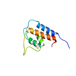
2K5S
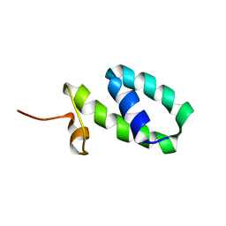 | | YmoA | | 分子名称: | Modulating protein ymoA | | 著者 | McFeeters, R.L, Byrd, R. | | 登録日 | 2008-06-30 | | 公開日 | 2008-12-09 | | 最終更新日 | 2024-05-29 | | 実験手法 | SOLUTION NMR | | 主引用文献 | The high-precision solution structure of Yersinia modulating
protein YmoA provides insight into interaction with H-NS
Biochemistry, 46, 2007
|
|
1N5B
 | |
1GA3
 | |
7KDO
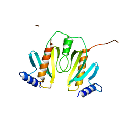
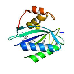 | | Crystal structure of Escherichia coli HPPK in complex with bisubstrate inhibitor HP-73 | | 分子名称: | 2-amino-4-hydroxy-6-hydroxymethyldihydropteridine pyrophosphokinase, 5'-S-[(2R,4R)-1-{2-[(2-amino-7,7-dimethyl-4-oxo-3,4,7,8-tetrahydropteridine-6-carbonyl)amino]ethyl}-2-carboxypiperidin-4-yl]-5'-thioadenosine | | 著者 | Shaw, G.X, Shi, G, Ji, X. | | 登録日 | 2020-10-09 | | 公開日 | 2020-12-02 | | 最終更新日 | 2023-10-18 | | 実験手法 | X-RAY DIFFRACTION (1.6 Å) | | 主引用文献 | Bisubstrate inhibitors of 6-hydroxymethyl-7,8-dihydropterin pyrophosphokinase: Transition state analogs for high affinity binding.
Bioorg.Med.Chem., 29, 2021
|
|
7KDR
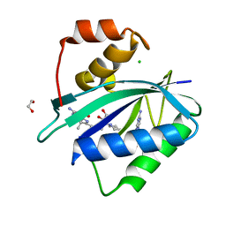 | | Crystal structure of Escherichia coli HPPK in complex with bisubstrate inhibitor HP-75 | | 分子名称: | 1,2-ETHANEDIOL, 2-amino-4-hydroxy-6-hydroxymethyldihydropteridine pyrophosphokinase, 5'-{[(2R,4R)-1-{2-[(2-amino-7,7-dimethyl-4-oxo-3,4,7,8-tetrahydropteridine-6-carbonyl)amino]ethyl}-2-carboxypiperidin-4-yl]sulfonyl}-5'-deoxyadenosine, ... | | 著者 | Shaw, G.X, Shi, G, Ji, X. | | 登録日 | 2020-10-09 | | 公開日 | 2020-12-02 | | 最終更新日 | 2023-10-18 | | 実験手法 | X-RAY DIFFRACTION (1.488 Å) | | 主引用文献 | Bisubstrate inhibitors of 6-hydroxymethyl-7,8-dihydropterin pyrophosphokinase: Transition state analogs for high affinity binding.
Bioorg.Med.Chem., 29, 2021
|
|


