2Z14
 
 | | Crystal structure of the N-terminal DUF1126 in human ef-hand domain containing 2 protein | | 分子名称: | EF-hand domain-containing family member C2 | | 著者 | Saito, K, Olsen, S, Kishishita, S, Nishino, A, Murayama, K, Terada, T, Shirouzu, M, Kigawa, T, Yokoyama, S, RIKEN Structural Genomics/Proteomics Initiative (RSGI) | | 登録日 | 2007-05-08 | | 公開日 | 2007-11-13 | | 最終更新日 | 2023-11-01 | | 実験手法 | X-RAY DIFFRACTION (1.68 Å) | | 主引用文献 | Crystal structure of the N-terminal DUF1126 in human ef-hand domain containing 2 protein
To be Published
|
|
2Z0G
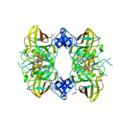
 | | The crystal structure of PII protein | | 分子名称: | CHLORIDE ION, Nitrogen regulatory protein P-II, PHOSPHATE ION | | 著者 | Sakai, H, Shinkai, A, Kitamura, Y, Kuramitsu, S, Yokoyama, S, RIKEN Structural Genomics/Proteomics Initiative (RSGI) | | 登録日 | 2007-05-07 | | 公開日 | 2008-05-13 | | 最終更新日 | 2023-11-01 | | 実験手法 | X-RAY DIFFRACTION (2.1 Å) | | 主引用文献 | The crystal structure of PII protein
To be Published
|
|
2Z0S
 | | Crystal structure of putative exosome complex RNA-binding protein | | 分子名称: | Probable exosome complex RNA-binding protein 1 | | 著者 | Murayama, K, Kato-Murayama, M, Takemoto, C, Terada, T, Shirouzu, M, Yokoyama, S, RIKEN Structural Genomics/Proteomics Initiative (RSGI) | | 登録日 | 2007-05-07 | | 公開日 | 2007-11-13 | | 最終更新日 | 2023-11-01 | | 実験手法 | X-RAY DIFFRACTION (3.2 Å) | | 主引用文献 | Crystal structure of putative exosome complex RNA-binding protein
To be Published
|
|
1WMW
 | | Crystal structure of geranulgeranyl diphosphate synthase from Thermus thermophilus | | 分子名称: | geranylgeranyl diphosphate synthetase | | 著者 | Suto, K, Nishio, K, Nodake, Y, Hamada, K, Kawamoto, M, Nakagawa, N, Kuramitu, S, Miura, K, RIKEN Structural Genomics/Proteomics Initiative (RSGI) | | 登録日 | 2004-07-21 | | 公開日 | 2005-07-21 | | 最終更新日 | 2024-03-13 | | 実験手法 | X-RAY DIFFRACTION (1.55 Å) | | 主引用文献 | Crystal structure of geranulgeranyl diphosphate synthase from Thermus thermophilus
To be Published
|
|
2YSP
 | | Solution structure of the C2H2 type zinc finger (region 507-539) of human Zinc finger protein 224 | | 分子名称: | ZINC ION, Zinc finger protein 224 | | 著者 | Takahashi, M, Kuwasako, K, Tsuda, K, Tanabe, W, Harada, T, Watanabe, S, Tochio, N, Muto, Y, Kigawa, T, Yokoyama, S, RIKEN Structural Genomics/Proteomics Initiative (RSGI) | | 登録日 | 2007-04-03 | | 公開日 | 2007-10-09 | | 最終更新日 | 2024-05-29 | | 実験手法 | SOLUTION NMR | | 主引用文献 | Solution structure of the C2H2 type zinc finger (region 507-539)of human Zinc finger protein 224
To be Published
|
|
2YT5
 | | Solution structure of the PHD domain of Metal-response element-binding transcription factor 2 | | 分子名称: | Metal-response element-binding transcription factor 2, ZINC ION | | 著者 | Masuda, K, Muto, Y, Isono, K, Watanabe, S, Harada, T, Kigawa, T, Koseki, H, Yokoyama, S, RIKEN Structural Genomics/Proteomics Initiative (RSGI) | | 登録日 | 2007-04-05 | | 公開日 | 2008-04-15 | | 最終更新日 | 2024-05-29 | | 実験手法 | SOLUTION NMR | | 主引用文献 | Solution structure of the PHD domain of Metal-response element-binding transcription factor 2
To be Published
|
|
2YTF
 | | Solution structure of the C2H2 type zinc finger (region 607-639) of human Zinc finger protein 268 | | 分子名称: | ZINC ION, Zinc finger protein 268 | | 著者 | Tomizawa, T, Tochio, N, Abe, H, Saito, K, Li, H, Sato, M, Koshiba, S, Kobayashi, N, Kigawa, T, Yokoyama, S, RIKEN Structural Genomics/Proteomics Initiative (RSGI) | | 登録日 | 2007-04-05 | | 公開日 | 2007-10-09 | | 最終更新日 | 2024-05-29 | | 実験手法 | SOLUTION NMR | | 主引用文献 | Solution structure of the C2H2 type zinc finger (region 607-639) of human Zinc finger protein 268
To be Published
|
|
2Z04
 | | Crystal structure of phosphoribosylaminoimidazole carboxylase ATPase subunit from Aquifex aeolicus | | 分子名称: | Phosphoribosylaminoimidazole carboxylase ATPase subunit, SULFATE ION | | 著者 | Okada, K, Tamura, S, Baba, S, Kanagawa, M, Kawai, G, Sampei, G, RIKEN Structural Genomics/Proteomics Initiative (RSGI) | | 登録日 | 2007-05-06 | | 公開日 | 2007-11-06 | | 最終更新日 | 2011-07-13 | | 実験手法 | X-RAY DIFFRACTION (2.35 Å) | | 主引用文献 | Crystal structure of phosphoribosylaminoimidazole carboxylase ATPase subunit from Aquifex aeolicus
To be Published
|
|
2YTQ
 | | Solution structure of the C2H2 type zinc finger (region 775-807) of human Zinc finger protein 268 | | 分子名称: | ZINC ION, Zinc finger protein 268 | | 著者 | Tochio, N, Tomizawa, T, Abe, H, Saito, K, Li, H, Sato, M, Koshiba, S, Kobayashi, N, Kigawa, T, Yokoyama, S, RIKEN Structural Genomics/Proteomics Initiative (RSGI) | | 登録日 | 2007-04-05 | | 公開日 | 2007-10-09 | | 最終更新日 | 2024-05-29 | | 実験手法 | SOLUTION NMR | | 主引用文献 | Solution structure of the C2H2 type zinc finger (region 775-807) of human Zinc finger protein 268
To be Published
|
|
2YU6
 | | Solution structure of the YTH domain in YTH domain-containing protein 2 | | 分子名称: | YTH domain-containing protein 2 | | 著者 | Endo, R, Muto, Y, Inoue, M, Kigawa, T, Shirouzu, M, Tarada, T, Yokoyama, S, RIKEN Structural Genomics/Proteomics Initiative (RSGI) | | 登録日 | 2007-04-05 | | 公開日 | 2007-10-09 | | 最終更新日 | 2024-05-29 | | 実験手法 | SOLUTION NMR | | 主引用文献 | Solution structure of the YTH domain in YTH domain-containing protein 2
To be Published
|
|
2Z0M
 | | Crystal structure of hypothetical ATP-dependent RNA helicase from Sulfolobus tokodaii | | 分子名称: | 337aa long hypothetical ATP-dependent RNA helicase deaD | | 著者 | Nakagawa, N, Kusano, S, Shirouzu, M, Chen, L, Fu, Z.-Q, Chrzas, J, Wang, B.-C, Yokoyama, S, Kuramitsu, S, RIKEN Structural Genomics/Proteomics Initiative (RSGI) | | 登録日 | 2007-05-07 | | 公開日 | 2007-11-13 | | 最終更新日 | 2023-11-01 | | 実験手法 | X-RAY DIFFRACTION (1.9 Å) | | 主引用文献 | Crystal structure of hypothetical ATP-dependent RNA helicase from Sulfolobus tokodaii
To be Published
|
|
2Z0Y
 | | Crystal structure of TTHA0657-SAM complex | | 分子名称: | Putative uncharacterized protein TTHA0657, S-ADENOSYLMETHIONINE | | 著者 | Murayama, K, Terada, T, Kuramitsu, S, Shirouzu, M, Yokoyama, S, RIKEN Structural Genomics/Proteomics Initiative (RSGI) | | 登録日 | 2007-05-07 | | 公開日 | 2007-11-13 | | 最終更新日 | 2023-11-15 | | 実験手法 | X-RAY DIFFRACTION (2.8 Å) | | 主引用文献 | Crystal structure of TTHA0657-SAM complex
To be Published
|
|
2YUR
 | | Solution structure of the Ring finger of human Retinoblastoma-binding protein 6 | | 分子名称: | Retinoblastoma-binding protein 6, ZINC ION | | 著者 | Abe, H, Miyamoto, K, Tochio, N, Tomizawa, T, Koshiba, S, Harada, T, Watanabe, S, Kigawa, T, Yokoyama, S, RIKEN Structural Genomics/Proteomics Initiative (RSGI) | | 登録日 | 2007-04-06 | | 公開日 | 2008-04-08 | | 最終更新日 | 2024-05-29 | | 実験手法 | SOLUTION NMR | | 主引用文献 | Solution structure of the Ring finger of human Retinoblastoma-binding protein 6
To be Published
|
|
2YV3
 | | Crystal Structure of Aspartate Semialdehyde Dehydrogenase from Thermus thermophilus HB8 | | 分子名称: | Aspartate-semialdehyde dehydrogenase | | 著者 | Kagawa, W, Fujikawa, N, Kurumizaka, H, Bessho, Y, Ellis, M.J, Antonyuk, S.V, Strange, R.W, Hasnain, S.S, Kuramitsu, S, Yokoyama, S, RIKEN Structural Genomics/Proteomics Initiative (RSGI) | | 登録日 | 2007-04-07 | | 公開日 | 2007-10-09 | | 最終更新日 | 2023-10-25 | | 実験手法 | X-RAY DIFFRACTION (2.7 Å) | | 主引用文献 | Crystal Structure of Aspartate Semialdehyde Dehydrogenase from Thermus thermophilus HB8
To be Published
|
|
2Z0Q
 | | Crystal structure of DH-PH domain of RhoGEF3(Xpln) | | 分子名称: | Rho guanine nucleotide exchange factor 3, SULFATE ION | | 著者 | Murayama, K, Kato-Murayama, M, Terada, T, Shirouzu, M, Yokoyama, S, RIKEN Structural Genomics/Proteomics Initiative (RSGI) | | 登録日 | 2007-05-07 | | 公開日 | 2008-05-13 | | 最終更新日 | 2012-12-12 | | 実験手法 | X-RAY DIFFRACTION (1.79 Å) | | 主引用文献 | Structure of the Rho-specific guanine nucleotide-exchange factor Xpln
Acta Crystallogr.,Sect.F, 68, 2012
|
|
2YVI
 | | Crystal structure of a death domain of human ankryn protein | | 分子名称: | Ankyrin-1, GLYCEROL | | 著者 | Ihsanawati, Bessho, Y, Chen, L, Liu, Z.J, Wang, B.C, Shirouzu, M, Yokoyama, S, RIKEN Structural Genomics/Proteomics Initiative (RSGI) | | 登録日 | 2007-04-12 | | 公開日 | 2008-04-15 | | 最終更新日 | 2024-03-13 | | 実験手法 | X-RAY DIFFRACTION (1.92 Å) | | 主引用文献 | Crystal structure of a death domain of human ankryn protein
To be Published
|
|
2Z16
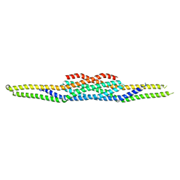
 | | Crystal structure of Matrix protein 1 from influenza A virus A/crow/Kyoto/T1/2004(H5N1) | | 分子名称: | Matrix protein 1 | | 著者 | Saijo, S, Kishishita, S, Uchikubo-Kamo, T, Terada, T, Shirouzu, M, Ito, H, Ito, T, Yokoyama, S, RIKEN Structural Genomics/Proteomics Initiative (RSGI) | | 登録日 | 2007-05-08 | | 公開日 | 2008-05-13 | | 最終更新日 | 2023-11-01 | | 実験手法 | X-RAY DIFFRACTION (2.02 Å) | | 主引用文献 | Crystal structure of Matrix protein 1 from influenza A virus A/crow/Kyoto/T1/2004(H5N1)
To be Published
|
|
2YW5
 | | Solution structure of the bromodomain from human bromodomain containing protein 3 | | 分子名称: | Bromodomain-containing protein 3 | | 著者 | Furue, M, Suzuki, S, Muto, Y, Inoue, M, Kigawa, T, Terada, T, Shirouzu, M, Yokoyama, S, RIKEN Structural Genomics/Proteomics Initiative (RSGI) | | 登録日 | 2007-04-19 | | 公開日 | 2008-04-22 | | 最終更新日 | 2024-05-29 | | 実験手法 | SOLUTION NMR | | 主引用文献 | Solution structure of the bromodomain from human bromodomain containing protein 3
To be Published
|
|
2YY0
 | | Crystal Structure of MS0802, c-Myc-1 binding protein domain from Homo sapiens | | 分子名称: | C-Myc-binding protein | | 著者 | Xie, Y, Wang, H, Ihsanawati, K.T, Kishishita, S, Takemoto, C, Shirozu, M, RIKEN Structural Genomics/Proteomics Initiative (RSGI) | | 登録日 | 2007-04-27 | | 公開日 | 2008-04-29 | | 最終更新日 | 2024-06-26 | | 実験手法 | X-RAY DIFFRACTION (2.4 Å) | | 主引用文献 | crystal structure of c-Myc-1 binding protein domain from Homo sapiens
To be Published
|
|
2YYA
 | | Crystal structure of GAR synthetase from Aquifex aeolicus | | 分子名称: | Phosphoribosylamine--glycine ligase | | 著者 | Baba, S, Kanagawa, M, Kuramitsu, S, Yokoyama, S, Kawai, G, Sampei, G, RIKEN Structural Genomics/Proteomics Initiative (RSGI) | | 登録日 | 2007-04-27 | | 公開日 | 2007-10-30 | | 最終更新日 | 2023-10-25 | | 実験手法 | X-RAY DIFFRACTION (2.4 Å) | | 主引用文献 | Crystal structures of glycinamide ribonucleotide synthetase, PurD, from thermophilic eubacteria
J.Biochem., 148, 2010
|
|
1WY2
 | |
1WZO
 | |
2Z1K
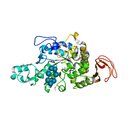 | | Crystal Structure of Ttha1563 from Thermus thermophilus HB8 | | 分子名称: | (Neo)pullulanase, Cycloheptakis-(1-4)-(alpha-D-glucopyranose), PHOSPHATE ION, ... | | 著者 | Niwa, H, Shimada, A, Matsunaga, E, Kuramitsu, S, Yokoyama, S, RIKEN Structural Genomics/Proteomics Initiative (RSGI) | | 登録日 | 2007-05-08 | | 公開日 | 2008-05-13 | | 最終更新日 | 2023-11-15 | | 実験手法 | X-RAY DIFFRACTION (2.3 Å) | | 主引用文献 | Crystal Structure of Ttha1563 from Thermus thermophilus HB8
To be Published
|
|
1WQU
 | | Solution structure of the human FES SH2 domain | | 分子名称: | Proto-oncogene tyrosine-protein kinase FES/FPS | | 著者 | Scott, A, Pantoja-Uceda, D, Koshiba, S, Inoue, M, Kigawa, T, Terada, T, Shirouzu, M, Tanaka, A, Sugano, S, Yokoyama, S, Guntert, P, RIKEN Structural Genomics/Proteomics Initiative (RSGI) | | 登録日 | 2004-10-02 | | 公開日 | 2005-06-14 | | 最終更新日 | 2024-05-29 | | 実験手法 | SOLUTION NMR | | 主引用文献 | Solution structure of the Src homology 2 domain from the human feline sarcoma oncogene Fes
J.Biomol.NMR, 31, 2005
|
|
1WDZ
 | | Crystal structure of RCB domain of IRSp53 | | 分子名称: | insulin receptor substrate p53 | | 著者 | Murayama, K, Suetsugu, S, Seto, A, Shirouzu, M, Takenawa, T, Yokoyama, S, RIKEN Structural Genomics/Proteomics Initiative (RSGI) | | 登録日 | 2004-05-21 | | 公開日 | 2005-06-07 | | 最終更新日 | 2024-03-13 | | 実験手法 | X-RAY DIFFRACTION (2.63 Å) | | 主引用文献 | Crystal structure of RCB domain of IRSp53
TO BE PUBLISHED
|
|


