8UG7
 
 | |
8UG8
 | |
8UG6
 | |
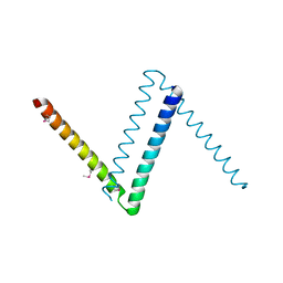
2M0Q
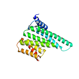
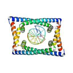
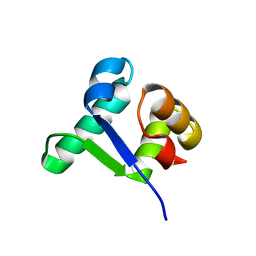
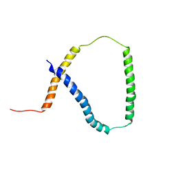 | | Solution NMR analysis of intact KCNE2 in detergent micelles demonstrate a straight transmembrane helix | | 分子名称: | Potassium voltage-gated channel subfamily E member 2 | | 著者 | Lai, C, Li, P, Chen, L, Zhang, L, Wu, F, Tian, C. | | 登録日 | 2012-11-01 | | 公開日 | 2014-04-30 | | 最終更新日 | 2024-05-15 | | 実験手法 | SOLUTION NMR | | 主引用文献 | Differential modulations of KCNQ1 by auxiliary proteins KCNE1 and KCNE2.
Sci Rep, 4, 2014
|
|
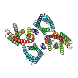
6AMK
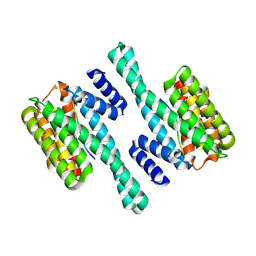
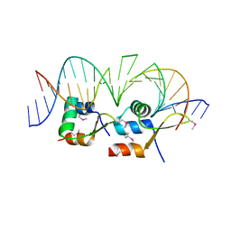 | | Structure of Streptomyces venezuelae BldC-whiI opt complex | | 分子名称: | DNA (5'-D(*AP*AP*TP*GP*TP*CP*CP*GP*AP*AP*TP*TP*AP*CP*CP*CP*GP*AP*AP*TP*TP*G)-3'), DNA (5'-D(*TP*TP*CP*AP*AP*TP*TP*CP*GP*GP*GP*TP*AP*AP*TP*TP*CP*GP*GP*GP*CP*A)-3'), Putative DNA-binding protein | | 著者 | Schumacher, M.A. | | 登録日 | 2017-08-09 | | 公開日 | 2018-03-28 | | 最終更新日 | 2018-11-07 | | 実験手法 | X-RAY DIFFRACTION (3.288 Å) | | 主引用文献 | The MerR-like protein BldC binds DNA direct repeats as cooperative multimers to regulate Streptomyces development.
Nat Commun, 9, 2018
|
|
6AMA
 | |
6BCJ
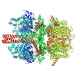
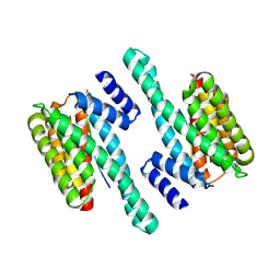
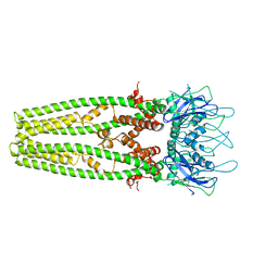
 | | cryo-EM structure of TRPM4 in apo state with short coiled coil at 3.1 angstrom resolution | | 分子名称: | SODIUM ION, Transient receptor potential cation channel subfamily M member 4 | | 著者 | Guo, J, She, J, Chen, Q, Bai, X, Jiang, Y. | | 登録日 | 2017-10-20 | | 公開日 | 2017-12-13 | | 最終更新日 | 2019-11-20 | | 実験手法 | ELECTRON MICROSCOPY (3.14 Å) | | 主引用文献 | Structures of the calcium-activated, non-selective cation channel TRPM4.
Nature, 552, 2017
|
|
6BCL
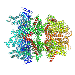
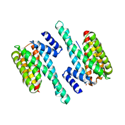
 | | cryo-EM structure of TRPM4 in apo state with long coiled coil at 3.5 angstrom resolution | | 分子名称: | SODIUM ION, Transient receptor potential cation channel subfamily M member 4 | | 著者 | Guo, J, She, J, Chen, Q, Bai, X, Jiang, Y. | | 登録日 | 2017-10-20 | | 公開日 | 2017-12-13 | | 最終更新日 | 2019-11-20 | | 実験手法 | ELECTRON MICROSCOPY (3.54 Å) | | 主引用文献 | Structures of the calcium-activated, non-selective cation channel TRPM4.
Nature, 552, 2017
|
|
6BCQ
 | | cryo-EM structure of TRPM4 in ATP bound state with long coiled coil at 3.3 angstrom resolution | | 分子名称: | ADENOSINE-5'-TRIPHOSPHATE, Transient receptor potential cation channel subfamily M member 4 | | 著者 | Guo, J, She, J, Chen, Q, Bai, X, Jiang, Y. | | 登録日 | 2017-10-20 | | 公開日 | 2017-12-13 | | 最終更新日 | 2019-11-20 | | 実験手法 | ELECTRON MICROSCOPY (3.25 Å) | | 主引用文献 | Structures of the calcium-activated, non-selective cation channel TRPM4.
Nature, 552, 2017
|
|
6BCO
 | | cryo-EM structure of TRPM4 in ATP bound state with short coiled coil at 2.9 angstrom resolution | | 分子名称: | ADENOSINE-5'-TRIPHOSPHATE, Transient receptor potential cation channel subfamily M member 4 | | 著者 | Guo, J, She, J, Chen, Q, Bai, X, Jiang, Y. | | 登録日 | 2017-10-20 | | 公開日 | 2017-12-13 | | 最終更新日 | 2019-11-20 | | 実験手法 | ELECTRON MICROSCOPY (2.88 Å) | | 主引用文献 | Structures of the calcium-activated, non-selective cation channel TRPM4.
Nature, 552, 2017
|
|
6BYK
 | |
6BZD
 | |
6BYJ
 | |
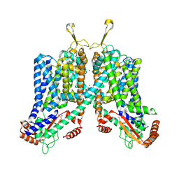
6C9A
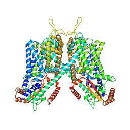 | | Cryo-EM structure of mouse TPC1 channel in the PtdIns(3,5)P2-bound state | | 分子名称: | (2R)-3-{[(S)-hydroxy{[(1S,2R,3R,4S,5S,6R)-2,4,6-trihydroxy-3,5-bis(phosphonooxy)cyclohexyl]oxy}phosphoryl]oxy}propane-1,2-diyl dioctanoate, 2-acetamido-2-deoxy-beta-D-glucopyranose, 2-acetamido-2-deoxy-beta-D-glucopyranose-(1-4)-2-acetamido-2-deoxy-beta-D-glucopyranose, ... | | 著者 | She, J, Guo, J, Chen, Q, Bai, X, Jiang, Y. | | 登録日 | 2018-01-25 | | 公開日 | 2018-04-04 | | 最終更新日 | 2020-07-29 | | 実験手法 | ELECTRON MICROSCOPY (3.2 Å) | | 主引用文献 | Structural insights into the voltage and phospholipid activation of the mammalian TPC1 channel.
Nature, 556, 2018
|
|
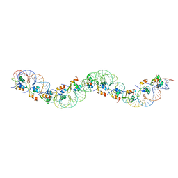
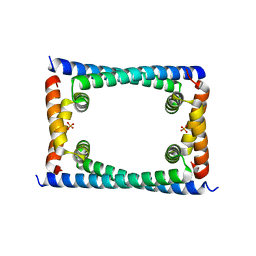
6CG8
 | | Structure of C. crescentus GapR-DNA | | 分子名称: | DNA (5'-D(*TP*TP*AP*AP*AP*AP*TP*TP*AP*AP*A)-3'), DNA (5'-D(*TP*TP*TP*AP*AP*TP*TP*TP*TP*AP*A)-3'), UPF0335 protein B7Z12_12435 | | 著者 | Schumacher, M.A. | | 登録日 | 2018-02-19 | | 公開日 | 2018-09-26 | | 最終更新日 | 2024-03-13 | | 実験手法 | X-RAY DIFFRACTION (2.299 Å) | | 主引用文献 | A Bacterial Chromosome Structuring Protein Binds Overtwisted DNA to Stimulate Type II Topoisomerases and Enable DNA Replication.
Cell, 175, 2018
|
|
6BYL
 | |
6CFY
 | | Bosea sp Root 381 apo GapR structure | | 分子名称: | UPF0335 protein ASE63_04290 | | 著者 | Schumacherr, M.A. | | 登録日 | 2018-02-18 | | 公開日 | 2018-09-12 | | 最終更新日 | 2018-10-17 | | 実験手法 | X-RAY DIFFRACTION (2.4 Å) | | 主引用文献 | A Bacterial Chromosome Structuring Protein Binds Overtwisted DNA to Stimulate Type II Topoisomerases and Enable DNA Replication.
Cell, 175, 2018
|
|
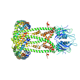
6C96
 | | Cryo-EM structure of mouse TPC1 channel in the apo state | | 分子名称: | 2-acetamido-2-deoxy-beta-D-glucopyranose, 2-acetamido-2-deoxy-beta-D-glucopyranose-(1-4)-2-acetamido-2-deoxy-beta-D-glucopyranose, SODIUM ION, ... | | 著者 | She, J, Guo, J, Chen, Q, Bai, X, Jiang, Y. | | 登録日 | 2018-01-25 | | 公開日 | 2018-04-04 | | 最終更新日 | 2020-07-29 | | 実験手法 | ELECTRON MICROSCOPY (3.4 Å) | | 主引用文献 | Structural insights into the voltage and phospholipid activation of the mammalian TPC1 channel.
Nature, 556, 2018
|
|
6CFX
 | | Bosea sp GapR solved in the presence of DNA | | 分子名称: | PHOSPHATE ION, UPF0335 protein ASE63_04290 | | 著者 | Schumacher, M.A. | | 登録日 | 2018-02-18 | | 公開日 | 2018-09-12 | | 最終更新日 | 2023-10-04 | | 実験手法 | X-RAY DIFFRACTION (2 Å) | | 主引用文献 | A Bacterial Chromosome Structuring Protein Binds Overtwisted DNA to Stimulate Type II Topoisomerases and Enable DNA Replication.
Cell, 175, 2018
|
|
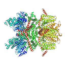
6D80
 | | Cryo-EM structure of the mitochondrial calcium uniporter from N. fischeri bound to saposin | | 分子名称: | CALCIUM ION, Mitochondrial calcium uniporter, Saposin A | | 著者 | Nguyen, N.X, Armache, J.-P, Cheng, Y, Bai, X.C. | | 登録日 | 2018-04-25 | | 公開日 | 2018-07-11 | | 最終更新日 | 2024-05-15 | | 実験手法 | ELECTRON MICROSCOPY (5 Å) | | 主引用文献 | Cryo-EM structure of a fungal mitochondrial calcium uniporter.
Nature, 559, 2018
|
|
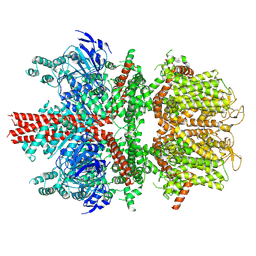
6D7W
 | | Cryo-EM structure of the mitochondrial calcium uniporter from N. fischeri at 3.8 Angstrom resolution | | 分子名称: | CALCIUM ION, Mitochondrial calcium uniporter | | 著者 | Nguyen, N.X, Armache, J.-P, Cheng, Y, Bai, X.C. | | 登録日 | 2018-04-25 | | 公開日 | 2018-07-11 | | 最終更新日 | 2024-05-15 | | 実験手法 | ELECTRON MICROSCOPY (3.8 Å) | | 主引用文献 | Cryo-EM structure of a fungal mitochondrial calcium uniporter.
Nature, 559, 2018
|
|
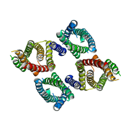
5TZF
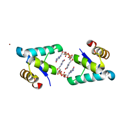
 | | Structure of the BldD CTD(D116A)-(c-di-GMP)2 intermediate, form 1 | | 分子名称: | 9,9'-[(2R,3R,3aS,5S,7aR,9R,10R,10aS,12S,14aR)-3,5,10,12-tetrahydroxy-5,12-dioxidooctahydro-2H,7H-difuro[3,2-d:3',2'-j][1,3,7,9,2,8]tetraoxadiphosphacyclododecine-2,9-diyl]bis(2-amino-1,9-dihydro-6H-purin-6-one), DNA-binding protein | | 著者 | Schumacher, M.A. | | 登録日 | 2016-11-21 | | 公開日 | 2017-04-19 | | 最終更新日 | 2023-10-04 | | 実験手法 | X-RAY DIFFRACTION (2.4 Å) | | 主引用文献 | The Streptomyces master regulator BldD binds c-di-GMP sequentially to create a functional BldD2-(c-di-GMP)4 complex.
Nucleic Acids Res., 45, 2017
|
|
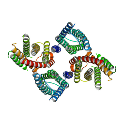
5TZG
 | | Structure of the BldD CTD(D116A)-(c-di-GMP)2, form 2 | | 分子名称: | 9,9'-[(2R,3R,3aS,5S,7aR,9R,10R,10aS,12S,14aR)-3,5,10,12-tetrahydroxy-5,12-dioxidooctahydro-2H,7H-difuro[3,2-d:3',2'-j][1,3,7,9,2,8]tetraoxadiphosphacyclododecine-2,9-diyl]bis(2-amino-1,9-dihydro-6H-purin-6-one), DNA-binding protein, ZINC ION | | 著者 | Schumacher, M.A. | | 登録日 | 2016-11-21 | | 公開日 | 2017-04-19 | | 最終更新日 | 2023-10-04 | | 実験手法 | X-RAY DIFFRACTION (2.5 Å) | | 主引用文献 | The Streptomyces master regulator BldD binds c-di-GMP sequentially to create a functional BldD2-(c-di-GMP)4 complex.
Nucleic Acids Res., 45, 2017
|
|
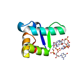
4IN4
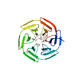 | | Crystal structure of cpd 15 bound to Keap1 Kelch domain | | 分子名称: | 2-({5-[(2,4-dimethylphenyl)sulfonyl]-6-oxo-1,6-dihydropyrimidin-2-yl}sulfanyl)-N-[2-(trifluoromethyl)phenyl]acetamide, Kelch-like ECH-associated protein 1, PHOSPHATE ION | | 著者 | Silvian, L, Marcotte, D. | | 登録日 | 2013-01-03 | | 公開日 | 2013-05-15 | | 最終更新日 | 2023-09-20 | | 実験手法 | X-RAY DIFFRACTION (2.59 Å) | | 主引用文献 | Small molecules inhibit the interaction of Nrf2 and the Keap1 Kelch domain through a non-covalent mechanism.
Bioorg.Med.Chem., 21, 2013
|
|
5TZD
 | | Structure of the WT S. venezulae BldD-(CTD-c-di-GMP)2 assembly intermediate | | 分子名称: | 9,9'-[(2R,3R,3aS,5S,7aR,9R,10R,10aS,12S,14aR)-3,5,10,12-tetrahydroxy-5,12-dioxidooctahydro-2H,7H-difuro[3,2-d:3',2'-j][1,3,7,9,2,8]tetraoxadiphosphacyclododecine-2,9-diyl]bis(2-amino-1,9-dihydro-6H-purin-6-one), DNA-binding protein | | 著者 | Schumacher, M. | | 登録日 | 2016-11-21 | | 公開日 | 2017-04-19 | | 最終更新日 | 2023-10-04 | | 実験手法 | X-RAY DIFFRACTION (1.749 Å) | | 主引用文献 | The Streptomyces master regulator BldD binds c-di-GMP sequentially to create a functional BldD2-(c-di-GMP)4 complex.
Nucleic Acids Res., 45, 2017
|
|


