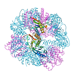
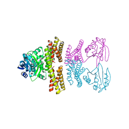
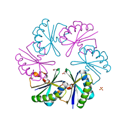
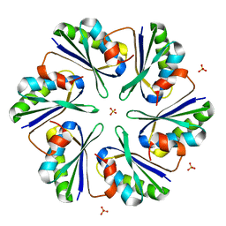
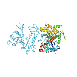
8UF0
 
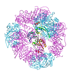 | | T33-ml23 - Designed Tetrahedral Protein Cage Using Machine Learning Algorithms | | 分子名称: | T33-ml23-redesigned-CutA-fold, T33-ml23-redesigned-tandem-BMC-T-fold | | 著者 | Castells-Graells, R, Meador, K, Sawaya, M.R, Yeates, T.O. | | 登録日 | 2023-10-03 | | 公開日 | 2023-11-15 | | 最終更新日 | 2024-06-19 | | 実験手法 | ELECTRON MICROSCOPY (2.02 Å) | | 主引用文献 | A suite of designed protein cages using machine learning and protein fragment-based protocols.
Structure, 32, 2024
|
|
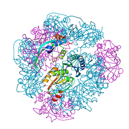
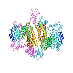
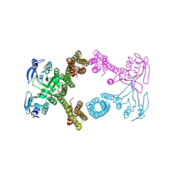
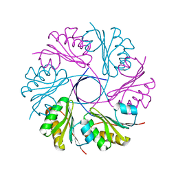
8UN1
 | | T33-ml23 Assembly Intermediate - Designed Tetrahedral Protein Cage Using Machine Learning Algorithms | | 分子名称: | T33-ml23-redesigned-CutA-fold, T33-ml23-redesigned-tandem-BMC-T-fold | | 著者 | Castells-Graells, R, Meador, K, Sawaya, M.R, Yeates, T.O. | | 登録日 | 2023-10-18 | | 公開日 | 2024-03-06 | | 最終更新日 | 2024-06-19 | | 実験手法 | ELECTRON MICROSCOPY (3.9 Å) | | 主引用文献 | A suite of designed protein cages using machine learning and protein fragment-based protocols.
Structure, 32, 2024
|
|
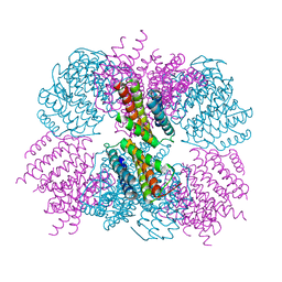
8UMR
 | | T33-ml35 Assembly Intermediate - Designed Tetrahedral Protein Cage Using Machine Learning Algorithms | | 分子名称: | T33-ml35-redesigned-CutA-fold, T33-ml35-redesigned-TPR-domain-fold | | 著者 | Castells-Graells, R, Meador, K, Sawaya, M.R, Yeates, T.O. | | 登録日 | 2023-10-18 | | 公開日 | 2024-03-06 | | 最終更新日 | 2024-06-19 | | 実験手法 | ELECTRON MICROSCOPY (4.42 Å) | | 主引用文献 | A suite of designed protein cages using machine learning and protein fragment-based protocols.
Structure, 32, 2024
|
|
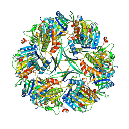
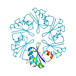
8UI2
 | | T33-ml28 - Designed Tetrahedral Protein Cage Using Machine Learning Algorithms | | 分子名称: | T33-ml28-redesigned-CutA-fold, T33-ml28-redesigned-tandem-BMC-T-fold | | 著者 | Castells-Graells, R, Meador, K, Sawaya, M.R, Yeates, T.O. | | 登録日 | 2023-10-09 | | 公開日 | 2024-03-06 | | 最終更新日 | 2024-06-19 | | 実験手法 | ELECTRON MICROSCOPY (2.73 Å) | | 主引用文献 | A suite of designed protein cages using machine learning and protein fragment-based protocols.
Structure, 32, 2024
|
|
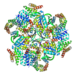
8UKM
 | | T33-ml30 - Designed Tetrahedral Protein Cage Using Machine Learning Algorithms | | 分子名称: | T33-ml30-redesigned-4-OT-fold, T33-ml30-redesigned-tandem-BMC-T-fold | | 著者 | Castells-Graells, R, Meador, K, Sawaya, M.R, Yeates, T.O. | | 登録日 | 2023-10-14 | | 公開日 | 2024-03-06 | | 最終更新日 | 2024-06-19 | | 実験手法 | ELECTRON MICROSCOPY (4.2 Å) | | 主引用文献 | A suite of designed protein cages using machine learning and protein fragment-based protocols.
Structure, 32, 2024
|
|
4ZSX
 | |
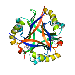
5CY5
 | | Crystal structure of the T33-51H designed self-assembling protein tetrahedron | | 分子名称: | T33-51H-A, T33-51H-B | | 著者 | Cannon, K.A, Cascio, D, Park, R, Boyken, S, King, N, Yeates, T.O. | | 登録日 | 2015-07-30 | | 公開日 | 2016-08-10 | | 最終更新日 | 2023-09-27 | | 実験手法 | X-RAY DIFFRACTION (3.4 Å) | | 主引用文献 | Design and structure of two new protein cages illustrate successes and ongoing challenges in protein engineering.
Protein Sci., 29, 2020
|
|
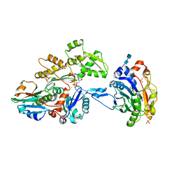
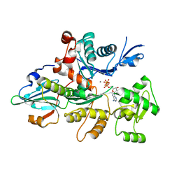
5D4P
 | | Structure of CPII bound to ADP and bicarbonate, from Thiomonas intermedia K12 | | 分子名称: | ADENOSINE-5'-DIPHOSPHATE, BICARBONATE ION, Putative Nitrogen regulatory protein P-II GlnB | | 著者 | Wheatley, N.M, Ngo, J, Cascio, D, Sawaya, M.R, Yeates, T.O. | | 登録日 | 2015-08-08 | | 公開日 | 2016-09-28 | | 最終更新日 | 2023-09-27 | | 実験手法 | X-RAY DIFFRACTION (2.2 Å) | | 主引用文献 | A PII-Like Protein Regulated by Bicarbonate: Structural and Biochemical Studies of the Carboxysome-Associated CPII Protein.
J.Mol.Biol., 428, 2016
|
|
4ZSZ
 | |
4ZSV
 | |
5D4O
 | | Structure of CPII, a nitrogen regulatory PII-like protein from Thiomonas intermedia K12, bound to ADP, AMP and bicarbonate. | | 分子名称: | ADENOSINE MONOPHOSPHATE, ADENOSINE-5'-DIPHOSPHATE, BICARBONATE ION, ... | | 著者 | Wheatley, N.M, Ngo, J, Cascio, D, Sawaya, M.R, Yeates, T.O. | | 登録日 | 2015-08-08 | | 公開日 | 2016-09-28 | | 最終更新日 | 2023-09-27 | | 実験手法 | X-RAY DIFFRACTION (1.8 Å) | | 主引用文献 | A PII-Like Protein Regulated by Bicarbonate: Structural and Biochemical Studies of the Carboxysome-Associated CPII Protein.
J.Mol.Biol., 428, 2016
|
|
5D4L
 | | Structure of the apo form of CPII from Thiomonas intermedia K12, a nitrogen regulatory PII-like protein | | 分子名称: | Nitrogen regulatory protein P-II | | 著者 | Wheatley, N.M, Ngo, J, Cascio, D, Sawaya, M.R, Yeates, T.O. | | 登録日 | 2015-08-08 | | 公開日 | 2016-09-28 | | 最終更新日 | 2023-09-27 | | 実験手法 | X-RAY DIFFRACTION (2.3 Å) | | 主引用文献 | A PII-Like Protein Regulated by Bicarbonate: Structural and Biochemical Studies of the Carboxysome-Associated CPII Protein.
J.Mol.Biol., 428, 2016
|
|
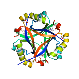
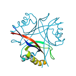
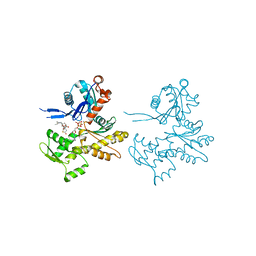
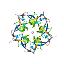
3CIM
 | | Carboxysome shell protein, CcmK2 C-terminal deletion mutant | | 分子名称: | Carbon dioxide-concentrating mechanism protein ccmK homolog 2, GLYCEROL, SULFATE ION | | 著者 | Tanaka, S, Sawaya, M.R, Yeates, T.O. | | 登録日 | 2008-03-11 | | 公開日 | 2009-02-03 | | 最終更新日 | 2024-02-21 | | 実験手法 | X-RAY DIFFRACTION (1.3 Å) | | 主引用文献 | Insights from multiple structures of the shell proteins from the beta-carboxysome.
Protein Sci., 18, 2009
|
|
3CGI
 | |
3CJC
 | | Actin dimer cross-linked by V. cholerae MARTX toxin and complexed with DNase I and Gelsolin-segment 1 | | 分子名称: | 2-acetamido-2-deoxy-beta-D-glucopyranose-(1-4)-2-acetamido-2-deoxy-beta-D-glucopyranose, ADENOSINE-5'-TRIPHOSPHATE, Actin, ... | | 著者 | Sawaya, M.R, Kudryashov, D.S, Pashkov, I, Reisler, E, Yeates, T.O. | | 登録日 | 2008-03-12 | | 公開日 | 2008-03-25 | | 最終更新日 | 2020-07-29 | | 実験手法 | X-RAY DIFFRACTION (3.9 Å) | | 主引用文献 | Connecting actin monomers by iso-peptide bond is a toxicity mechanism of the Vibrio cholerae MARTX toxin.
Proc.Natl.Acad.Sci.USA, 105, 2008
|
|
3DNC
 | | Carboxysome shell protein, CcmK2 C-terminal deletion mutant, with a closer spacing between hexamers | | 分子名称: | Carbon dioxide-concentrating mechanism protein ccmK homolog 2, GLYCEROL, SULFATE ION | | 著者 | Tanaka, S, Sawaya, M.R, Yeates, T.O. | | 登録日 | 2008-07-01 | | 公開日 | 2009-01-20 | | 最終更新日 | 2023-08-30 | | 実験手法 | X-RAY DIFFRACTION (2.05 Å) | | 主引用文献 | Insights from multiple structures of the shell proteins from the beta-carboxysome.
Protein Sci., 18, 2009
|
|
3CJB
 | | Actin dimer cross-linked by V. cholerae MARTX toxin and complexed with Gelsolin-segment 1 | | 分子名称: | ADENOSINE-5'-TRIPHOSPHATE, Actin, alpha skeletal muscle, ... | | 著者 | Sawaya, M.R, Kudryashov, D.S, Pashkov, I, Reisler, E, Yeates, T.O. | | 登録日 | 2008-03-12 | | 公開日 | 2008-03-25 | | 最終更新日 | 2024-02-21 | | 実験手法 | X-RAY DIFFRACTION (3.21 Å) | | 主引用文献 | Connecting actin monomers by iso-peptide bond is a toxicity mechanism of the Vibrio cholerae MARTX toxin.
Proc.Natl.Acad.Sci.USA, 105, 2008
|
|
2PY8
 | | RbcX | | 分子名称: | 2-{2-[2-(2-{2-[2-(2-ETHOXY-ETHOXY)-ETHOXY]-ETHOXY}-ETHOXY)-ETHOXY]-ETHOXY}-ETHANOL, CHLORIDE ION, Hypothetical protein rbcX | | 著者 | Tanaka, S, Sawaya, M.R, Kerfeld, C.A, Yeates, T.O. | | 登録日 | 2007-05-15 | | 公開日 | 2007-10-09 | | 最終更新日 | 2024-02-21 | | 実験手法 | X-RAY DIFFRACTION (2.45 Å) | | 主引用文献 | Structure of the RuBisCO chaperone RbcX from Synechocystis sp. PCC6803.
Acta Crystallogr.,Sect.D, 63, 2007
|
|
3DN9
 | |
2Q1N
 | | Actin Dimer Cross-linked Between Residues 41 and 374 | | 分子名称: | Actin, alpha skeletal muscle, CALCIUM ION, ... | | 著者 | Sawaya, M.R, Pashkov, I, Kudryashov, D.S, Adisetiyo, H, Reisler, E, Yeates, T.O. | | 登録日 | 2007-05-25 | | 公開日 | 2007-06-05 | | 最終更新日 | 2023-08-30 | | 実験手法 | X-RAY DIFFRACTION (2.7 Å) | | 主引用文献 | Multiple crystal structures of actin dimers and their implications for interactions in the actin filament.
Acta Crystallogr.,Sect.D, 64, 2008
|
|
2Q36
 | | Actin Dimer Cross-linked between Residues 191 and 374 and complexed with Kabiramide C | | 分子名称: | ADENOSINE-5'-TRIPHOSPHATE, Actin, alpha skeletal muscle, ... | | 著者 | Sawaya, M.R, Pashkov, I, Kudryashov, D.S, Reisler, E, Yeates, T.O. | | 登録日 | 2007-05-29 | | 公開日 | 2007-06-05 | | 最終更新日 | 2023-08-30 | | 実験手法 | X-RAY DIFFRACTION (2.5 Å) | | 主引用文献 | Multiple crystal structures of actin dimers and their implications for interactions in the actin filament.
Acta Crystallogr.,Sect.D, 64, 2008
|
|
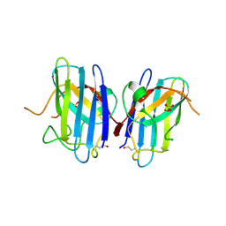
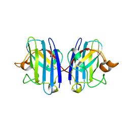
2QW7
 | | Carboxysome Subunit, CcmL | | 分子名称: | Carbon dioxide concentrating mechanism protein ccmL, GLYCEROL | | 著者 | Tanaka, S, Sawaya, M.R, Kerfeld, C.A, Yeates, T.O. | | 登録日 | 2007-08-09 | | 公開日 | 2008-03-04 | | 最終更新日 | 2024-02-21 | | 実験手法 | X-RAY DIFFRACTION (2.4 Å) | | 主引用文献 | Atomic-level models of the bacterial carboxysome shell.
Science, 319, 2008
|
|
1OZU
 | | Crystal Structure of Familial ALS Mutant S134N of human Cu,Zn Superoxide Dismutase (CuZnSOD) to 1.3A resolution | | 分子名称: | SULFATE ION, Superoxide dismutase [Cu-Zn], ZINC ION | | 著者 | Elam, J.S, Taylor, A.B, Strange, R, Antonyuk, S, Doucette, P.A, Rodriguez, J.A, Hasnain, S.S, Hayward, L.J, Valentine, J.S, Yeates, T.O, Hart, P.J. | | 登録日 | 2003-04-09 | | 公開日 | 2003-05-27 | | 最終更新日 | 2023-08-16 | | 実験手法 | X-RAY DIFFRACTION (1.3 Å) | | 主引用文献 | Amyloid-like Filaments and Water-filled Nanotubes Formed by SOD1 Mutant Proteins Linked to Familial ALS
Nat.Struct.Biol., 10, 2003
|
|
1OZT
 | | Crystal Structure of apo-H46R Familial ALS Mutant human Cu,Zn Superoxide Dismutase (CuZnSOD) to 2.5A resolution | | 分子名称: | Superoxide dismutase [Cu-Zn] | | 著者 | Elam, J.S, Taylor, A.B, Strange, R, Antonyuk, S, Doucette, P.A, Rodriguez, J.A, Hasnain, S.S, Hayward, L.J, Valentine, J.S, Yeates, T.O, Hart, P.J. | | 登録日 | 2003-04-09 | | 公開日 | 2003-05-27 | | 最終更新日 | 2023-08-16 | | 実験手法 | X-RAY DIFFRACTION (2.5 Å) | | 主引用文献 | Amyloid-like Filaments and Water-filled Nanotubes Formed by SOD1 Mutant Proteins Linked to Familial ALS
Nat.Struct.Biol., 10, 2003
|
|
1OY0
 | | The crystal Structure of the First Enzyme of Pantothenate Biosynthetic Pathway, Ketopantoate Hydroxymethyltransferase from Mycobacterium Tuberculosis Shows a Decameric Assembly and Terminal Helix-Swapping | | 分子名称: | Ketopantoate hydroxymethyltransferase, MAGNESIUM ION | | 著者 | Chaudhuri, B.N, Sawaya, M.R, Kim, C.Y, Waldo, G.S, Park, M.S, Terwilliger, T.C, Yeates, T.O, TB Structural Genomics Consortium (TBSGC) | | 登録日 | 2003-04-03 | | 公開日 | 2003-07-15 | | 最終更新日 | 2024-02-14 | | 実験手法 | X-RAY DIFFRACTION (2.8 Å) | | 主引用文献 | The Crystal Structure of the First Enzyme in the Pantothenate Biosynthetic Pathway,
Ketopantoate Hydroxymethyltransferase, from M. tuberculosis
Structure, 11, 2003
|
|


