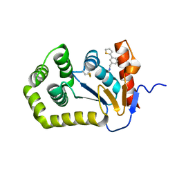
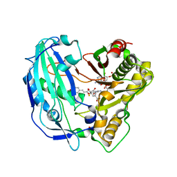
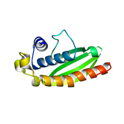
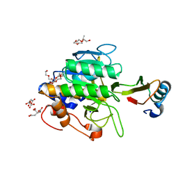
1Y3B
 
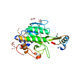 | | Crystal structure of the complex of subtilisin BPN' with chymotrypsin inhibitor 2 E60S mutant | | 分子名称: | CALCIUM ION, CITRIC ACID, POLYETHYLENE GLYCOL (N=34), ... | | 著者 | Radisky, E.S, Lu, C.J, Kwan, G, Koshland Jr, D.E. | | 登録日 | 2004-11-24 | | 公開日 | 2005-05-17 | | 最終更新日 | 2023-08-23 | | 実験手法 | X-RAY DIFFRACTION (1.8 Å) | | 主引用文献 | Role of the intramolecular hydrogen bond network in the inhibitory power of chymotrypsin inhibitor 2
Biochemistry, 44, 2005
|
|
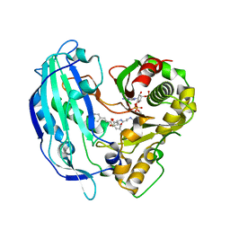
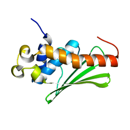
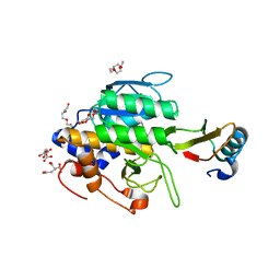
1Y48
 | | Crystal structure of the complex of subtilisin BPN' with chymotrypsin inhibitor 2 R65A mutant | | 分子名称: | CALCIUM ION, CITRIC ACID, POLYETHYLENE GLYCOL (N=34), ... | | 著者 | Radisky, E.S, Lu, C.J, Kwan, G, Koshland Jr, D.E. | | 登録日 | 2004-11-30 | | 公開日 | 2005-05-17 | | 最終更新日 | 2023-08-23 | | 実験手法 | X-RAY DIFFRACTION (1.84 Å) | | 主引用文献 | Role of the intramolecular hydrogen bond network in the inhibitory power of chymotrypsin inhibitor 2
Biochemistry, 44, 2005
|
|
5GUJ
 | |
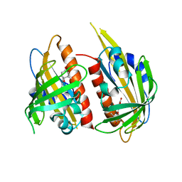
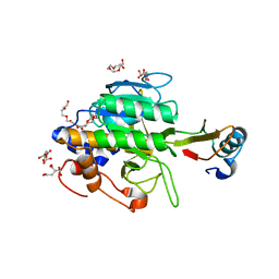
1Y34
 | | Crystal structure of the complex of subtilisin BPN' with chymotrypsin inhibitor 2 E60A mutant | | 分子名称: | CALCIUM ION, CITRIC ACID, POLYETHYLENE GLYCOL (N=34), ... | | 著者 | Radisky, E.S, Lu, C.J, Kwan, G, Koshland Jr, D.E. | | 登録日 | 2004-11-23 | | 公開日 | 2005-05-17 | | 最終更新日 | 2023-08-23 | | 実験手法 | X-RAY DIFFRACTION (1.55 Å) | | 主引用文献 | Role of the intramolecular hydrogen bond network in the inhibitory power of chymotrypsin inhibitor 2
Biochemistry, 44, 2005
|
|
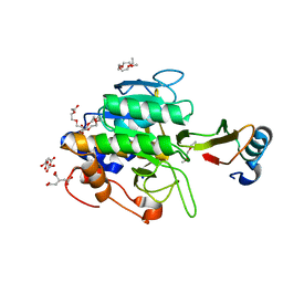
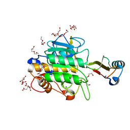
1Y4A
 | | Crystal structure of the complex of subtilisin BPN' with chymotrypsin inhibitor 2 M59R/E60S mutant | | 分子名称: | CALCIUM ION, CITRIC ACID, POLYETHYLENE GLYCOL (N=34), ... | | 著者 | Radisky, E.S, Lu, C.J, Kwan, G, Koshland Jr, D.E. | | 登録日 | 2004-11-30 | | 公開日 | 2005-05-17 | | 最終更新日 | 2023-08-23 | | 実験手法 | X-RAY DIFFRACTION (1.6 Å) | | 主引用文献 | Role of the intramolecular hydrogen bond network in the inhibitory power of chymotrypsin inhibitor 2
Biochemistry, 44, 2005
|
|
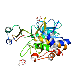
6BQX
 | | Crystal structure of Escherichia coli DsbA in complex with N-methyl-1-(4-phenoxyphenyl)methanamine | | 分子名称: | N-methyl-1-(4-phenoxyphenyl)methanamine, Thiol:disulfide interchange protein DsbA | | 著者 | Heras, B, Totsika, M, Paxman, J.J, Wang, G, Scanlon, M.J. | | 登録日 | 2017-11-29 | | 公開日 | 2017-12-27 | | 最終更新日 | 2020-01-01 | | 実験手法 | X-RAY DIFFRACTION (1.992 Å) | | 主引用文献 | Inhibition of Diverse DsbA Enzymes in Multi-DsbA Encoding Pathogens.
Antioxid. Redox Signal., 29, 2018
|
|
6BR4
 | | Crystal structure of Escherichia coli DsbA in complex with {N}-methyl-1-(3-thiophen-2-ylphenyl)methanamine | | 分子名称: | COPPER (II) ION, Thiol:disulfide interchange protein DsbA, ~{N}-methyl-1-(3-thiophen-2-ylphenyl)methanamine | | 著者 | Heras, B, Totsika, M, Paxman, J.J, Wang, G, Scanlon, M.J, Martin, J.L. | | 登録日 | 2017-11-29 | | 公開日 | 2017-12-27 | | 最終更新日 | 2020-01-01 | | 実験手法 | X-RAY DIFFRACTION (1.99 Å) | | 主引用文献 | Inhibition of Diverse DsbA Enzymes in Multi-DsbA Encoding Pathogens.
Antioxid. Redox Signal., 29, 2018
|
|
7CHH
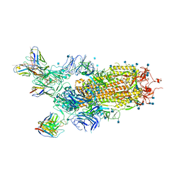 | | Cryo-EM structure of the SARS-CoV-2 S-6P in complex with BD-368-2 Fabs | | 分子名称: | 2-acetamido-2-deoxy-beta-D-glucopyranose, 2-acetamido-2-deoxy-beta-D-glucopyranose-(1-4)-2-acetamido-2-deoxy-beta-D-glucopyranose, BD-368-2 Fab heavy chain, ... | | 著者 | Xiao, J, Zhu, Q, Wang, G. | | 登録日 | 2020-07-05 | | 公開日 | 2020-09-16 | | 最終更新日 | 2020-11-25 | | 実験手法 | ELECTRON MICROSCOPY (3.49 Å) | | 主引用文献 | Structurally Resolved SARS-CoV-2 Antibody Shows High Efficacy in Severely Infected Hamsters and Provides a Potent Cocktail Pairing Strategy.
Cell, 183, 2020
|
|
4ME3
 | | 1.8 Angstrom Crystal Structure of the N-terminal Domain of an Archaeal MCM | | 分子名称: | DNA replication licensing factor MCM related protein, ZINC ION | | 著者 | Fu, Y, Slaymaker, I.M, Wang, G, Chen, X.S. | | 登録日 | 2013-08-24 | | 公開日 | 2014-01-08 | | 最終更新日 | 2024-02-28 | | 実験手法 | X-RAY DIFFRACTION (1.794 Å) | | 主引用文献 | The 1.8- angstrom Crystal Structure of the N-Terminal Domain of an Archaeal MCM as a Right-Handed Filament.
J.Mol.Biol., 426, 2014
|
|
4HNM
 | | Crystal structure of human catenin-beta-like 1 56 kDa fragment | | 分子名称: | Beta-catenin-like protein 1 | | 著者 | Du, Z, Huang, X, Wang, G, Wu, Y. | | 登録日 | 2012-10-19 | | 公開日 | 2013-07-31 | | 最終更新日 | 2013-08-21 | | 実験手法 | X-RAY DIFFRACTION (2.9001 Å) | | 主引用文献 | The structure of full-length human CTNNBL1 reveals a distinct member of the armadillo-repeat protein family.
Acta Crystallogr.,Sect.D, 69, 2013
|
|
5FAL
 | | Crystal structure of PvHCT in complex with CoA and p-coumaroyl-shikimate | | 分子名称: | (3~{R},4~{R},5~{R})-5-[(~{E})-3-(4-hydroxyphenyl)prop-2-enoyl]oxy-3,4-bis(oxidanyl)cyclohexene-1-carboxylic acid, COENZYME A, GLYCEROL, ... | | 著者 | Pereira, J.H, Moriarty, N.W, Eudes, A, Yogiswara, S, Wang, G, Benites, V.T, Baidoo, E.E.K, Lee, T.S, Keasling, J.D, Loque, D, Adams, P.D. | | 登録日 | 2015-12-11 | | 公開日 | 2016-02-24 | | 最終更新日 | 2024-03-06 | | 実験手法 | X-RAY DIFFRACTION (1.861 Å) | | 主引用文献 | Exploiting the Substrate Promiscuity of Hydroxycinnamoyl-CoA:Shikimate Hydroxycinnamoyl Transferase to Reduce Lignin.
Plant Cell.Physiol., 57, 2016
|
|
4HM9
 | | Crystal structure of full-length human catenin-beta-like 1 | | 分子名称: | Beta-catenin-like protein 1 | | 著者 | Du, Z, Huang, X, Wang, G, Wu, Y. | | 登録日 | 2012-10-18 | | 公開日 | 2013-07-31 | | 最終更新日 | 2024-02-28 | | 実験手法 | X-RAY DIFFRACTION (3.1001 Å) | | 主引用文献 | The structure of full-length human CTNNBL1 reveals a distinct member of the armadillo-repeat protein family.
Acta Crystallogr.,Sect.D, 69, 2013
|
|
5FAN
 | | Crystal structure of PvHCT in complex with p-coumaroyl-CoA and protocatechuate | | 分子名称: | 3,4-DIHYDROXYBENZOIC ACID, Hydroxycinnamoyl-CoA shikimate/quinate hydroxycinnamoyltransferase 2, p-coumaroyl-CoA | | 著者 | Pereira, J.H, Moriarty, N.W, Eudes, A, Yogiswara, S, Wang, G, Benites, V.T, Baidoo, E.E.K, Lee, T.S, Keasling, J.D, Loque, D, Adams, P.D. | | 登録日 | 2015-12-11 | | 公開日 | 2016-02-24 | | 最終更新日 | 2024-03-06 | | 実験手法 | X-RAY DIFFRACTION (1.901 Å) | | 主引用文献 | Exploiting the Substrate Promiscuity of Hydroxycinnamoyl-CoA:Shikimate Hydroxycinnamoyl Transferase to Reduce Lignin.
Plant Cell.Physiol., 57, 2016
|
|
1TM7
 | | crystal structure of the complex of subtilisin BPN' with chymotrypsin inhibitor 2 M59Y mutant | | 分子名称: | CALCIUM ION, CITRIC ACID, PENTAETHYLENE GLYCOL, ... | | 著者 | Radisky, E.S, Kwan, G, Karen Lu, C.J, Koshland Jr, D.E. | | 登録日 | 2004-06-10 | | 公開日 | 2004-11-09 | | 最終更新日 | 2023-08-23 | | 実験手法 | X-RAY DIFFRACTION (1.59 Å) | | 主引用文献 | Binding, Proteolytic, and Crystallographic Analyses of Mutations at the Protease-Inhibitor Interface of the Subtilisin BPN'/Chymotrypsin Inhibitor 2 Complex(,).
Biochemistry, 43, 2004
|
|
4L9C
 | | Crystal structure of the FP domain of human F-box protein Fbxo7 (native) | | 分子名称: | F-box only protein 7, GLYCEROL | | 著者 | Du, Z, Huang, X, Shang, J, Yang, Y, Wang, G. | | 登録日 | 2013-06-18 | | 公開日 | 2014-01-15 | | 最終更新日 | 2024-02-28 | | 実験手法 | X-RAY DIFFRACTION (2.1 Å) | | 主引用文献 | Structure of the FP domain of Fbxo7 reveals a novel mode of protein-protein interaction.
Acta Crystallogr.,Sect.D, 70, 2014
|
|
4L9H
 | | Crystal structure of the FP domain of human F-box protein Fbxo7(SeMet) | | 分子名称: | F-box only protein 7 | | 著者 | Du, Z, Huang, X, Shang, J, Yang, Y, Wang, G. | | 登録日 | 2013-06-18 | | 公開日 | 2014-01-15 | | 最終更新日 | 2014-10-08 | | 実験手法 | X-RAY DIFFRACTION (2 Å) | | 主引用文献 | Structure of the FP domain of Fbxo7 reveals a novel mode of protein-protein interaction.
Acta Crystallogr.,Sect.D, 70, 2014
|
|
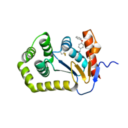
4JDA
 | | Complex structure of abscisic acid receptor PYL3 with (-)-ABA | | 分子名称: | (2Z,4E)-5-[(1R)-1-hydroxy-2,6,6-trimethyl-4-oxocyclohex-2-en-1-yl]-3-methylpenta-2,4-dienoic acid, Abscisic acid receptor PYL3 | | 著者 | Zhang, X, Wang, G, Chen, Z. | | 登録日 | 2013-02-24 | | 公開日 | 2013-07-24 | | 最終更新日 | 2023-11-08 | | 実験手法 | X-RAY DIFFRACTION (2.65 Å) | | 主引用文献 | Structural Insights into the Abscisic Acid Stereospecificity by the ABA Receptors PYR/PYL/RCAR
Plos One, 8, 2013
|
|
1TO1
 | | crystal structure of the complex of subtilisin BPN' with chymotrypsin inhibitor 2 Y61A mutant | | 分子名称: | CALCIUM ION, CITRIC ACID, PENTAETHYLENE GLYCOL, ... | | 著者 | Radisky, E.S, Kwan, G, Karen Lu, C.J, Koshland Jr, D.E. | | 登録日 | 2004-06-11 | | 公開日 | 2004-11-09 | | 最終更新日 | 2023-08-23 | | 実験手法 | X-RAY DIFFRACTION (1.68 Å) | | 主引用文献 | Binding, Proteolytic, and Crystallographic Analyses of Mutations at the Protease-Inhibitor Interface of the Subtilisin BPN'/Chymotrypsin Inhibitor 2 Complex(,).
Biochemistry, 43, 2004
|
|
1TO2
 | | crystal structure of the complex of subtilisin BPN' with chymotrypsin inhibitor 2 M59K, in pH 9 cryosoak | | 分子名称: | CALCIUM ION, CITRIC ACID, POLYETHYLENE GLYCOL (N=34), ... | | 著者 | Radisky, E.S, Kwan, G, Karen Lu, C.J, Koshland Jr, D.E. | | 登録日 | 2004-06-11 | | 公開日 | 2004-11-09 | | 最終更新日 | 2023-08-23 | | 実験手法 | X-RAY DIFFRACTION (1.3 Å) | | 主引用文献 | Binding, Proteolytic, and Crystallographic Analyses of Mutations at the Protease-Inhibitor Interface of the Subtilisin BPN'/Chymotrypsin Inhibitor 2 Complex(,).
Biochemistry, 43, 2004
|
|
1TM4
 | | crystal structure of the complex of subtilsin BPN'with chymotrypsin inhibitor 2 M59G mutant | | 分子名称: | CALCIUM ION, CITRIC ACID, PENTAETHYLENE GLYCOL, ... | | 著者 | Radisky, E.S, Kwan, G, Karen Lu, C.J, Koshland Jr, D.E. | | 登録日 | 2004-06-10 | | 公開日 | 2004-11-09 | | 最終更新日 | 2023-08-23 | | 実験手法 | X-RAY DIFFRACTION (1.7 Å) | | 主引用文献 | Binding, Proteolytic, and Crystallographic Analyses of Mutations at the Protease-Inhibitor Interface of the Subtilisin BPN'/Chymotrypsin Inhibitor 2 Complex(,).
Biochemistry, 43, 2004
|
|
1TM5
 | | crystal structure of the complex of subtilisin BPN' with chymotrypsin inhibitor 2 M59A mutant | | 分子名称: | CALCIUM ION, CITRIC ACID, PENTAETHYLENE GLYCOL, ... | | 著者 | Radisky, E.S, Kwan, G, Karen Lu, C.J, Koshland Jr, D.E. | | 登録日 | 2004-06-10 | | 公開日 | 2004-11-09 | | 最終更新日 | 2023-08-23 | | 実験手法 | X-RAY DIFFRACTION (1.45 Å) | | 主引用文献 | Binding, Proteolytic, and Crystallographic Analyses of Mutations at the Protease-Inhibitor Interface of the Subtilisin BPN'/Chymotrypsin Inhibitor 2 Complex(,).
Biochemistry, 43, 2004
|
|
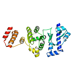
1TM1
 | | CRYSTAL STRUCTURE OF THE COMPLEX OF SUBTILISIN BPN' WITH CHYMOTRYPSIN INHIBITOR 2 | | 分子名称: | CALCIUM ION, CITRIC ACID, PENTAETHYLENE GLYCOL, ... | | 著者 | Radisky, E.S, Kwan, G, Karen Lu, C.J, Koshland Jr, D.E. | | 登録日 | 2004-06-10 | | 公開日 | 2004-11-09 | | 最終更新日 | 2023-08-23 | | 実験手法 | X-RAY DIFFRACTION (1.7 Å) | | 主引用文献 | Binding, Proteolytic, and Crystallographic Analyses of Mutations at the Protease-Inhibitor Interface of the Subtilisin BPN'/Chymotrypsin Inhibitor 2 Complex(,).
Biochemistry, 43, 2004
|
|
1TM3
 | | crystal structure of the complex of subtilisin BPN' with chymotrypsin inhibitor 2 M59k mutant | | 分子名称: | CALCIUM ION, CITRIC ACID, PENTAETHYLENE GLYCOL, ... | | 著者 | Radisky, E.S, Kwan, G, Karen Lu, C.J, Koshland Jr, D.E. | | 登録日 | 2004-06-10 | | 公開日 | 2004-11-09 | | 最終更新日 | 2023-08-23 | | 実験手法 | X-RAY DIFFRACTION (1.57 Å) | | 主引用文献 | Binding, Proteolytic, and Crystallographic Analyses of Mutations at the Protease-Inhibitor Interface of the Subtilisin BPN'/Chymotrypsin Inhibitor 2 Complex(,).
Biochemistry, 43, 2004
|
|
1TMG
 | | crystal structure of the complex of subtilisin BPN' with chymotrypsin inhibitor 2 M59F mutant | | 分子名称: | CALCIUM ION, CITRIC ACID, PENTAETHYLENE GLYCOL, ... | | 著者 | Radisky, E.S, Kwan, G, Karen Lu, C.J, Koshland Jr, D.E. | | 登録日 | 2004-06-10 | | 公開日 | 2004-11-09 | | 最終更新日 | 2023-08-23 | | 実験手法 | X-RAY DIFFRACTION (1.67 Å) | | 主引用文献 | Binding, Proteolytic, and Crystallographic Analyses of Mutations at the Protease-Inhibitor Interface of the Subtilisin BPN'/Chymotrypsin Inhibitor 2 Complex(,).
Biochemistry, 43, 2004
|
|
4NLH
 | |


