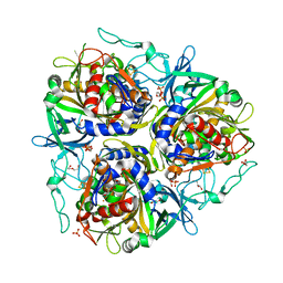
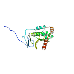
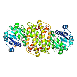
7VJU
 
 | |
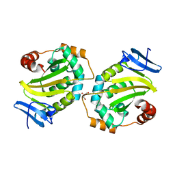
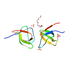
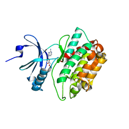
3C0G
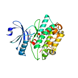 | | CASK CaM-Kinase Domain- 3'-AMP complex, P1 form | | 分子名称: | Peripheral plasma membrane protein CASK, [(2R,3S,4R,5R)-5-(6-aminopurin-9-yl)-4-hydroxy-2-(hydroxymethyl)oxolan-3-yl] dihydrogen phosphate | | 著者 | Wahl, M.C. | | 登録日 | 2008-01-20 | | 公開日 | 2008-04-29 | | 最終更新日 | 2024-04-03 | | 実験手法 | X-RAY DIFFRACTION (2.19 Å) | | 主引用文献 | CASK Functions as a Mg2+-independent neurexin kinase
Cell(Cambridge,Mass.), 133, 2008
|
|
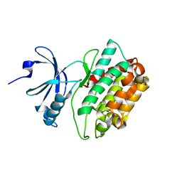
3C0H
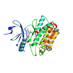 | | CASK CaM-Kinase Domain- AMPPNP complex, P1 form | | 分子名称: | ADENOSINE MONOPHOSPHATE, Peripheral plasma membrane protein CASK | | 著者 | Wahl, M.C. | | 登録日 | 2008-01-20 | | 公開日 | 2008-04-29 | | 最終更新日 | 2024-04-03 | | 実験手法 | X-RAY DIFFRACTION (2.3 Å) | | 主引用文献 | CASK Functions as a Mg2+-independent neurexin kinase
Cell(Cambridge,Mass.), 133, 2008
|
|
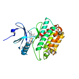
3C0I
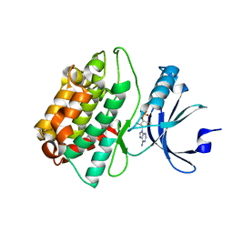 | | CASK CaM-Kinase Domain- 3'-AMP complex, P212121 form | | 分子名称: | Peripheral plasma membrane protein CASK, [(2R,3S,4R,5R)-5-(6-aminopurin-9-yl)-4-hydroxy-2-(hydroxymethyl)oxolan-3-yl] dihydrogen phosphate | | 著者 | Wahl, M.C. | | 登録日 | 2008-01-20 | | 公開日 | 2008-04-29 | | 最終更新日 | 2024-04-03 | | 実験手法 | X-RAY DIFFRACTION (1.85 Å) | | 主引用文献 | CASK Functions as a Mg2+-independent neurexin kinase
Cell(Cambridge,Mass.), 133, 2008
|
|
5DOE
 | | Crystal structure of the Human Cytomegalovirus UL53 (residues 72-292) | | 分子名称: | Virion egress protein UL31 homolog, ZINC ION | | 著者 | Lye, M.F, El Omari, K, Filman, D.J, Hogle, J.M. | | 登録日 | 2015-09-11 | | 公開日 | 2015-11-25 | | 最終更新日 | 2024-03-06 | | 実験手法 | X-RAY DIFFRACTION (3 Å) | | 主引用文献 | Unexpected features and mechanism of heterodimer formation of a herpesvirus nuclear egress complex.
Embo J., 34, 2015
|
|
5DOC
 | | Crystal structure of the Human Cytomegalovirus UL53 subunit of the NEC | | 分子名称: | GLYCEROL, Virion egress protein UL31 homolog, ZINC ION | | 著者 | Lye, M.F, El Omari, K, Filman, D.J, Hogle, J.M. | | 登録日 | 2015-09-11 | | 公開日 | 2015-11-25 | | 最終更新日 | 2024-03-06 | | 実験手法 | X-RAY DIFFRACTION (1.94 Å) | | 主引用文献 | Unexpected features and mechanism of heterodimer formation of a herpesvirus nuclear egress complex.
Embo J., 34, 2015
|
|
5DOB
 | | Crystal structure of the Human Cytomegalovirus Nuclear Egress Complex (NEC) | | 分子名称: | CALCIUM ION, Virion egress protein UL31 homolog, Virion egress protein UL34 homolog, ... | | 著者 | Lye, M.F, El Omari, K, Filman, D.J, Hogle, J.M. | | 登録日 | 2015-09-11 | | 公開日 | 2015-11-25 | | 最終更新日 | 2024-03-06 | | 実験手法 | X-RAY DIFFRACTION (2.47 Å) | | 主引用文献 | Unexpected features and mechanism of heterodimer formation of a herpesvirus nuclear egress complex.
Embo J., 34, 2015
|
|
5G6S
 | | Imine reductase from Aspergillus oryzae in complex with NADP(H) and (R)-rasagiline | | 分子名称: | IMINE REDUCTASE, NADPH DIHYDRO-NICOTINAMIDE-ADENINE-DINUCLEOTIDE PHOSPHATE, RASAGILINE | | 著者 | Man, H, Aleku, G, Turner, N.J, Grogan, G. | | 登録日 | 2016-06-23 | | 公開日 | 2017-05-31 | | 最終更新日 | 2024-01-10 | | 実験手法 | X-RAY DIFFRACTION (2.35 Å) | | 主引用文献 | A reductive aminase from Aspergillus oryzae.
Nat Chem, 9, 2017
|
|
5G6R
 | | Imine reductase from Aspergillus oryzae | | 分子名称: | IMINE REDUCTASE, NADPH DIHYDRO-NICOTINAMIDE-ADENINE-DINUCLEOTIDE PHOSPHATE | | 著者 | Man, H, Aleku, G, Turner, N.J, Grogan, G. | | 登録日 | 2016-06-23 | | 公開日 | 2017-05-31 | | 最終更新日 | 2024-01-10 | | 実験手法 | X-RAY DIFFRACTION (1.82 Å) | | 主引用文献 | A reductive aminase from Aspergillus oryzae.
Nat Chem, 9, 2017
|
|
2MCW
 | | Solid-state NMR structure of piscidin 3 in aligned 3:1 phosphatidylcholine/phosphoglycerol lipid bilayers | | 分子名称: | Piscidin-3 | | 著者 | Fu, R, Tian, Y, Perrin Jr, B.S, Grant, C.V, Pastor, R.W, Cotten, M.L. | | 登録日 | 2013-08-27 | | 公開日 | 2014-01-22 | | 最終更新日 | 2014-03-19 | | 実験手法 | SOLID-STATE NMR | | 主引用文献 | High-resolution structures and orientations of antimicrobial peptides piscidin 1 and piscidin 3 in fluid bilayers reveal tilting, kinking, and bilayer immersion.
J.Am.Chem.Soc., 136, 2014
|
|
2MCV
 | | Solid-state NMR structure of piscidin 1 in aligned 1:1 phosphatidylethanolamine/phosphoglycerol lipid bilayers | | 分子名称: | Moronecidin | | 著者 | Fu, R, Tian, Y, Perrin Jr, B.S, Grant, C.V, Hayden, R.M, Pastor, R.W, Cotten, M.L. | | 登録日 | 2013-08-27 | | 公開日 | 2014-01-22 | | 最終更新日 | 2014-03-19 | | 実験手法 | SOLID-STATE NMR | | 主引用文献 | High-resolution structures and orientations of antimicrobial peptides piscidin 1 and piscidin 3 in fluid bilayers reveal tilting, kinking, and bilayer immersion.
J.Am.Chem.Soc., 136, 2014
|
|
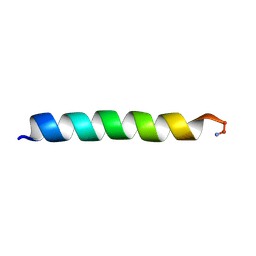
7YZS
 | |
7YZU
 | | Crystal structure of the sulfoquinovosyl binding protein SmoF complexed with SQMe | | 分子名称: | Sulfoquinovosyl binding protein, [(2S,3S,4S,5R,6S)-6-methoxy-3,4,5-tris(oxidanyl)oxan-2-yl]methanesulfonic acid | | 著者 | Snow, A.J.D, Davies, G.J. | | 登録日 | 2022-02-21 | | 公開日 | 2022-04-13 | | 最終更新日 | 2024-05-01 | | 実験手法 | X-RAY DIFFRACTION (1.59 Å) | | 主引用文献 | The sulfoquinovosyl glycerol binding protein SmoF binds and accommodates plant sulfolipids.
Curr Res Struct Biol, 4, 2022
|
|
2MCX
 | | Solid-state NMR structure of piscidin 3 in aligned 1:1 phosphatidylethanolamine/phosphoglycerol lipid bilayers | | 分子名称: | Piscidin-3 | | 著者 | Fu, R, Tian, Y, Perrin Jr, B.S, Grant, C.V, Wieczorek, W.E, Pastor, R.W, Cotten, M.L. | | 登録日 | 2013-08-27 | | 公開日 | 2014-01-22 | | 最終更新日 | 2021-08-18 | | 実験手法 | SOLID-STATE NMR | | 主引用文献 | High-resolution structures and orientations of antimicrobial peptides piscidin 1 and piscidin 3 in fluid bilayers reveal tilting, kinking, and bilayer immersion.
J.Am.Chem.Soc., 136, 2014
|
|
2MCU
 | | Solid-state NMR structure of piscidin 1 in aligned 3:1 phosphatidylcholine/phosphoglycerol lipid bilayers | | 分子名称: | Moronecidin | | 著者 | Fu, R, Tian, Y, Perrin Jr, B.S, Grant, C.V, Pastor, R.W, Cotten, M.L. | | 登録日 | 2013-08-27 | | 公開日 | 2014-01-22 | | 最終更新日 | 2014-03-19 | | 実験手法 | SOLID-STATE NMR | | 主引用文献 | High-resolution structures and orientations of antimicrobial peptides piscidin 1 and piscidin 3 in fluid bilayers reveal tilting, kinking, and bilayer immersion.
J.Am.Chem.Soc., 136, 2014
|
|
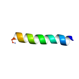
2FPF
 | |
2FPD
 | |
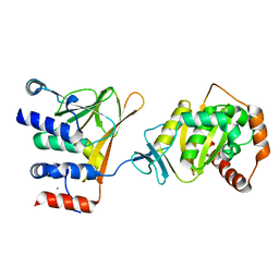
2FPE
 | | Conserved dimerization of the ib1 src-homology 3 domain | | 分子名称: | C-jun-amino-terminal kinase interacting protein 1, HEXAETHYLENE GLYCOL, SULFATE ION, ... | | 著者 | Guenat, S, Dar, I, Bonny, C, Kastrup, J.S, Gajhede, M, Kristensen, O. | | 登録日 | 2006-01-16 | | 公開日 | 2006-02-28 | | 最終更新日 | 2023-11-15 | | 実験手法 | X-RAY DIFFRACTION (1.75 Å) | | 主引用文献 | A unique set of SH3-SH3 interactions controls IB1 homodimerization
Embo J., 25, 2006
|
|
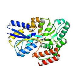
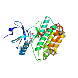
3MFT
 | |
3MFS
 | | CASK-4M CaM Kinase Domain, AMPPNP | | 分子名称: | PHOSPHOAMINOPHOSPHONIC ACID-ADENYLATE ESTER, Peripheral plasma membrane protein CASK | | 著者 | Wahl, M.C, Mukherjee, K. | | 登録日 | 2010-04-03 | | 公開日 | 2010-04-28 | | 最終更新日 | 2023-09-06 | | 実験手法 | X-RAY DIFFRACTION (2.1 Å) | | 主引用文献 | Evolution of CASK into a Mg2+-sensitive kinase.
Sci.Signal., 3, 2010
|
|
3MFU
 | | CASK-4M CaM Kinase Domain, AMPPNP-Mn2+ | | 分子名称: | MANGANESE (II) ION, PHOSPHOAMINOPHOSPHONIC ACID-ADENYLATE ESTER, Peripheral plasma membrane protein CASK | | 著者 | Wahl, M.C, Mukherjee, K. | | 登録日 | 2010-04-03 | | 公開日 | 2010-04-28 | | 最終更新日 | 2023-09-06 | | 実験手法 | X-RAY DIFFRACTION (2.3 Å) | | 主引用文献 | Evolution of CASK into a Mg2+-sensitive kinase.
Sci.Signal., 3, 2010
|
|
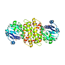
3MFR
 | | CASK-4M CaM Kinase Domain, native | | 分子名称: | PHOSPHOAMINOPHOSPHONIC ACID-ADENYLATE ESTER, Peripheral plasma membrane protein CASK | | 著者 | Wahl, M.C, Mukherjee, K. | | 登録日 | 2010-04-03 | | 公開日 | 2010-04-28 | | 最終更新日 | 2023-09-06 | | 実験手法 | X-RAY DIFFRACTION (2 Å) | | 主引用文献 | Evolution of CASK into a Mg2+-sensitive kinase.
Sci.Signal., 3, 2010
|
|


