6I59
 
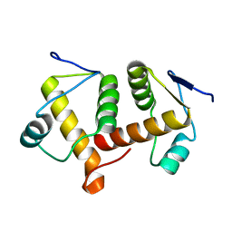
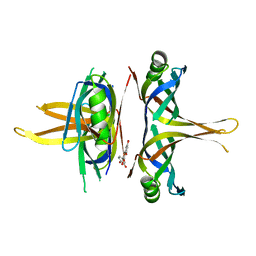
 | | Long wavelength native-SAD phasing of Sen1 helicase | | 分子名称: | 1,2-ETHANEDIOL, ADENOSINE-5'-DIPHOSPHATE, DI(HYDROXYETHYL)ETHER, ... | | 著者 | Basu, S, Olieric, V, Matsugaki, N, Kawano, Y, Takashi, T, Huang, C.Y, Leonarski, F, Yamada, Y, Vera, L, Olieric, N, Basquin, J, Wojdyla, J.A, Diederichs, K, Yamamoto, M, Bunk, O, Wang, M. | | 登録日 | 2018-11-13 | | 公開日 | 2019-03-13 | | 最終更新日 | 2024-05-15 | | 実験手法 | X-RAY DIFFRACTION (2.95 Å) | | 主引用文献 | Long-wavelength native-SAD phasing: opportunities and challenges.
Iucrj, 6, 2019
|
|
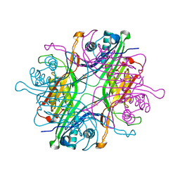
5XGT
 | |
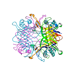
5YNZ
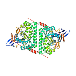
 | | Crystal structure of the dihydroorotase domain (K1556A) of human CAD | | 分子名称: | CAD protein, ZINC ION | | 著者 | Huang, Y.H, Chen, K.L, Cheng, J.H, Huang, C.Y. | | 登録日 | 2017-10-26 | | 公開日 | 2018-10-24 | | 最終更新日 | 2023-11-22 | | 実験手法 | X-RAY DIFFRACTION (2.774 Å) | | 主引用文献 | Crystal structures of monometallic dihydropyrimidinase and the human dihydroorotase domain K1556A mutant reveal no lysine carbamylation within the active site
Biochem. Biophys. Res. Commun., 505, 2018
|
|
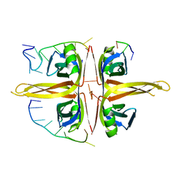
5YUO
 | |
6AEQ
 | |
6AEP
 | |
7F2V
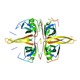
 | | Urate oxidase from Thermobispora bispora in apo form | | 分子名称: | DIMETHYL SULFOXIDE, SULFATE ION, Uricase | | 著者 | Chiu, Y.C, Hsu, T.S, Huang, C.Y, Hsu, C.H. | | 登録日 | 2021-06-14 | | 公開日 | 2022-05-04 | | 最終更新日 | 2023-11-29 | | 実験手法 | X-RAY DIFFRACTION (1.6 Å) | | 主引用文献 | Structural and biochemical insights into a hyperthermostable urate oxidase from Thermobispora bispora for hyperuricemia and gout therapy.
Int.J.Biol.Macromol., 188, 2021
|
|
7F2W
 | | TbUox in complex with uric acid | | 分子名称: | URIC ACID, Uricase | | 著者 | Chiu, Y.C, Hsu, T.S, Huang, C.Y, Hsu, C.H. | | 登録日 | 2021-06-14 | | 公開日 | 2022-05-04 | | 最終更新日 | 2023-11-29 | | 実験手法 | X-RAY DIFFRACTION (2.16 Å) | | 主引用文献 | Structural and biochemical insights into a hyperthermostable urate oxidase from Thermobispora bispora for hyperuricemia and gout therapy.
Int.J.Biol.Macromol., 188, 2021
|
|
6JDG
 | |
6IRQ
 | |
6KLK
 | |
6L0I
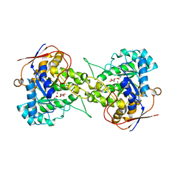
 | | Crystal structure of dihydroorotase in complex with malate at pH6.5 from Saccharomyces cerevisiae | | 分子名称: | (2S)-2-hydroxybutanedioic acid, Dihydroorotase, ZINC ION | | 著者 | Guan, H.H, Huang, Y.H, Huang, C.Y, Chen, C.J. | | 登録日 | 2019-09-26 | | 公開日 | 2020-12-02 | | 最終更新日 | 2023-11-22 | | 実験手法 | X-RAY DIFFRACTION (2.2 Å) | | 主引用文献 | Structural basis for the interaction modes of dihydroorotase with the anticancer drugs 5-fluorouracil and 5-aminouracil.
Biochem.Biophys.Res.Commun., 551, 2021
|
|
6L0F
 | | Crystal structure of dihydroorotase in complex with 5-Aminouracil from Saccharomyces cerevisiae | | 分子名称: | 5-AMINO-1H-PYRIMIDINE-2,4-DIONE, Dihydroorotase, ZINC ION | | 著者 | Guan, H.H, Huang, Y.H, Huang, C.Y, Chen, C.J. | | 登録日 | 2019-09-26 | | 公開日 | 2020-12-02 | | 最終更新日 | 2023-11-22 | | 実験手法 | X-RAY DIFFRACTION (3.26 Å) | | 主引用文献 | Structural basis for the interaction modes of dihydroorotase with the anticancer drugs 5-fluorouracil and 5-aminouracil.
Biochem.Biophys.Res.Commun., 551, 2021
|
|
6L0K
 | | Crystal structure of dihydroorotase in complex with malate at pH9 from Saccharomyces cerevisiae | | 分子名称: | (2S)-2-hydroxybutanedioic acid, Dihydroorotase, ZINC ION | | 著者 | Guan, H.H, Huang, Y.H, Huang, C.Y, Chen, C.J. | | 登録日 | 2019-09-26 | | 公開日 | 2020-12-02 | | 最終更新日 | 2023-11-22 | | 実験手法 | X-RAY DIFFRACTION (3.3 Å) | | 主引用文献 | Structural basis for the interaction modes of dihydroorotase with the anticancer drugs 5-fluorouracil and 5-aminouracil.
Biochem.Biophys.Res.Commun., 551, 2021
|
|
6L0J
 | | Crystal structure of Dihydroorotase in complex with malate at pH7.5 from Saccharomyces cerevisiae | | 分子名称: | (2S)-2-hydroxybutanedioic acid, Dihydroorotase, ZINC ION | | 著者 | Guan, H.H, Huang, Y.H, Huang, C.Y, Chen, C.J. | | 登録日 | 2019-09-26 | | 公開日 | 2020-12-02 | | 最終更新日 | 2023-11-22 | | 実験手法 | X-RAY DIFFRACTION (1.933 Å) | | 主引用文献 | Structural Analysis of Saccharomyces cerevisiae Dihydroorotase Reveals Molecular Insights into the Tetramerization Mechanism
Molecules, 2021
|
|
6L0H
 | | Crystal structure of dihydroorotase in complex with malate at pH7 from Saccharomyces cerevisiae | | 分子名称: | (2S)-2-hydroxybutanedioic acid, Dihydroorotase, ZINC ION | | 著者 | Guan, H.H, Huang, Y.H, Huang, C.Y, Chen, C.J. | | 登録日 | 2019-09-26 | | 公開日 | 2020-12-02 | | 最終更新日 | 2023-11-22 | | 実験手法 | X-RAY DIFFRACTION (2.054 Å) | | 主引用文献 | Structural basis for the interaction modes of dihydroorotase with the anticancer drugs 5-fluorouracil and 5-aminouracil.
Biochem.Biophys.Res.Commun., 551, 2021
|
|
6I5C
 | | Long wavelength native-SAD phasing of Tubulin-Stathmin-TTL complex | | 分子名称: | 2-(N-MORPHOLINO)-ETHANESULFONIC ACID, CALCIUM ION, CHLORIDE ION, ... | | 著者 | Basu, S, Olieric, V, Matsugaki, N, Kawano, Y, Takashi, T, Huang, C.Y, Leonarski, F, Yamada, Y, Vera, L, Olieric, N, Basquin, J, Wojdyla, J.A, Diederichs, K, Yamamoto, M, Bunk, O, Wang, M. | | 登録日 | 2018-11-13 | | 公開日 | 2019-03-13 | | 最終更新日 | 2024-05-15 | | 実験手法 | X-RAY DIFFRACTION (2.95 Å) | | 主引用文献 | Long-wavelength native-SAD phasing: opportunities and challenges.
Iucrj, 6, 2019
|
|
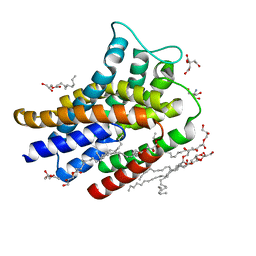
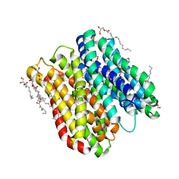
7YM1
 | | Structure of SsbA protein in complex with the anticancer drug 5-fluorouracil | | 分子名称: | 5-FLUOROURACIL, GLYCEROL, Single-stranded DNA-binding protein | | 著者 | Huang, Y.H, Yang, P.C, Chiang, W.Y, Lin, E.S, Huang, C.Y. | | 登録日 | 2022-07-27 | | 公開日 | 2023-08-02 | | 最終更新日 | 2024-02-14 | | 実験手法 | X-RAY DIFFRACTION (2.36 Å) | | 主引用文献 | Crystal Structure of DNA Replication Protein SsbA Complexed with the Anticancer Drug 5-Fluorouracil.
Int J Mol Sci, 24, 2023
|
|
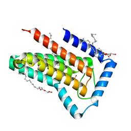
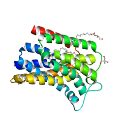
7VUM
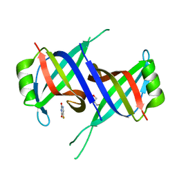
 | | Crystal structure of SSB complexed with que | | 分子名称: | 3,5,7,3',4'-PENTAHYDROXYFLAVONE, Single-stranded DNA-binding protein | | 著者 | Lin, E.S, Huang, Y.H, Huang, C.Y. | | 登録日 | 2021-11-03 | | 公開日 | 2022-03-09 | | 最終更新日 | 2023-11-29 | | 実験手法 | X-RAY DIFFRACTION (2.319 Å) | | 主引用文献 | A Complexed Crystal Structure of a Single-Stranded DNA-Binding Protein with Quercetin and the Structural Basis of Flavonol Inhibition Specificity.
Int J Mol Sci, 23, 2022
|
|
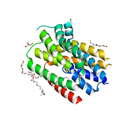
6J0I
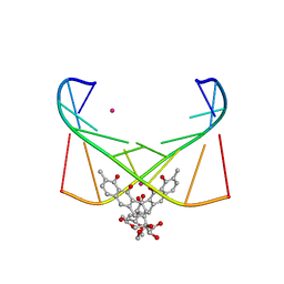 | | Structure of [Co2+-(Chromomycin A3)2]-d(TTGGCGAA)2 complex | | 分子名称: | 1,2-HYDRO-1-OXY-3,4-HYDRO-3-(1-METHOXY-2-OXY-3,4-DIHYDROXYPENTYL)-8,9-DIHYROXY-7-METHYLANTHRACENE, 2,6-dideoxy-4-O-methyl-alpha-D-galactopyranose-(1-3)-(2R,3R,6R)-6-hydroxy-2-methyltetrahydro-2H-pyran-3-yl acetate, 3-C-methyl-4-O-acetyl-alpha-L-Olivopyranose-(1-3)-(2R,5S,6R)-6-methyltetrahydro-2H-pyran-2,5-diol-(1-3)-(2R,5S,6R)-6-methyltetrahydro-2H-pyran-2,5-diol, ... | | 著者 | Satange, R.B, Chuang, C.Y, Hou, M.H. | | 登録日 | 2018-12-24 | | 公開日 | 2019-07-24 | | 最終更新日 | 2023-11-22 | | 実験手法 | X-RAY DIFFRACTION (2.5 Å) | | 主引用文献 | Polymorphic G:G mismatches act as hotspots for inducing right-handed Z DNA by DNA intercalation.
Nucleic Acids Res., 47, 2019
|
|
6FMV
 | | IMISX-EP of Hg-BacA soaking SIRAS | | 分子名称: | (2R)-2,3-dihydroxypropyl (9Z)-octadec-9-enoate, 2-AMINO-2-HYDROXYMETHYL-PROPANE-1,3-DIOL, CITRATE ANION, ... | | 著者 | Huang, C.-Y, Olieric, V, Howe, N, Warshamanage, R, Weinert, T, Panepucci, E, Vogeley, L, Basu, S, Diederichs, K, Caffrey, M, Wang, M. | | 登録日 | 2018-02-02 | | 公開日 | 2018-09-05 | | 最終更新日 | 2024-06-19 | | 実験手法 | X-RAY DIFFRACTION (2.3 Å) | | 主引用文献 | In situ serial crystallography for rapid de novo membrane protein structure determination.
Commun Biol, 1, 2018
|
|
6FMX
 | | IMISX-EP of W-PgpB | | 分子名称: | (2R)-2,3-dihydroxypropyl (9Z)-octadec-9-enoate, Phosphatidylglycerophosphatase B, TUNGSTATE(VI)ION | | 著者 | Huang, C.-Y, Olieric, V, Howe, N, Warshamanage, R, Weinert, T, Panepucci, E, Vogeley, L, Basu, S, Diederichs, K, Caffrey, M, Wang, M. | | 登録日 | 2018-02-02 | | 公開日 | 2018-09-05 | | 最終更新日 | 2024-05-08 | | 実験手法 | X-RAY DIFFRACTION (1.79 Å) | | 主引用文献 | In situ serial crystallography for rapid de novo membrane protein structure determination.
Commun Biol, 1, 2018
|
|
6FMR
 | | IMISX-EP of Se-PepTSt | | 分子名称: | (2S)-2,3-DIHYDROXYPROPYL(7Z)-PENTADEC-7-ENOATE, Di-or tripeptide:H+ symporter, PHOSPHATE ION | | 著者 | Huang, C.-Y, Olieric, V, Howe, N, Warshamanage, R, Weinert, T, Panepucci, E, Vogeley, L, Basu, S, Diederichs, K, Caffrey, M, Wang, M. | | 登録日 | 2018-02-02 | | 公開日 | 2018-08-29 | | 最終更新日 | 2018-10-10 | | 実験手法 | X-RAY DIFFRACTION (2.7 Å) | | 主引用文献 | In situ serial crystallography for rapid de novo membrane protein structure determination.
Commun Biol, 1, 2018
|
|
6FMW
 | | IMISX-EP of Hg-BacA cocrystallization | | 分子名称: | (2R)-2,3-dihydroxypropyl (9Z)-octadec-9-enoate, 2-AMINO-2-HYDROXYMETHYL-PROPANE-1,3-DIOL, MERCURY (II) ION, ... | | 著者 | Huang, C.-Y, Olieric, V, Howe, N, Warshamanage, R, Weinert, T, Panepucci, E, Vogeley, L, Basu, S, Diederichs, K, Caffrey, M, Wang, M. | | 登録日 | 2018-02-02 | | 公開日 | 2018-09-05 | | 最終更新日 | 2024-06-19 | | 実験手法 | X-RAY DIFFRACTION (2.6 Å) | | 主引用文献 | In situ serial crystallography for rapid de novo membrane protein structure determination.
Commun Biol, 1, 2018
|
|
6FMT
 | | IMISX-EP of Hg-BacA Soaking SAD | | 分子名称: | (2R)-2,3-dihydroxypropyl (9Z)-octadec-9-enoate, 2-AMINO-2-HYDROXYMETHYL-PROPANE-1,3-DIOL, MERCURY (II) ION, ... | | 著者 | Huang, C.-Y, Olieric, V, Howe, N, Warshamanage, R, Weinert, T, Panepucci, E, Vogeley, L, Basu, S, Diederichs, K, Caffrey, M, Wang, M. | | 登録日 | 2018-02-02 | | 公開日 | 2018-09-05 | | 最終更新日 | 2024-06-19 | | 実験手法 | X-RAY DIFFRACTION (3 Å) | | 主引用文献 | In situ serial crystallography for rapid de novo membrane protein structure determination.
Commun Biol, 1, 2018
|
|


