4QC4
 
 | |
4Q9P
 | |
7WDP
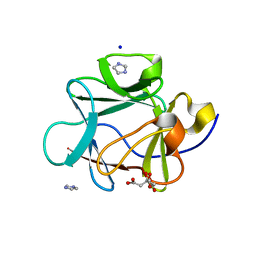
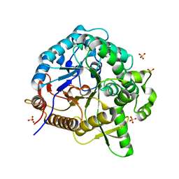
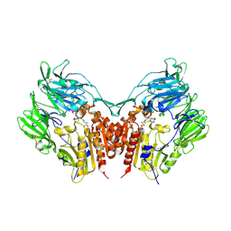
 | | Crystal structures of MeBglD2 in complex with various saccharides | | 分子名称: | Beta-glucosidase, SULFATE ION, alpha-D-glucopyranose, ... | | 著者 | Watanabe, M, Matsuzawa, T, Nakamichi, Y, Akita, H, Yaoi, K. | | 登録日 | 2021-12-22 | | 公開日 | 2022-11-02 | | 最終更新日 | 2023-11-29 | | 実験手法 | X-RAY DIFFRACTION (2.39 Å) | | 主引用文献 | Crystal structure of metagenomic beta-glycosidase MeBglD2 in complex with various saccharides
Appl.Microbiol.Biotechnol., 106, 2022
|
|
7WDS
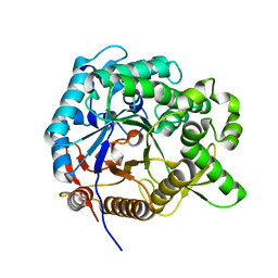
 | | Crystal structures of MeBglD2 in complex with various saccharides | | 分子名称: | Beta-glucosidase, SULFATE ION, beta-D-xylopyranose | | 著者 | Watanabe, M, Matsuzawa, T, Nakamichi, Y, Akita, H, Yaoi, K. | | 登録日 | 2021-12-22 | | 公開日 | 2022-11-02 | | 最終更新日 | 2023-11-29 | | 実験手法 | X-RAY DIFFRACTION (1.68 Å) | | 主引用文献 | Crystal structure of metagenomic beta-glycosidase MeBglD2 in complex with various saccharides
Appl.Microbiol.Biotechnol., 106, 2022
|
|
7WDV
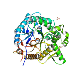
 | | Crystal structures of MeBglD2 in complex with various saccharides | | 分子名称: | Beta-glucosidase, SULFATE ION, beta-D-glucopyranose, ... | | 著者 | Watanabe, M, Matsuzawa, T, Nakamichi, Y, Akita, H, Yaoi, K. | | 登録日 | 2021-12-22 | | 公開日 | 2022-11-02 | | 最終更新日 | 2023-11-29 | | 実験手法 | X-RAY DIFFRACTION (1.812 Å) | | 主引用文献 | Crystal structure of metagenomic beta-glycosidase MeBglD2 in complex with various saccharides
Appl.Microbiol.Biotechnol., 106, 2022
|
|
7WDN
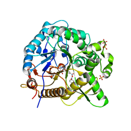
 | | Crystal structures of MeBglD2 in complex with various saccharides | | 分子名称: | alpha-D-glucopyranose, beta-glucosidase | | 著者 | Watanabe, M, Matsuzawa, T, Nakamichi, Y, Akita, H, Yaoi, K. | | 登録日 | 2021-12-22 | | 公開日 | 2023-01-04 | | 最終更新日 | 2024-05-29 | | 実験手法 | X-RAY DIFFRACTION (1.8 Å) | | 主引用文献 | Crystal structure of metagenomic beta-glycosidase MeBglD2 in complex with various saccharides.
Appl.Microbiol.Biotechnol., 106, 2022
|
|
7WDO
 | | Crystal structures of MeBglD2 in complex with various saccharides | | 分子名称: | Beta-glucosidase, SULFATE ION, beta-D-glucopyranose, ... | | 著者 | Watanabe, M, Matsuzawa, T, Nakamichi, Y, Akita, H, Yaoi, K. | | 登録日 | 2021-12-22 | | 公開日 | 2022-11-02 | | 最終更新日 | 2023-11-29 | | 実験手法 | X-RAY DIFFRACTION (2.21 Å) | | 主引用文献 | Crystal structure of metagenomic beta-glycosidase MeBglD2 in complex with various saccharides
Appl.Microbiol.Biotechnol., 106, 2022
|
|
7WDR
 | | Crystal structures of MeBglD2 in complex with various saccharides | | 分子名称: | 4-nitrophenyl beta-D-glucopyranoside, Beta-glucosidase, SULFATE ION | | 著者 | Watanabe, M, Matsuzawa, T, Nakamichi, Y, Akita, H, Yaoi, K. | | 登録日 | 2021-12-22 | | 公開日 | 2022-11-02 | | 最終更新日 | 2023-11-29 | | 実験手法 | X-RAY DIFFRACTION (2 Å) | | 主引用文献 | Crystal structure of metagenomic beta-glycosidase MeBglD2 in complex with various saccharides
Appl.Microbiol.Biotechnol., 106, 2022
|
|
4OW4
 | |
4QAL
 | |
3HOM
 | |
3VU3
 | |
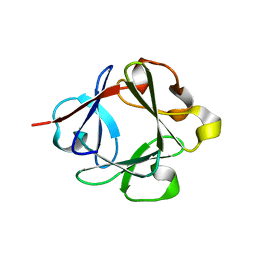
4D8H
 | | Crystal structure of Symfoil-4P/PV2: de novo designed beta-trefoil architecture with symmetric primary structure, primitive version 2 (6xLeu / PV1) | | 分子名称: | 2-AMINO-2-HYDROXYMETHYL-PROPANE-1,3-DIOL, SULFATE ION, de novo protein | | 著者 | Blaber, M, Longo, L. | | 登録日 | 2012-01-10 | | 公開日 | 2013-01-16 | | 最終更新日 | 2024-02-28 | | 実験手法 | X-RAY DIFFRACTION (1.901 Å) | | 主引用文献 | Simplified protein design biased for prebiotic amino acids yields a foldable, halophilic protein.
Proc.Natl.Acad.Sci.USA, 110, 2013
|
|
7DE8
 | |
3Q7Y
 | |
3Q7W
 | |
3Q7X
 | | Crystal structure of Symfoil-4P/PV1: de novo designed beta-trefoil architecture with symmetric primary structure, primitive version 1 | | 分子名称: | 2-AMINO-2-HYDROXYMETHYL-PROPANE-1,3-DIOL, SULFATE ION, de novo designed beta-trefoil architecture with symmetric primary structure | | 著者 | Blaber, M, Lee, J. | | 登録日 | 2011-01-05 | | 公開日 | 2012-01-11 | | 最終更新日 | 2024-02-21 | | 実験手法 | X-RAY DIFFRACTION (1.4 Å) | | 主引用文献 | Simplified protein design biased for prebiotic amino acids yields a foldable, halophilic protein.
Proc.Natl.Acad.Sci.USA, 110, 2013
|
|
2LGC
 | |
2N64
 | |
3BA5
 | |
3BAD
 | |
3BA7
 | |
3BA4
 | |
3B9U
 | |
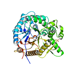
3W2T
 | | Crystal structure of human depiptidyl peptidase IV (DPP-4) in complex with vildagliptin | | 分子名称: | 2-acetamido-2-deoxy-beta-D-glucopyranose, 2-acetamido-2-deoxy-beta-D-glucopyranose-(1-4)-2-acetamido-2-deoxy-beta-D-glucopyranose, 2-{[(1r,3s,5R,7S)-3-hydroxytricyclo[3.3.1.1~3,7~]decan-1-yl]amino}-1-{(2S)-2-[(E)-iminomethyl]pyrrolidin-1-yl}ethan-1-o ne, ... | | 著者 | Kishida, H, Nabeno, M, Miyaguchi, I, Tanaka, Y, Katou, R, Akahoshi, F. | | 登録日 | 2012-12-04 | | 公開日 | 2013-05-15 | | 最終更新日 | 2024-10-30 | | 実験手法 | X-RAY DIFFRACTION (2.36 Å) | | 主引用文献 | A comparative study of the binding modes of recently launched dipeptidyl peptidase IV inhibitors in the active site
Biochem.Biophys.Res.Commun., 434, 2013
|
|


