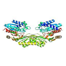
3DUL
 
 | |
3DUW
 | |
3CZK
 | |
3CZE
 | |
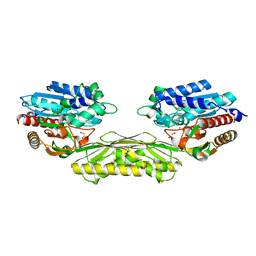
2HK1
 | | Crystal structure of D-psicose 3-epimerase (DPEase) in the presence of D-fructose | | 分子名称: | D-PSICOSE 3-EPIMERASE, D-fructose, MANGANESE (II) ION | | 著者 | Kim, K, Kim, H.J, Oh, D.K, Cha, S.S, Rhee, S. | | 登録日 | 2006-07-03 | | 公開日 | 2006-08-29 | | 最終更新日 | 2017-10-18 | | 実験手法 | X-RAY DIFFRACTION (2.3 Å) | | 主引用文献 | Crystal Structure of d-Psicose 3-epimerase from Agrobacterium tumefaciens and its Complex with True Substrate d-Fructose: A Pivotal Role of Metal in Catalysis, an Active Site for the Non-phosphorylated Substrate, and its Conformational Changes
J.Mol.Biol., 361, 2006
|
|
2HK0
 | | Crystal structure of D-psicose 3-epimerase (DPEase) in the absence of substrate | | 分子名称: | D-PSICOSE 3-EPIMERASE | | 著者 | Kim, K, Kim, H.J, Oh, D.K, Cha, S.S, Rhee, S. | | 登録日 | 2006-07-03 | | 公開日 | 2006-08-29 | | 最終更新日 | 2017-10-18 | | 実験手法 | X-RAY DIFFRACTION (2 Å) | | 主引用文献 | Crystal Structure of d-Psicose 3-epimerase from Agrobacterium tumefaciens and its Complex with True Substrate d-Fructose: A Pivotal Role of Metal in Catalysis, an Active Site for the Non-phosphorylated Substrate, and its Conformational Changes
J.Mol.Biol., 361, 2006
|
|
4PXB
 | | The crystal structure of AtUAH in complex with (S)-ureidoglycolate | | 分子名称: | (2S)-(carbamoylamino)(hydroxy)ethanoic acid, MANGANESE (II) ION, Ureidoglycolate hydrolase | | 著者 | Shin, I, Rhee, S. | | 登録日 | 2014-03-23 | | 公開日 | 2014-07-23 | | 最終更新日 | 2024-05-29 | | 実験手法 | X-RAY DIFFRACTION (1.903 Å) | | 主引用文献 | Structural insights into the substrate specificity of (s)-ureidoglycolate amidohydrolase and its comparison with allantoate amidohydrolase.
J.Mol.Biol., 426, 2014
|
|
4PXC
 | | The crystal structure of AtUAH in complex with (S)-hydroxyglycine | | 分子名称: | (2S)-amino(hydroxy)ethanoic acid, MANGANESE (II) ION, Ureidoglycolate hydrolase | | 著者 | Shin, I, Rhee, S. | | 登録日 | 2014-03-23 | | 公開日 | 2014-07-23 | | 最終更新日 | 2024-05-29 | | 実験手法 | X-RAY DIFFRACTION (1.893 Å) | | 主引用文献 | Structural insights into the substrate specificity of (s)-ureidoglycolate amidohydrolase and its comparison with allantoate amidohydrolase.
J.Mol.Biol., 426, 2014
|
|
4PXD
 | | The crystal structure of EcAAH in complex with allantoate | | 分子名称: | ALLANTOATE ION, Allantoate amidohydrolase, MANGANESE (II) ION | | 著者 | Shin, I, Rhee, S. | | 登録日 | 2014-03-23 | | 公開日 | 2014-07-23 | | 最終更新日 | 2023-11-08 | | 実験手法 | X-RAY DIFFRACTION (2.2 Å) | | 主引用文献 | Structural insights into the substrate specificity of (s)-ureidoglycolate amidohydrolase and its comparison with allantoate amidohydrolase.
J.Mol.Biol., 426, 2014
|
|
4PXE
 | | The crystal structure of AtUAH in complex with glyoxylate | | 分子名称: | GLYOXYLIC ACID, MANGANESE (II) ION, Ureidoglycolate hydrolase | | 著者 | Shin, I, Rhee, S. | | 登録日 | 2014-03-23 | | 公開日 | 2014-07-23 | | 最終更新日 | 2024-05-29 | | 実験手法 | X-RAY DIFFRACTION (1.449 Å) | | 主引用文献 | Structural insights into the substrate specificity of (s)-ureidoglycolate amidohydrolase and its comparison with allantoate amidohydrolase.
J.Mol.Biol., 426, 2014
|
|
6KIA
 | |
6KI9
 | | Apo structure of FabMG, novel types of Enoyl-acyl carrier protein reductase | | 分子名称: | 1,2-ETHANEDIOL, FabMG, novel types of Enoyl-acyl carrier protein reductase, ... | | 著者 | Kim, S, Rhee, S. | | 登録日 | 2019-07-17 | | 公開日 | 2020-05-20 | | 最終更新日 | 2024-03-27 | | 実験手法 | X-RAY DIFFRACTION (1.64 Å) | | 主引用文献 | A triclosan-resistance protein from the soil metagenome is a novel enoyl-acyl carrier protein reductase: Structure-guided functional analysis.
Febs J., 287, 2020
|
|
4NNC
 | | Ternary complex of ObcA with C4-CoA adduct and oxalate | | 分子名称: | (3S)-3-[2-[3-[[(2R)-4-[[[(2R,3S,4R,5R)-5-(6-aminopurin-9-yl)-4-oxidanyl-3-phosphonooxy-oxolan-2-yl]methoxy-oxidanyl-phosphoryl]oxy-oxidanyl-phosphoryl]oxy-3,3-dimethyl-2-oxidanyl-butanoyl]amino]propanoylamino]ethylsulfanyl]-3-oxidanyl-butanoic acid, COBALT (II) ION, OBCA, ... | | 著者 | Oh, J.T, Goo, E, Hwang, I, Rhee, S. | | 登録日 | 2013-11-17 | | 公開日 | 2014-03-19 | | 最終更新日 | 2023-09-20 | | 実験手法 | X-RAY DIFFRACTION (2.279 Å) | | 主引用文献 | Structural Basis for Bacterial Quorum Sensing-mediated Oxalogenesis.
J.Biol.Chem., 289, 2014
|
|
4RSX
 | |
6ILR
 | |
6ILS
 | | Structure of Arabidopsis thaliana Ribokinase complexed with Ribose and ATP | | 分子名称: | ADENOSINE-5'-TRIPHOSPHATE, Ribokinase, SODIUM ION, ... | | 著者 | Kang, P, Oh, J, Rhee, S. | | 登録日 | 2018-10-19 | | 公開日 | 2019-03-13 | | 最終更新日 | 2023-11-22 | | 実験手法 | X-RAY DIFFRACTION (1.8 Å) | | 主引用文献 | Crystal structure and mutational analyses of ribokinase from Arabidopsis thaliana.
J. Struct. Biol., 206, 2019
|
|
6ILT
 | | Structure of Arabidopsis thaliana Ribokinase complexed with ATP and Magnesium ion | | 分子名称: | ADENOSINE-5'-TRIPHOSPHATE, MAGNESIUM ION, Ribokinase, ... | | 著者 | Kang, P, Oh, J, Rhee, S. | | 登録日 | 2018-10-19 | | 公開日 | 2019-03-13 | | 最終更新日 | 2023-11-22 | | 実験手法 | X-RAY DIFFRACTION (2.2 Å) | | 主引用文献 | Crystal structure and mutational analyses of ribokinase from Arabidopsis thaliana.
J. Struct. Biol., 206, 2019
|
|
1BL0
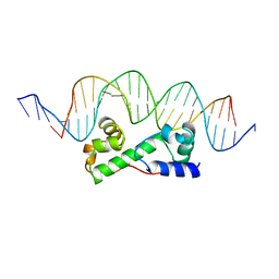 | | MULTIPLE ANTIBIOTIC RESISTANCE PROTEIN (MARA)/DNA COMPLEX | | 分子名称: | DNA (5'-D(*CP*CP*GP*AP*TP*GP*CP*CP*AP*CP*GP*TP*TP*TP*TP*GP*CP*TP*AP*AP*AP*TP* CP*C)-3'), DNA (5'-D(*GP*GP*GP*GP*AP*TP*TP*TP*AP*GP*CP*AP*AP*AP*AP*CP*GP*TP*GP*GP*CP*AP* TP*C)-3'), PROTEIN (MULTIPLE ANTIBIOTIC RESISTANCE PROTEIN) | | 著者 | Davies, S, Rhee, R.G, Martin, J.L, Rosner, D.R. | | 登録日 | 1998-07-22 | | 公開日 | 1998-09-02 | | 最終更新日 | 2024-02-07 | | 実験手法 | X-RAY DIFFRACTION (2.3 Å) | | 主引用文献 | A novel DNA-binding motif in MarA: the first structure for an AraC family transcriptional activator.
Proc.Natl.Acad.Sci.USA, 95, 1998
|
|
1BKS
 | | TRYPTOPHAN SYNTHASE (E.C.4.2.1.20) FROM SALMONELLA TYPHIMURIUM | | 分子名称: | PYRIDOXAL-5'-PHOSPHATE, SODIUM ION, TRYPTOPHAN SYNTHASE | | 著者 | Hyde, C.C. | | 登録日 | 1998-07-10 | | 公開日 | 1999-03-23 | | 最終更新日 | 2023-08-02 | | 実験手法 | X-RAY DIFFRACTION (2.2 Å) | | 主引用文献 | Exchange of K+ or Cs+ for Na+ induces local and long-range changes in the three-dimensional structure of the tryptophan synthase alpha2beta2 complex.
Biochemistry, 35, 1996
|
|
8K05
 | |
8K06
 | | Pseudouridine 5'-monophosphate glycosylase from Arabidopsis thaliana -- PSU, R5P bound K185A mutant | | 分子名称: | 5-O-phosphono-beta-D-ribofuranose, MANGANESE (II) ION, PSEUDOURIDINE-5'-MONOPHOSPHATE, ... | | 著者 | Lee, J.Y, Kim, S.H, Rhee, S.K. | | 登録日 | 2023-07-07 | | 公開日 | 2024-05-15 | | 実験手法 | X-RAY DIFFRACTION (1.845 Å) | | 主引用文献 | Structure and function of the pseudouridine 5'-monophosphate glycosylase PUMY from Arabidopsis thaliana.
Rna Biol., 21, 2024
|
|
8K07
 | |
3S8R
 | | Crystal Structures of Glutaryl 7-Aminocephalosporanic Acid Acylase: Insight into Autoproteolytic Activation | | 分子名称: | GLYCEROL, Glutaryl-7-aminocephalosporanic-acid acylase | | 著者 | Kim, J.K, Yang, I.S, Park, S.S, Kim, K.H. | | 登録日 | 2011-05-30 | | 公開日 | 2011-07-06 | | 最終更新日 | 2024-03-20 | | 実験手法 | X-RAY DIFFRACTION (2.5 Å) | | 主引用文献 | Crystal structures of glutaryl 7-aminocephalosporanic acid acylase: insight into autoproteolytic activation.
Biochemistry, 42, 2003
|
|
2Q37
 | |
2MUK
 | |


