1J7Z
 
 | |
1J81
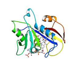
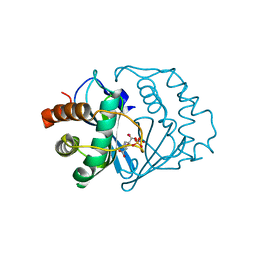
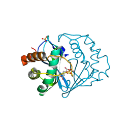
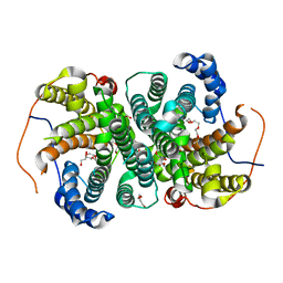
 | | Osmolyte Stabilization of RNase | | 分子名称: | RIBONUCLEASE PANCREATIC, SULFATE ION | | 著者 | Ratnaparkhi, G.S, Varadarajan, R. | | 登録日 | 2001-05-19 | | 公開日 | 2001-06-06 | | 最終更新日 | 2017-11-29 | | 実験手法 | X-RAY DIFFRACTION (2.2 Å) | | 主引用文献 | Osmolytes stabilize ribonuclease S by stabilizing its fragments S protein and S peptide to compact folding-competent states.
J.Biol.Chem., 276, 2001
|
|
1CJQ
 | |
1CJR
 | |
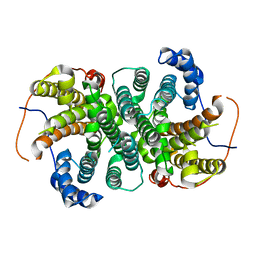
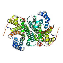
1A19
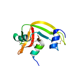
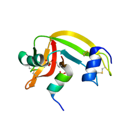
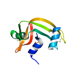
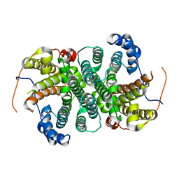
 | | BARSTAR (FREE), C82A MUTANT | | 分子名称: | BARSTAR | | 著者 | Ratnaparkhi, G.S, Varadarajan, R. | | 登録日 | 1997-12-25 | | 公開日 | 1998-04-08 | | 最終更新日 | 2024-05-22 | | 実験手法 | X-RAY DIFFRACTION (2.76 Å) | | 主引用文献 | Discrepancies between the NMR and X-ray structures of uncomplexed barstar: analysis suggests that packing densities of protein structures determined by NMR are unreliable.
Biochemistry, 37, 1998
|
|
1DAJ
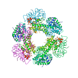
 | | COMPARISON OF TERNARY COMPLEXES OF PNEUMOCYSTIS CARINII AND WILD TYPE HUMAN DIHYDROFOLATE REDUCTASE WITH COENZYME NADPH AND A NOVEL CLASSICAL ANTITUMOR FURO[2,3D]PYRIMIDINE ANTIFOLATE | | 分子名称: | DIHYDROFOLATE REDUCTASE, N-[4-[(2,4-DIAMINOFURO[2,3D]PYRIMIDIN-5-YL)METHYL]METHYLAMINO]-BENZOYL]-L-GLUTAMATE, NADPH DIHYDRO-NICOTINAMIDE-ADENINE-DINUCLEOTIDE PHOSPHATE | | 著者 | Cody, V, Galitsky, N, Luft, J.R, Pangborn, W, Gangjee, A, Devraj, R, Queener, S.F, Blakley, R.L. | | 登録日 | 1997-07-29 | | 公開日 | 1997-12-24 | | 最終更新日 | 2024-02-07 | | 実験手法 | X-RAY DIFFRACTION (2.3 Å) | | 主引用文献 | Comparison of ternary complexes of Pneumocystis carinii and wild-type human dihydrofolate reductase with coenzyme NADPH and a novel classical antitumor furo[2,3-d]pyrimidine antifolate.
Acta Crystallogr.,Sect.D, 53, 1997
|
|
1Z3M
 | | Crystal structure of mutant Ribonuclease S (F8Nva) | | 分子名称: | Ribonuclease pancreatic, S-Peptide, S-protein, ... | | 著者 | Das, M, Vasudeva Rao, B, Ghosh, S, Varadarajan, R. | | 登録日 | 2005-03-14 | | 公開日 | 2005-03-29 | | 最終更新日 | 2023-11-15 | | 実験手法 | X-RAY DIFFRACTION (2 Å) | | 主引用文献 | Attempts to delineate the relative contributions of changes in hydrophobicity and packing to changes in stability of ribonuclease S mutants.
Biochemistry, 44, 2005
|
|
1Z3P
 | | X-Ray crystal structure of a mutant Ribonuclease S (M13Nva) | | 分子名称: | Ribonuclease pancreatic, S-Peptide, S-Protein, ... | | 著者 | Das, M, Rao, B.V, Ghosh, S, Varadarajan, R. | | 登録日 | 2005-03-14 | | 公開日 | 2005-03-29 | | 最終更新日 | 2023-10-25 | | 実験手法 | X-RAY DIFFRACTION (2 Å) | | 主引用文献 | Attempts to delineate the relative contributions of changes in hydrophobicity and packing to changes in stability of ribonuclease S mutants.
Biochemistry, 44, 2005
|
|
4L1D
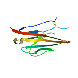 | | Voltage-gated sodium channel beta3 subunit Ig domain | | 分子名称: | Sodium channel subunit beta-3 | | 著者 | Namadurai, S, Weimhofer, M, Rajappa, R, Stott, K, Klingauf, J, Chirgadze, D.Y, Jackson, A.P. | | 登録日 | 2013-06-03 | | 公開日 | 2014-03-05 | | 最終更新日 | 2023-09-20 | | 実験手法 | X-RAY DIFFRACTION (2.5 Å) | | 主引用文献 | Crystal Structure and Molecular Imaging of the Nav Channel beta 3 Subunit Indicates a Trimeric Assembly.
J.Biol.Chem., 289, 2014
|
|
1Z3L
 | | X-Ray Crystal Structure of a Mutant Ribonuclease S (F8Anb) | | 分子名称: | Ribonuclease pancreatic, S-Peptide, S-Protein, ... | | 著者 | Das, M, Vasudeva Rao, B, Ghosh, S, Varadarajan, R. | | 登録日 | 2005-03-14 | | 公開日 | 2005-03-29 | | 最終更新日 | 2023-11-15 | | 実験手法 | X-RAY DIFFRACTION (1.8 Å) | | 主引用文献 | Attempts to delineate the relative contributions of changes in hydrophobicity and packing to changes in stability of ribonuclease S mutants.
Biochemistry, 44, 2005
|
|
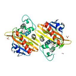
2FQO
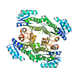
 | | Crystal structure of B. subtilis LuxS in complex with (2S)-2-Amino-4-[(2R,3R)-2,3-dihydroxy-3-N- hydroxycarbamoyl-propylmercapto]butyric acid | | 分子名称: | (2S)-2-AMINO-4-[(2R,3R)-2,3-DIHYDROXY-3-N-HYDROXYCARBAMOYL-PROPYLMERCAPTO]BUTYRIC ACID, COBALT (II) ION, S-ribosylhomocysteine lyase, ... | | 著者 | Shen, G, Rajan, R, Zhu, J, Bell, C.E, Pei, D. | | 登録日 | 2006-01-18 | | 公開日 | 2006-05-30 | | 最終更新日 | 2023-08-30 | | 実験手法 | X-RAY DIFFRACTION (1.87 Å) | | 主引用文献 | Design and Synthesis of Substrate and Intermediate Analogue Inhibitors of S-Ribosylhomocysteinase
J.Med.Chem., 49, 2006
|
|
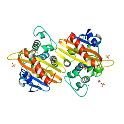
2FQT
 | | Crystal structure of B.subtilis LuxS in complex with (2S)-2-Amino-4-[(2R,3S)-2,3-dihydroxy-3-N-hydroxycarbamoyl-propylmercapto]butyric acid | | 分子名称: | (2S)-2-AMINO-4-[(2R,3S)-2,3-DIHYDROXY-3-N-HYDROXYCARBAMOYL-PROPYLMERCAPTO]BUTYRIC ACID, COBALT (II) ION, S-ribosylhomocysteine lyase, ... | | 著者 | Shen, G, Rajan, R, Zhu, J, Bell, C.E, Pei, D. | | 登録日 | 2006-01-18 | | 公開日 | 2006-05-30 | | 最終更新日 | 2023-08-30 | | 実験手法 | X-RAY DIFFRACTION (1.79 Å) | | 主引用文献 | Design and Synthesis of Substrate Analogue Inhibitors of S-Ribosylhomocysteinase (LuxS)
J.Med.Chem., 49, 2006
|
|
2RNS
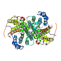
 | | REFINEMENT OF THE CRYSTAL STRUCTURE OF RIBONUCLEASE S. COMPARISON WITH AND BETWEEN THE VARIOUS RIBONUCLEASE A STRUCTURES | | 分子名称: | RIBONUCLEASE S, SULFATE ION | | 著者 | Kim, E.E, Varadarajan, R, Wyckoff, H.W, Richards, F.M. | | 登録日 | 1992-02-19 | | 公開日 | 1994-01-31 | | 最終更新日 | 2019-08-14 | | 実験手法 | X-RAY DIFFRACTION (1.6 Å) | | 主引用文献 | Refinement of the crystal structure of ribonuclease S. Comparison with and between the various ribonuclease A structures.
Biochemistry, 31, 1992
|
|
1RNU
 | | REFINEMENT OF THE CRYSTAL STRUCTURE OF RIBONUCLEASE S. COMPARISON WITH AND BETWEEN THE VARIOUS RIBONUCLEASE A STRUCTURES | | 分子名称: | RIBONUCLEASE S, SULFATE ION | | 著者 | Kim, E.E, Varadarajan, R, Wyckoff, H.W, Richards, F.M. | | 登録日 | 1992-02-19 | | 公開日 | 1994-01-31 | | 最終更新日 | 2019-08-14 | | 実験手法 | X-RAY DIFFRACTION (1.6 Å) | | 主引用文献 | Refinement of the crystal structure of ribonuclease S. Comparison with and between the various ribonuclease A structures.
Biochemistry, 31, 1992
|
|
1RNV
 | | REFINEMENT OF THE CRYSTAL STRUCTURE OF RIBONUCLEASE S. COMPARISON WITH AND BETWEEN THE VARIOUS RIBONUCLEASE A STRUCTURES | | 分子名称: | RIBONUCLEASE S, SULFATE ION | | 著者 | Kim, E.E, Varadarajan, R, Wyckoff, H.W, Richards, F.M. | | 登録日 | 1992-02-19 | | 公開日 | 1994-01-31 | | 最終更新日 | 2019-08-14 | | 実験手法 | X-RAY DIFFRACTION (1.6 Å) | | 主引用文献 | Refinement of the crystal structure of ribonuclease S. Comparison with and between the various ribonuclease A structures.
Biochemistry, 31, 1992
|
|
4RJT
 | |
6THU
 | |
6T65
 | |
6TJA
 | | Crystal structure of the SVS_A2 protein (W79F,G83L mutant) from ancestral sequence reconstruction at 2.27 A resolution | | 分子名称: | DI(HYDROXYETHYL)ETHER, SVS_variant_AS1 | | 著者 | Rudraraju, R, Schnell, R, Schneider, G. | | 登録日 | 2019-11-25 | | 公開日 | 2020-12-09 | | 最終更新日 | 2024-01-24 | | 実験手法 | X-RAY DIFFRACTION (2.27 Å) | | 主引用文献 | Engineering of Ancestors as a Tool to Elucidate Structure, Mechanism, and Specificity of Extant Terpene Cyclase.
J.Am.Chem.Soc., 143, 2021
|
|
6TIV
 | | Crystal structure of the SVS_A2 protein (205-DREMH-209 /205-AQDLE-209 mutant) from ancestral sequence reconstruction at 2.38 A resolution | | 分子名称: | 1,2-ETHANEDIOL, DI(HYDROXYETHYL)ETHER, SVS variant AT2, ... | | 著者 | Rudraraju, R, Schnell, R, Schneider, G. | | 登録日 | 2019-11-22 | | 公開日 | 2020-12-02 | | 最終更新日 | 2024-01-24 | | 実験手法 | X-RAY DIFFRACTION (2.38 Å) | | 主引用文献 | Engineering of Ancestors as a Tool to Elucidate Structure, Mechanism, and Specificity of Extant Terpene Cyclase.
J.Am.Chem.Soc., 143, 2021
|
|
6TJZ
 | |
6TBD
 | |
7B3S
 | | OXA-10 beta-lactamase with S67Dha modification | | 分子名称: | Beta-lactamase OXA-10, CARBON DIOXIDE, SODIUM ION, ... | | 著者 | Lang, P.A, Brem, J, Schofield, C.J. | | 登録日 | 2020-12-01 | | 公開日 | 2022-01-12 | | 最終更新日 | 2024-01-31 | | 実験手法 | X-RAY DIFFRACTION (1.85 Å) | | 主引用文献 | Studies on enmetazobactam clarify mechanisms of widely used beta-lactamase inhibitors.
Proc.Natl.Acad.Sci.USA, 119, 2022
|
|
7B3U
 | | OXA-10 beta-lactamase with covalent modification | | 分子名称: | Beta-lactamase OXA-10, DI(HYDROXYETHYL)ETHER, SODIUM ION, ... | | 著者 | Lang, P.A, Brem, J, Schofield, C.J. | | 登録日 | 2020-12-01 | | 公開日 | 2022-01-12 | | 最終更新日 | 2024-01-31 | | 実験手法 | X-RAY DIFFRACTION (1.598 Å) | | 主引用文献 | Studies on enmetazobactam clarify mechanisms of widely used beta-lactamase inhibitors.
Proc.Natl.Acad.Sci.USA, 119, 2022
|
|
7B3R
 | |


