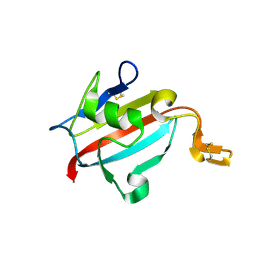
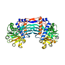
3BC9
 
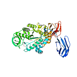 | | Alpha-amylase B in complex with acarbose | | 分子名称: | 4,6-dideoxy-4-{[(1S,4R,5S,6S)-4,5,6-trihydroxy-3-(hydroxymethyl)cyclohex-2-en-1-yl]amino}-alpha-D-glucopyranose-(1-4)-alpha-D-glucopyranose-(1-4)-alpha-D-glucopyranose, 4,6-dideoxy-alpha-D-xylo-hexopyranose-(1-4)-alpha-D-glucopyranose, 4,6-dideoxy-alpha-D-xylo-hexopyranose-(1-4)-alpha-D-glucopyranose-(1-4)-beta-D-glucopyranose, ... | | 著者 | Tan, T.-C, Mijts, B.N, Swaminathan, K, Patel, B.K.C, Divne, C. | | 登録日 | 2007-11-12 | | 公開日 | 2008-04-22 | | 最終更新日 | 2024-03-13 | | 実験手法 | X-RAY DIFFRACTION (1.35 Å) | | 主引用文献 | Crystal Structure of the Polyextremophilic alpha-Amylase AmyB from Halothermothrix orenii: Details of a Productive Enzyme-Substrate Complex and an N Domain with a Role in Binding Raw Starch
J.Mol.Biol., 378, 2008
|
|
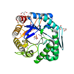
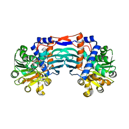
3BCF
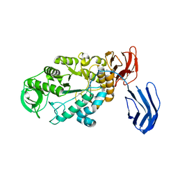 | | Alpha-amylase B from Halothermothrix orenii | | 分子名称: | Alpha amylase, catalytic region, CALCIUM ION, ... | | 著者 | Tan, T.-C, Mijts, B.N, Swaminathan, K, Patel, B.K.C, Divne, C. | | 登録日 | 2007-11-12 | | 公開日 | 2008-04-22 | | 最終更新日 | 2023-11-01 | | 実験手法 | X-RAY DIFFRACTION (2.3 Å) | | 主引用文献 | Crystal Structure of the Polyextremophilic alpha-Amylase AmyB from Halothermothrix orenii: Details of a Productive Enzyme-Substrate Complex and an N Domain with a Role in Binding Raw Starch
J.Mol.Biol., 378, 2008
|
|
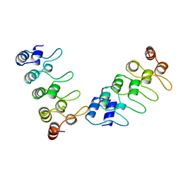
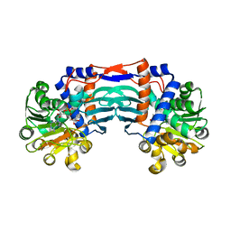
3B23
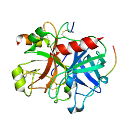 | | Crystal structure of thrombin-variegin complex: Insights of a novel mechanism of inhibition and design of tunable thrombin inhibitors | | 分子名称: | Thrombin heavy chain, Thrombin light chain, Variegin | | 著者 | Koh, C.Y, Kumar, S, Swaminathan, K, Kini, R.M. | | 登録日 | 2011-07-20 | | 公開日 | 2011-11-23 | | 最終更新日 | 2023-11-01 | | 実験手法 | X-RAY DIFFRACTION (2.4 Å) | | 主引用文献 | Crystal structure of thrombin in complex with s-variegin: insights of a novel mechanism of inhibition and design of tunable thrombin inhibitors
Plos One, 6, 2011
|
|
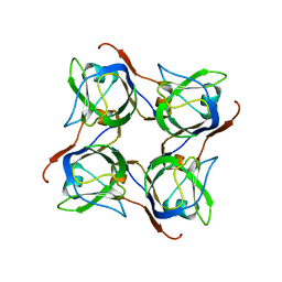
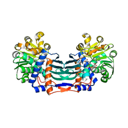
1HI4
 | | Eosinophil-derived Neurotoxin (EDN) - Adenosien-3'-5'-Diphosphate Complex | | 分子名称: | ADENOSINE-3'-5'-DIPHOSPHATE, EOSINOPHIL-DERIVED NEUROTOXIN | | 著者 | Leonidas, D.D, Boix, E, Prill, R, Suzuki, M, Turton, R, Minson, K, Swaminathan, G.J, Youle, R.J, Acharya, K.R. | | 登録日 | 2001-01-02 | | 公開日 | 2001-05-31 | | 最終更新日 | 2018-05-30 | | 実験手法 | X-RAY DIFFRACTION (1.8 Å) | | 主引用文献 | Mapping the Ribonucleolytic Active Site of Eosinophil-Derived Neurotoxin (Edn): High Resolution Crystal Structures of Edn Complexes with Adenylic Nucleotide Inhibitors
J.Biol.Chem., 276, 2001
|
|
1HI2
 | | Eosinophil-derived Neurotoxin (EDN) - Sulphate Complex | | 分子名称: | EOSINOPHIL-DERIVED NEUROTOXIN, SULFATE ION | | 著者 | Leonidas, D.D, Boix, E, Prill, R, Suzuki, M, Turton, R, Minson, K, Swaminathan, G.J, Youle, R.J, Acharya, K.R. | | 登録日 | 2001-01-02 | | 公開日 | 2001-05-31 | | 最終更新日 | 2011-07-13 | | 実験手法 | X-RAY DIFFRACTION (1.6 Å) | | 主引用文献 | Mapping the Ribonucleolytic Active Site of Eosinophil-Derived Neurotoxin (Edn): High Resolution Crystal Structures of Edn Complexes with Adenylic Nucleotide Inhibitors
J.Biol.Chem., 276, 2001
|
|
1HI5
 | | Eosinophil-derived Neurotoxin (EDN) - Adenosine-5'-Diphosphate Complex | | 分子名称: | ADENOSINE-5'-DIPHOSPHATE, EOSINOPHIL-DERIVED NEUROTOXIN | | 著者 | Leonidas, D.D, Boix, E, Prill, R, Suzuki, M, Turton, R, Minson, K, Swaminathan, G.J, Youle, R.J, Acharya, K.R. | | 登録日 | 2001-01-02 | | 公開日 | 2001-05-31 | | 最終更新日 | 2018-05-30 | | 実験手法 | X-RAY DIFFRACTION (1.8 Å) | | 主引用文献 | Mapping the Ribonucleolytic Active Site of Eosinophil-Derived Neurotoxin (Edn): High Resolution Crystal Structures of Edn Complexes with Adenylic Nucleotide Inhibitors
J.Biol.Chem., 276, 2001
|
|
1PWO
 | |
1HI3
 | | Eosinophil-derived Neurotoxin (EDN) - Adenosine 2'-5'-Diphosphate Complex | | 分子名称: | ADENOSINE-2'-5'-DIPHOSPHATE, EOSINOPHIL-DERIVED NEUROTOXIN | | 著者 | Leonidas, D.D, Boix, E, Prill, R, Suzuki, M, Turton, R, Minson, K, Swaminathan, G.J, Youle, R.J, Acharya, K.R. | | 登録日 | 2001-01-02 | | 公開日 | 2001-05-31 | | 最終更新日 | 2018-05-30 | | 実験手法 | X-RAY DIFFRACTION (1.8 Å) | | 主引用文献 | Mapping the Ribonucleolytic Active Site of Eosinophil-Derived Neurotoxin (Edn): High Resolution Crystal Structures of Edn Complexes with Adenylic Nucleotide Inhibitors
J.Biol.Chem., 276, 2001
|
|
3VUP
 | | Beta-1,4-mannanase from the common sea hare Aplysia kurodai | | 分子名称: | 2-AMINO-2-HYDROXYMETHYL-PROPANE-1,3-DIOL, Beta-1,4-mannanase, SULFATE ION | | 著者 | Mizutani, K, Tsuchiya, S, Toyoda, M, Nanbu, Y, Tominaga, K, Yuasa, K, Takahashi, N, Tsuji, A, Mikami, B. | | 登録日 | 2012-07-04 | | 公開日 | 2012-10-17 | | 実験手法 | X-RAY DIFFRACTION (1.05 Å) | | 主引用文献 | Structure of beta-1,4-mannanase from the common sea hare Aplysia kurodai at 1.05 A resolution.
Acta Crystallogr.,Sect.F, 68, 2012
|
|
1U79
 | | Crystal structure of AtFKBP13 | | 分子名称: | FKBP-type peptidyl-prolyl cis-trans isomerase 3 | | 著者 | Gopalan, G, Swaminathan, K. | | 登録日 | 2004-08-03 | | 公開日 | 2004-09-28 | | 最終更新日 | 2023-10-25 | | 実験手法 | X-RAY DIFFRACTION (1.85 Å) | | 主引用文献 | Structural analysis uncovers a role for redox in regulating FKBP13, an immunophilin of the chloroplast thylakoid lumen
Proc.Natl.Acad.Sci.Usa, 101, 2004
|
|
3WFL
 | | Crtstal structure of glycoside hydrolase family 5 beta-mannanase from Talaromyces trachyspermus | | 分子名称: | 2-AMINO-2-HYDROXYMETHYL-PROPANE-1,3-DIOL, 2-acetamido-2-deoxy-beta-D-glucopyranose, GLYCEROL, ... | | 著者 | Suzuki, K, Ichinose, H, Kamino, K, Ogasawara, W, Kaneko, S, Fushinobu, S. | | 登録日 | 2013-07-19 | | 公開日 | 2014-07-23 | | 最終更新日 | 2023-11-08 | | 実験手法 | X-RAY DIFFRACTION (1.6 Å) | | 主引用文献 | Purification, cloning, functional expression, structure, and characterization of a thermostable beta-mannanase from Talaromyces trachyspermus and its efficiency in production of mannooligosaccharides from coffee wastes
To be Published
|
|
1IHB
 | |
2C45
 | |
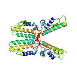
5YU4
 | | Structural basis for recognition of L-lysine, L-ornithine, and L-2,4-diamino butyric acid by lysine cyclodeaminase | | 分子名称: | 2,4-DIAMINOBUTYRIC ACID, Lysine cyclodeaminase, NICOTINAMIDE-ADENINE-DINUCLEOTIDE, ... | | 著者 | Min, K.J, Yoon, H.J, Matsuura, A, Kim, Y.H, Lee, H.H. | | 登録日 | 2017-11-20 | | 公開日 | 2018-05-02 | | 最終更新日 | 2024-03-27 | | 実験手法 | X-RAY DIFFRACTION (2.144 Å) | | 主引用文献 | Structural Basis for Recognition of L-lysine, L-ornithine, and L-2,4-diamino Butyric Acid by Lysine Cyclodeaminase.
Mol. Cells, 41, 2018
|
|
5YU3
 | | Structural basis for recognition of L-lysine, L-ornithine, and L-2,4-diamino butyric acid by lysine cyclodeaminase | | 分子名称: | Lysine cyclodeaminase, NICOTINAMIDE-ADENINE-DINUCLEOTIDE, PROLINE, ... | | 著者 | Min, K.J, Yoon, H.J, Matsuura, A, Kim, Y.H, Lee, H.H. | | 登録日 | 2017-11-20 | | 公開日 | 2018-05-02 | | 最終更新日 | 2024-03-27 | | 実験手法 | X-RAY DIFFRACTION (1.79 Å) | | 主引用文献 | Structural Basis for Recognition of L-lysine, L-ornithine, and L-2,4-diamino Butyric Acid by Lysine Cyclodeaminase.
Mol. Cells, 41, 2018
|
|
5YU0
 | | Structural basis for recognition of L-lysine, L-ornithine, and L-2,4-diamino butyric acid by lysine cyclodeaminase | | 分子名称: | Lysine cyclodeaminase, NICOTINAMIDE-ADENINE-DINUCLEOTIDE, SODIUM ION | | 著者 | Min, K.J, Yoon, H.J, Matsuura, A, Kim, Y.H, Lee, H.H. | | 登録日 | 2017-11-20 | | 公開日 | 2018-05-02 | | 最終更新日 | 2024-03-27 | | 実験手法 | X-RAY DIFFRACTION (1.92 Å) | | 主引用文献 | Structural Basis for Recognition of L-lysine, L-ornithine, and L-2,4-diamino Butyric Acid by Lysine Cyclodeaminase.
Mol. Cells, 41, 2018
|
|
5YU1
 | | Structural basis for recognition of L-lysine, L-ornithine, and L-2,4-diamino butyric acid by lysine cyclodeaminase | | 分子名称: | (2S)-piperidine-2-carboxylic acid, Lysine cyclodeaminase, NICOTINAMIDE-ADENINE-DINUCLEOTIDE, ... | | 著者 | Min, K.J, Yoon, H.J, Matsuura, A, Kim, Y.H, Lee, H.H. | | 登録日 | 2017-11-20 | | 公開日 | 2018-05-02 | | 最終更新日 | 2024-03-27 | | 実験手法 | X-RAY DIFFRACTION (1.923 Å) | | 主引用文献 | Structural Basis for Recognition of L-lysine, L-ornithine, and L-2,4-diamino Butyric Acid by Lysine Cyclodeaminase.
Mol. Cells, 41, 2018
|
|
6JN8
 | | Structure of H216A mutant open form peptidoglycan peptidase | | 分子名称: | Peptidase M23, SULFATE ION, ZINC ION | | 著者 | Min, K.J, An, D.R, Yoon, H.J, Suh, S.W, Lee, H.H. | | 登録日 | 2019-03-13 | | 公開日 | 2020-01-15 | | 最終更新日 | 2024-05-29 | | 実験手法 | X-RAY DIFFRACTION (2.106 Å) | | 主引用文献 | Peptidoglycan reshaping by a noncanonical peptidase for helical cell shape in Campylobacter jejuni.
Nat Commun, 11, 2020
|
|
6JMZ
 | | Structure of H247A mutant open form peptidoglycan peptidase | | 分子名称: | Peptidase M23, ZINC ION | | 著者 | Min, K.J, An, D.R, Yoon, H.J, Suh, S.W, Lee, H.H. | | 登録日 | 2019-03-13 | | 公開日 | 2020-01-15 | | 最終更新日 | 2024-05-29 | | 実験手法 | X-RAY DIFFRACTION (1.92 Å) | | 主引用文献 | Peptidoglycan reshaping by a noncanonical peptidase for helical cell shape in Campylobacter jejuni.
Nat Commun, 11, 2020
|
|
6JMX
 | | Structure of open form of peptidoglycan peptidase | | 分子名称: | D(-)-TARTARIC ACID, GLYCEROL, Peptidase M23, ... | | 著者 | Min, K.J, An, D.R, Yoon, H.J, Suh, S.W, Lee, H.H. | | 登録日 | 2019-03-13 | | 公開日 | 2020-01-15 | | 最終更新日 | 2024-05-29 | | 実験手法 | X-RAY DIFFRACTION (1.859 Å) | | 主引用文献 | Peptidoglycan reshaping by a noncanonical peptidase for helical cell shape in Campylobacter jejuni.
Nat Commun, 11, 2020
|
|
6JN1
 | | Structure of H247A mutant peptidoglycan peptidase complex with penta peptide | | 分子名称: | C0O-DAL-DAL, Peptidase M23, ZINC ION | | 著者 | Min, K.J, An, D.R, Yoon, H.J, Suh, S.W, Lee, H.H. | | 登録日 | 2019-03-13 | | 公開日 | 2020-01-15 | | 最終更新日 | 2023-11-15 | | 実験手法 | X-RAY DIFFRACTION (2.382 Å) | | 主引用文献 | Peptidoglycan reshaping by a noncanonical peptidase for helical cell shape in Campylobacter jejuni.
Nat Commun, 11, 2020
|
|
6JN0
 | | Structure of H247A mutant peptidoglycan peptidase complex with tetra-tri peptide | | 分子名称: | C0O-DAL-API, Peptidase M23, ZINC ION | | 著者 | Min, K.J, An, D.R, Yoon, H.J, Suh, S.W, Lee, H.H. | | 登録日 | 2019-03-13 | | 公開日 | 2020-01-15 | | 最終更新日 | 2024-03-20 | | 実験手法 | X-RAY DIFFRACTION (2.164 Å) | | 主引用文献 | Peptidoglycan reshaping by a noncanonical peptidase for helical cell shape in Campylobacter jejuni.
Nat Commun, 11, 2020
|
|
6JN7
 | | Structure of H216A mutant closed form peptidoglycan peptidase | | 分子名称: | D(-)-TARTARIC ACID, Peptidase M23, ZINC ION | | 著者 | Min, K.J, An, D.R, Yoon, H.J, Suh, S.W, Lee, H.H. | | 登録日 | 2019-03-13 | | 公開日 | 2020-01-15 | | 最終更新日 | 2024-05-29 | | 実験手法 | X-RAY DIFFRACTION (2.04 Å) | | 主引用文献 | Peptidoglycan reshaping by a noncanonical peptidase for helical cell shape in Campylobacter jejuni.
Nat Commun, 11, 2020
|
|
6JMY
 | | Structure of wild type closed form of peptidoglycan peptidase | | 分子名称: | CITRIC ACID, Peptidase M23, ZINC ION | | 著者 | Min, K.J, An, D.R, Yoon, H.J, Suh, S.W, Lee, H.H. | | 登録日 | 2019-03-13 | | 公開日 | 2020-01-15 | | 最終更新日 | 2024-05-29 | | 実験手法 | X-RAY DIFFRACTION (1.661 Å) | | 主引用文献 | Peptidoglycan reshaping by a noncanonical peptidase for helical cell shape in Campylobacter jejuni.
Nat Commun, 11, 2020
|
|
6K3F
 | |


