Loading
PDBj
メニューPDBj@FacebookPDBj@TwitterPDBj@YouTubewwPDB FoundationwwPDB
RCSB PDBPDBeBMRBAdv. SearchSearch help
Search by PDB author
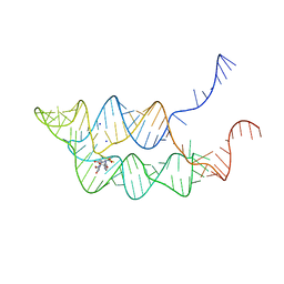
3SUH
DownloadVisualize
BU of 3suh by Molmil
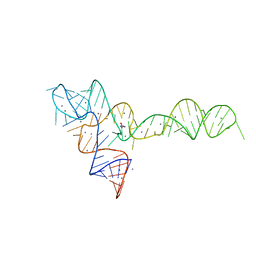
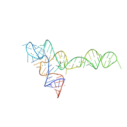
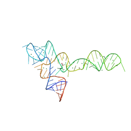
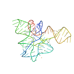
Crystal structure of THF riboswitch, bound with 5-formyl-THF
分子名称: N-[4-({[(6S)-2-amino-5-formyl-4-oxo-3,4,5,6,7,8-hexahydropteridin-6-yl]methyl}amino)benzoyl]-L-glutamic acid, Riboswitch, SODIUM ION
著者Huang, L, Serganov, A, Patel, D.J.
登録日2011-07-11
公開日2011-09-14
最終更新日2023-09-13
実験手法X-RAY DIFFRACTION (2.65 Å)
主引用文献Long-range pseudoknot interactions dictate the regulatory response in the tetrahydrofolate riboswitch.
Proc.Natl.Acad.Sci.USA, 108, 2011
3OXE
DownloadVisualize
BU of 3oxe by Molmil
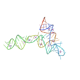
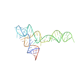
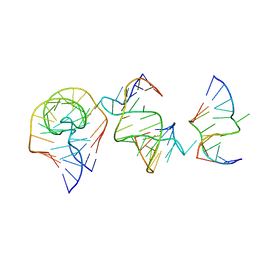
crystal structure of glycine riboswitch, Mn2+ soaked
分子名称: GLYCINE, MAGNESIUM ION, MANGANESE (II) ION, ...
著者Huang, L, Serganov, A, Patel, D.J.
登録日2010-09-21
公開日2010-12-29
最終更新日2023-09-06
実験手法X-RAY DIFFRACTION (2.899 Å)
主引用文献Structural insights into ligand recognition by a sensing domain of the cooperative glycine riboswitch.
Mol.Cell, 40, 2010
3OWZ
DownloadVisualize
BU of 3owz by Molmil
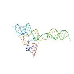
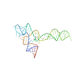
Crystal structure of glycine riboswitch, soaked in Iridium
分子名称: Domain II of glycine riboswitch, GLYCINE, IRIDIUM HEXAMMINE ION, ...
著者Huang, L, Serganov, A, Patel, D.J.
登録日2010-09-20
公開日2010-12-29
最終更新日2024-02-21
実験手法X-RAY DIFFRACTION (2.949 Å)
主引用文献Structural insights into ligand recognition by a sensing domain of the cooperative glycine riboswitch.
Mol.Cell, 40, 2010
3OXB
DownloadVisualize
BU of 3oxb by Molmil
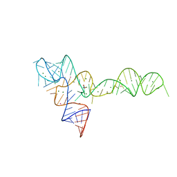
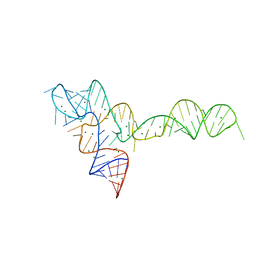
Crystal structure of glycine riboswitch with single mutation
分子名称: Domain II of glycine riboswitch, MAGNESIUM ION
著者Huang, L, Serganov, A, Patel, D.J.
登録日2010-09-21
公開日2010-12-29
最終更新日2023-09-06
実験手法X-RAY DIFFRACTION (2.947 Å)
主引用文献Structural insights into ligand recognition by a sensing domain of the cooperative glycine riboswitch.
Mol.Cell, 40, 2010
3OXM
DownloadVisualize
BU of 3oxm by Molmil
crystal structure of glycine riboswitch, Tl-Acetate soaked
分子名称: GLYCINE, MAGNESIUM ION, THALLIUM (I) ION, ...
著者Huang, L, Serganov, A, Patel, D.J.
登録日2010-09-21
公開日2010-12-29
最終更新日2023-09-06
実験手法X-RAY DIFFRACTION (2.95 Å)
主引用文献Structural insights into ligand recognition by a sensing domain of the cooperative glycine riboswitch.
Mol.Cell, 40, 2010
3OXJ
DownloadVisualize
BU of 3oxj by Molmil
crystal structure of glycine riboswitch, soaked in Ba2+
分子名称: BARIUM ION, GLYCINE, MAGNESIUM ION, ...
著者Huang, L, Serganov, A, Patel, D.J.
登録日2010-09-21
公開日2010-12-29
最終更新日2024-03-13
実験手法X-RAY DIFFRACTION (3.2 Å)
主引用文献Structural insights into ligand recognition by a sensing domain of the cooperative glycine riboswitch.
Mol.Cell, 40, 2010
3OX0
DownloadVisualize
BU of 3ox0 by Molmil
Crystal structure of glycine riboswitch, unbound state
分子名称: Domain II of glycine riboswitch, MAGNESIUM ION
著者Huang, L, Serganov, A, Patel, D.J.
登録日2010-09-20
公開日2010-12-29
最終更新日2023-09-06
実験手法X-RAY DIFFRACTION (3.049 Å)
主引用文献Structural insights into ligand recognition by a sensing domain of the cooperative glycine riboswitch.
Mol.Cell, 40, 2010
3OWI
DownloadVisualize
BU of 3owi by Molmil
Crystal structure of the glycine riboswitch bound to glycine
分子名称: Domain II of glycine riboswitch, GLYCINE, MAGNESIUM ION
著者Huang, L, Serganov, A, Patel, D.J.
登録日2010-09-19
公開日2010-12-29
最終更新日2023-09-06
実験手法X-RAY DIFFRACTION (2.845 Å)
主引用文献Structural insights into ligand recognition by a sensing domain of the cooperative glycine riboswitch.
Mol.Cell, 40, 2010
3OXD
DownloadVisualize
BU of 3oxd by Molmil
Crystal structure of glycine riboswitch with two mutations
分子名称: MAGNESIUM ION, domain II of glycine riboswitch
著者Huang, L, Serganov, A, Patel, D.J.
登録日2010-09-21
公開日2010-12-29
最終更新日2023-09-06
実験手法X-RAY DIFFRACTION (2.999 Å)
主引用文献Structural insights into ligand recognition by a sensing domain of the cooperative glycine riboswitch.
Mol.Cell, 40, 2010
3OWW
DownloadVisualize
BU of 3oww by Molmil
Crystal structure of the glycine riboswitch bound to glycine
分子名称: GLYCINE, MAGNESIUM ION, domain II of glycine riboswitch
著者Huang, L, Serganov, A, Patel, D.J.
登録日2010-09-20
公開日2010-12-29
最終更新日2023-09-06
実験手法X-RAY DIFFRACTION (2.802 Å)
主引用文献Structural insights into ligand recognition by a sensing domain of the cooperative glycine riboswitch.
Mol.Cell, 40, 2010
6HBT
DownloadVisualize
BU of 6hbt by Molmil
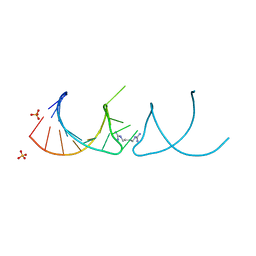
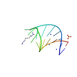
The structure of the G. violaceus guanidine II riboswitch P2 stem-loop with arcaine
分子名称: 1-(4-carbamimidamidobutyl)guanidine, RNA (5'-R(*GP*GP*UP*GP*GP*GP*GP*AP*CP*GP*AP*CP*CP*CP*CP*AP*(CBV)P*C)-3'), SULFATE ION
著者Huang, L, Lilley, D.M.J.
登録日2018-08-13
公開日2019-02-20
最終更新日2024-05-15
実験手法X-RAY DIFFRACTION (1.66 Å)
主引用文献Structure-guided design of a high-affinity ligand for a riboswitch.
Rna, 25, 2019
6HBX
DownloadVisualize
BU of 6hbx by Molmil
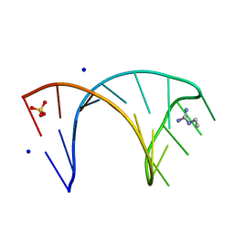
The structure of the G. violaceus guanidine II riboswitch P2 stem-loop with ethylguanidine
分子名称: N-ETHYLGUANIDINE, RNA (5'-R(*GP*GP*UP*GP*GP*GP*GP*AP*CP*GP*AP*CP*CP*CP*CP*AP*(CBV)P*C)-3'), SODIUM ION, ...
著者Huang, L, Lilley, D.M.J.
登録日2018-08-13
公開日2019-02-20
最終更新日2024-05-15
実験手法X-RAY DIFFRACTION (1.54 Å)
主引用文献Structure-guided design of a high-affinity ligand for a riboswitch.
Rna, 25, 2019
6HCT
DownloadVisualize
BU of 6hct by Molmil
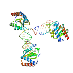
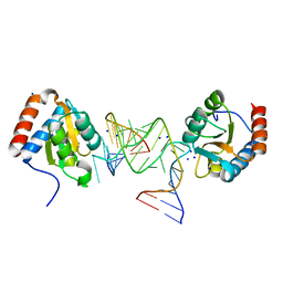
Crystal structure of Archeoglobus fulgidus L7Ae bound to its cognate UTR k-turn
分子名称: 50S ribosomal protein L7Ae, RNA (5'-R(*GP*CP*CP*GP*AP*UP*GP*AP*AP*UP*G)-3'), RNA (5'-R(*GP*CP*CP*GP*AP*UP*GP*AP*AP*UP*GP*CP*AP*UP*GP*AP*AP*GP*C)-3'), ...
著者Huang, L, Lilley, D.M.J.
登録日2018-08-16
公開日2018-10-31
最終更新日2024-01-17
実験手法X-RAY DIFFRACTION (3.091 Å)
主引用文献The role of RNA structure in translational regulation by L7Ae protein in archaea.
RNA, 25, 2019
6HC5
DownloadVisualize
BU of 6hc5 by Molmil
The structure of the G. violaceus guanidine II riboswitch P2 stem-loop with audouine
分子名称: 1-(5-carbamimidamidopentyl)guanidine, RNA (5'-R(*GP*GP*UP*GP*GP*GP*GP*AP*CP*GP*AP*CP*CP*CP*CP*AP*(CBV)P*C)-3'), SODIUM ION, ...
著者Huang, L, Lilley, D.M.J.
登録日2018-08-14
公開日2019-02-20
最終更新日2024-05-15
実験手法X-RAY DIFFRACTION (1.413 Å)
主引用文献Structure-guided design of a high-affinity ligand for a riboswitch.
Rna, 25, 2019
7XY1
DownloadVisualize
BU of 7xy1 by Molmil
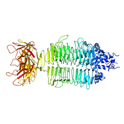
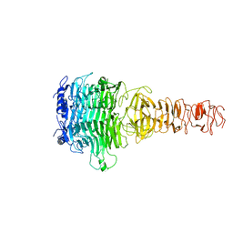
Cryo-EM structure of Klebsiella phage Kp9 type I tail fiber gp42 in vitro
分子名称: Tail fiber protein
著者Huang, L, Xiang, Y.
登録日2022-05-31
公開日2023-06-07
最終更新日2024-07-03
実験手法ELECTRON MICROSCOPY (3.3 Å)
主引用文献Structure and assembly of the Klebsiella pneumoniae phage tail fibers
To Be Published
7XYC
DownloadVisualize
BU of 7xyc by Molmil
CryoEM structure of Klebsiella phage Kp7 type II tail fiber gp52 in vitro
分子名称: phage tail fiber
著者Huang, L, Xiang, Y.
登録日2022-06-01
公開日2023-06-07
最終更新日2024-07-03
実験手法ELECTRON MICROSCOPY (3.8 Å)
主引用文献Structure and assembly of the Klebsiella pneumoniae phage tail fibers
To Be Published
7Y3T
DownloadVisualize
BU of 7y3t by Molmil
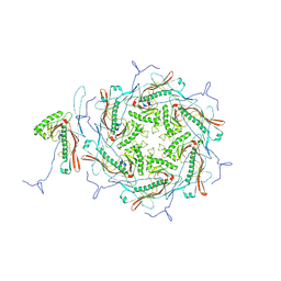
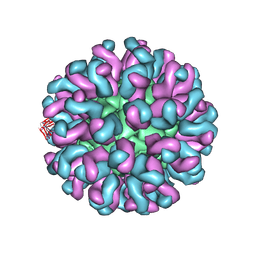
CryoEM structure of Klebsiella phage Kp7 icosahedral head
分子名称: phage major capsid protein
著者Huang, L, Xiang, Y.
登録日2022-06-12
公開日2023-06-14
最終更新日2024-07-03
実験手法ELECTRON MICROSCOPY (3.9 Å)
主引用文献Structure and assembly of the Klebsiella pneumoniae phage tail fibers
To Be Published
7Y22
DownloadVisualize
BU of 7y22 by Molmil
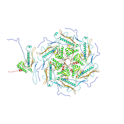
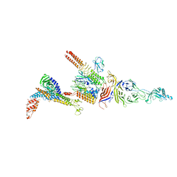
CryoEM structure of Klebsiella phage Kp7 tail complex applied with C6 symmetry
分子名称: phage connector protein, phage tail tubular protein A, phage tail tubular protein B, ...
著者Huang, L, Xiang, Y.
登録日2022-06-09
公開日2023-06-14
最終更新日2024-07-03
実験手法ELECTRON MICROSCOPY (4 Å)
主引用文献Structure and assembly of the Klebsiella pneumoniae phage tail fibers
To Be Published
7Y23
DownloadVisualize
BU of 7y23 by Molmil
CryoEM structure of Klebsiella phage Kp9 icosahedral head
分子名称: phage capsid protein
著者Huang, L, Xiang, Y.
登録日2022-06-09
公開日2023-06-14
最終更新日2024-07-03
実験手法ELECTRON MICROSCOPY (3.2 Å)
主引用文献Structure and assembly of the Klebsiella pneumoniae phage tail fibers
To Be Published
7Y1C
DownloadVisualize
BU of 7y1c by Molmil
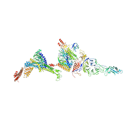
CryoEM structure of Klebsiella phage Kp9 tail complex applied with C6 symmetry
分子名称: phage connector protein, phage tail tubular protein A, phage tail tubular protein B, ...
著者Huang, L, Xiang, Y.
登録日2022-06-08
公開日2023-06-14
最終更新日2024-07-03
実験手法ELECTRON MICROSCOPY (3.13 Å)
主引用文献Structure and assembly of the Klebsiella pneumoniae phage tail fibers
To Be Published
7Y5S
DownloadVisualize
BU of 7y5s by Molmil
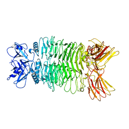
CryoEM structure of Klebsiella phage Kp7 type I tail fiber gp51 in vitro
分子名称: phage tail fiber
著者Huang, L, Xiang, Y.
登録日2022-06-17
公開日2023-06-21
最終更新日2024-07-03
実験手法ELECTRON MICROSCOPY (3 Å)
主引用文献Structure and assembly of the Klebsiella pneumoniae phage tail fibers
To Be Published
7EAG
DownloadVisualize
BU of 7eag by Molmil
Crystal structure of the RAGATH-18 k-turn
分子名称: RNA (5'-R(*GP*UP*CP*UP*AP*UP*GP*AP*AP*GP*GP*CP*UP*GP*GP*AP*GP*AP*C)-3')
著者Huang, L, Lilley, D.M.J.
登録日2021-03-07
公開日2021-06-02
最終更新日2023-11-29
実験手法X-RAY DIFFRACTION (2.5 Å)
主引用文献Structure and folding of four putative kink turns identified in structured RNA species in a test of structural prediction rules.
Nucleic Acids Res., 49, 2021
7EAF
DownloadVisualize
BU of 7eaf by Molmil
Crystal structure of SAM-I riboswitch with the Actinomyces-1 k-turn
分子名称: BARIUM ION, RNA (94-MER), S-ADENOSYLMETHIONINE, ...
著者Huang, L, Lilley, D.M.J.
登録日2021-03-07
公開日2021-06-02
最終更新日2023-11-29
実験手法X-RAY DIFFRACTION (2.85 Å)
主引用文献Structure and folding of four putative kink turns identified in structured RNA species in a test of structural prediction rules.
Nucleic Acids Res., 49, 2021
6L62
DownloadVisualize
BU of 6l62 by Molmil
Neutralization mechanism of a monoclonal antibody targeting a porcine circovirus type 2 Cap protein conformational epitope
分子名称: Capsid protein, Heavy chain of Fab fragment, Light chain of Fab fragment
著者Sun, Z, Huang, L, Xia, D, Wei, Y, Sun, E, Zhu, H, Bian, H, Wu, H, Feng, L, Wang, J, Liu, C.
登録日2019-10-25
公開日2020-02-12
最終更新日2020-04-29
実験手法ELECTRON MICROSCOPY (7.2 Å)
主引用文献Neutralization Mechanism of a Monoclonal Antibody Targeting a Porcine Circovirus Type 2 Cap Protein Conformational Epitope.
J.Virol., 94, 2020
6Q8U
DownloadVisualize
BU of 6q8u by Molmil
Structure of the standard kink turn HmKt-7 variant A2bm6A bound with AfL7Ae protein
分子名称: 50S ribosomal protein L7Ae, RNA (5'-R(*CP*GP*GP*CP*GP*AP*AP*GP*(6MZ)P*AP*CP*CP*GP*GP*GP*GP*AP*GP*CP*CP*G)-3'), SODIUM ION
著者Huang, L, Lilley, D.M.J.
登録日2018-12-16
公開日2019-07-03
最終更新日2024-01-24
実験手法X-RAY DIFFRACTION (1.99 Å)
主引用文献Effect of methylation of adenine N6on kink turn structure depends on location.
Rna Biol., 16, 2019

224201

件を2024-08-28に公開中

PDB statisticsPDBj update infoContact PDBjnumon