Loading
PDBj
メニューPDBj@FacebookPDBj@TwitterPDBj@YouTubewwPDB FoundationwwPDB
RCSB PDBPDBeBMRBAdv. SearchSearch help
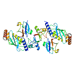
Search by PDB author
5CLR
DownloadVisualize
BU of 5clr by Molmil
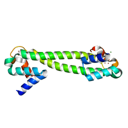
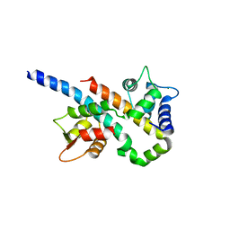
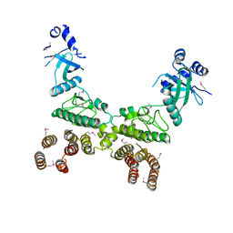
Crystal structure of LegK4_APO Kinase
分子名称: LegK4
著者Flayhan, A, Terradot, L.
登録日2015-07-16
公開日2015-10-14
実験手法X-RAY DIFFRACTION (3.706 Å)
主引用文献The structure of Legionella pneumophila LegK4 type four secretion system (T4SS) effector reveals a novel dimeric eukaryotic-like kinase.
Sci Rep, 5, 2015
7EPQ
DownloadVisualize
BU of 7epq by Molmil
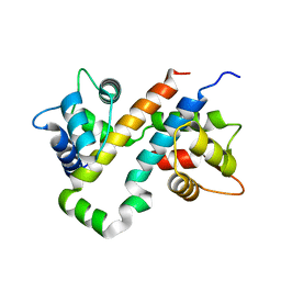
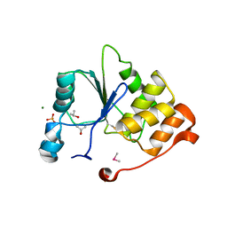
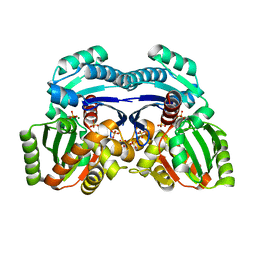
Crystal structure of exopolyphosphatase (PPX) from Porphyromonas gingivalis in complex with sulfate and magnesium ions
分子名称: MAGNESIUM ION, Putative exopolyphosphatase, SULFATE ION
著者Zhang, A.
登録日2021-04-27
公開日2022-05-11
最終更新日2023-11-29
実験手法X-RAY DIFFRACTION (2.2 Å)
主引用文献The structure of exopolyphosphatase (PPX) from Porphyromonas gingivalis in complex with substrate analogs and magnesium ions reveals the basis for polyphosphate processivity.
J.Struct.Biol., 213, 2021
1Q08
DownloadVisualize
BU of 1q08 by Molmil
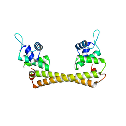
Crystal structure of the Zn(II) form of E. coli ZntR, a zinc-sensing transcriptional regulator, at 1.9 A resolution (space group P212121)
分子名称: MAGNESIUM ION, PHOSPHATE ION, ZINC ION, ...
著者Changela, A, Chen, K, Xue, Y, Holschen, J, Outten, C.E, O'Halloran, T.V, Mondragon, A.
登録日2003-07-15
公開日2003-09-16
最終更新日2024-02-14
実験手法X-RAY DIFFRACTION (1.9 Å)
主引用文献Molecular basis of metal-ion selectivity and zeptomolar sensitivity by CueR
Science, 301, 2003
1Q07
DownloadVisualize
BU of 1q07 by Molmil
Crystal structure of the Au(I) form of E. coli CueR, a copper efflux regulator
分子名称: GOLD ION, Transcriptional regulator cueR
著者Changela, A, Chen, K, Xue, Y, Holschen, J, Outten, C.E, O'Halloran, T.V, Mondragon, A.
登録日2003-07-15
公開日2003-09-16
最終更新日2024-02-14
実験手法X-RAY DIFFRACTION (2.5 Å)
主引用文献Molecular basis of metal-ion selectivity and zeptomolar sensitivity by CueR
Science, 301, 2003
7F80
DownloadVisualize
BU of 7f80 by Molmil
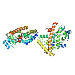
Co-crystal structure of Inhibitor compound MA-211 in complex with human PPARdelta LBD
分子名称: (3R)-3-methyl-6-[2-[[5-methyl-2-[4-(trifluoromethyl)phenyl]imidazol-1-yl]methyl]phenoxy]hexanoic acid, Peroxisome proliferator-activated receptor delta
著者Lakshminarasimhan, A, Rani, S.T, Senaiar, R.S, Krishnamurthy, N.
登録日2021-06-30
公開日2022-07-06
最終更新日2023-11-29
実験手法X-RAY DIFFRACTION (2.8 Å)
主引用文献Co-crystal structure of Inhibitor compound in complex with human PPARdelta LBD
To Be Published
3SJG
DownloadVisualize
BU of 3sjg by Molmil
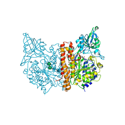
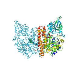
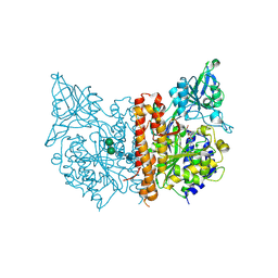
Human glutamate carboxypeptidase II (E424A inactive mutant ) in complex with N-acetyl-aspartyl-aminooctanoic acid
分子名称: (2S)-2-[(N-acetyl-L-alpha-aspartyl)amino]nonanoic acid, 2-acetamido-2-deoxy-beta-D-glucopyranose, 2-acetamido-2-deoxy-beta-D-glucopyranose-(1-4)-2-acetamido-2-deoxy-beta-D-glucopyranose, ...
著者Plechanovova, A, Byun, Y, Alquicer, G, Skultetyova, L, Mlcochova, P, Nemcova, A, Kim, H, Navratil, M, Mease, R, Lubkowski, J, Pomper, M, Konvalinka, J, Rulisek, L, Barinka, C.
登録日2011-06-21
公開日2011-10-05
最終更新日2020-07-29
実験手法X-RAY DIFFRACTION (1.65 Å)
主引用文献Novel Substrate-Based Inhibitors of Human Glutamate Carboxypeptidase II with Enhanced Lipophilicity.
J.Med.Chem., 54, 2011
3SJE
DownloadVisualize
BU of 3sje by Molmil
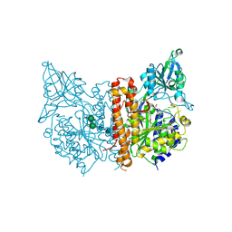
X-ray structure of human glutamate carboxypeptidase II (the E424A inactive mutant) in complex with N-acetyl-aspartyl-aminononanoic acid
分子名称: (2S)-2-[(N-acetyl-L-alpha-aspartyl)amino]nonanoic acid, 2-acetamido-2-deoxy-beta-D-glucopyranose, 2-acetamido-2-deoxy-beta-D-glucopyranose-(1-4)-2-acetamido-2-deoxy-beta-D-glucopyranose, ...
著者Plechanovova, A, Byun, Y, Alquicer, G, Skultetyova, L, Mlcochova, P, Nemcova, A, Kim, H, Navratil, M, Mease, R, Lubkowski, J, Pomper, M, Konvalinka, J, Rulisek, L, Barinka, C.
登録日2011-06-21
公開日2011-10-05
最終更新日2020-07-29
実験手法X-RAY DIFFRACTION (1.7 Å)
主引用文献Novel Substrate-Based Inhibitors of Human Glutamate Carboxypeptidase II with Enhanced Lipophilicity.
J.Med.Chem., 54, 2011
3SJX
DownloadVisualize
BU of 3sjx by Molmil
X-ray structure of human glutamate carboxypeptidase II (the E424A inactive mutant) in complex with N-acetyl-aspartyl-methionine
分子名称: 2-acetamido-2-deoxy-beta-D-glucopyranose, 2-acetamido-2-deoxy-beta-D-glucopyranose-(1-4)-2-acetamido-2-deoxy-beta-D-glucopyranose, CALCIUM ION, ...
著者Plechanovova, A, Byun, Y, Alquicer, G, Skultetyova, L, Mlcochova, P, Nemcova, A, Kim, H, Navratil, M, Mease, R, Lubkowski, J, Pomper, M, Konvalinka, J, Rulisek, L, Barinka, C.
登録日2011-06-22
公開日2011-10-05
最終更新日2020-07-29
実験手法X-RAY DIFFRACTION (1.66 Å)
主引用文献Novel Substrate-Based Inhibitors of Human Glutamate Carboxypeptidase II with Enhanced Lipophilicity.
J.Med.Chem., 54, 2011
6B8M
DownloadVisualize
BU of 6b8m by Molmil
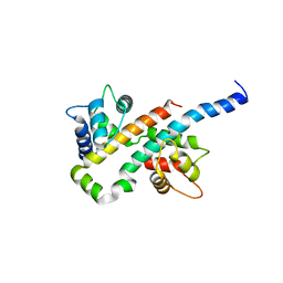
Crystal Structure of the Ca2+/CaM:Kv7.4 (KCNQ4) AB Domain Complex, 1 mM CaCl2 soak
分子名称: CALCIUM ION, Calmodulin-1, Potassium voltage-gated channel subfamily KQT member 4, ...
著者Chang, A, Minor, D.L.
登録日2017-10-09
公開日2018-03-14
最終更新日2023-10-04
実験手法X-RAY DIFFRACTION (2.3 Å)
主引用文献A Calmodulin C-Lobe Ca
Neuron, 97, 2018
6B8Q
DownloadVisualize
BU of 6b8q by Molmil
Crystal Structure of the Mg2+/CaM:Kv7.5 (KCNQ5) AB domain complex
分子名称: Calmodulin-1, MAGNESIUM ION, Potassium voltage-gated channel subfamily KQT member 5
著者Chang, A, Abderemane-Ali, F, Minor, D.L.
登録日2017-10-09
公開日2018-03-14
最終更新日2023-10-04
実験手法X-RAY DIFFRACTION (2.6 Å)
主引用文献A Calmodulin C-Lobe Ca
Neuron, 97, 2018
6B8P
DownloadVisualize
BU of 6b8p by Molmil
Crystal Structure of the Mg2+/CaM:Kv7.4 (KCNQ4) AB domain complex
分子名称: Calmodulin-1, MAGNESIUM ION, Potassium voltage-gated channel subfamily KQT member 4, ...
著者Chang, A, Minor, D.L.
登録日2017-10-09
公開日2018-03-14
最終更新日2023-10-04
実験手法X-RAY DIFFRACTION (2.2 Å)
主引用文献A Calmodulin C-Lobe Ca
Neuron, 97, 2018
6B8L
DownloadVisualize
BU of 6b8l by Molmil
Crystal Structure of the Apo/CaM:Kv7.4 (KCNQ4) AB Domain Complex
分子名称: Calmodulin-1, Potassium voltage-gated channel subfamily KQT member 4, SULFATE ION
著者Chang, A, Abderemane-Ali, F, Minor, D.L.
登録日2017-10-09
公開日2018-03-14
最終更新日2024-03-13
実験手法X-RAY DIFFRACTION (2.3 Å)
主引用文献A Calmodulin C-Lobe Ca2+-Dependent Switch Governs Kv7 Channel Function
Neuron, 97, 2018
3SJF
DownloadVisualize
BU of 3sjf by Molmil
X-ray structure of human glutamate carboxypeptidase II in complex with a urea-based inhibitor (A25)
分子名称: 2-acetamido-2-deoxy-beta-D-glucopyranose, 2-acetamido-2-deoxy-beta-D-glucopyranose-(1-4)-2-acetamido-2-deoxy-beta-D-glucopyranose, CALCIUM ION, ...
著者Plechanovova, A, Byun, Y, Alquicer, G, Skultetyova, L, Mlcochova, P, Nemcova, A, Kim, H, Navratil, M, Mease, R, Lubkowski, J, Pomper, M, Konvalinka, J, Rulisek, L, Barinka, C.
登録日2011-06-21
公開日2011-10-05
最終更新日2020-07-29
実験手法X-RAY DIFFRACTION (1.65 Å)
主引用文献Novel Substrate-Based Inhibitors of Human Glutamate Carboxypeptidase II with Enhanced Lipophilicity.
J.Med.Chem., 54, 2011
1YN9
DownloadVisualize
BU of 1yn9 by Molmil
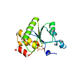
Crystal structure of baculovirus RNA 5'-phosphatase complexed with phosphate
分子名称: PHOSPHATE ION, polynucleotide 5'-phosphatase
著者Changela, A, Martins, A, Shuman, S, Mondragon, A.
登録日2005-01-24
公開日2005-02-22
最終更新日2023-08-23
実験手法X-RAY DIFFRACTION (1.5 Å)
主引用文献Crystal structure of baculovirus RNA triphosphatase complexed with phosphate
J.Biol.Chem., 280, 2005
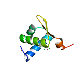
1XMK
DownloadVisualize
BU of 1xmk by Molmil
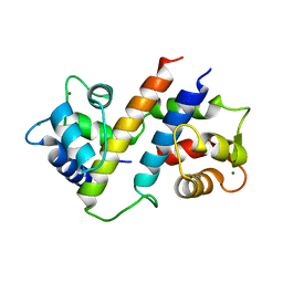
The Crystal structure of the Zb domain from the RNA editing enzyme ADAR1
分子名称: CADMIUM ION, CHLORIDE ION, Double-stranded RNA-specific adenosine deaminase, ...
著者Athanasiadis, A, Placido, D, Maas, S, Brown II, B.A, Lowenhaupt, K, Rich, A.
登録日2004-10-03
公開日2005-08-02
最終更新日2024-02-14
実験手法X-RAY DIFFRACTION (0.97 Å)
主引用文献The Crystal Structure of the Z[beta] Domain of the RNA-editing Enzyme ADAR1 Reveals Distinct Conserved Surfaces Among Z-domains.
J.Mol.Biol., 351, 2005
6B8N
DownloadVisualize
BU of 6b8n by Molmil
Crystal Structure of the Ca2+/CaM:Kv7.4 (KCNQ4) AB Domain Complex, 10 uM CaCl2 soak
分子名称: CALCIUM ION, Calmodulin-1, Potassium voltage-gated channel subfamily KQT member 4, ...
著者Chang, A, Minor, D.L.
登録日2017-10-09
公開日2018-03-14
最終更新日2023-10-04
実験手法X-RAY DIFFRACTION (2.201 Å)
主引用文献A Calmodulin C-Lobe Ca
Neuron, 97, 2018
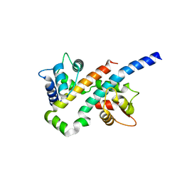
4AP4
DownloadVisualize
BU of 4ap4 by Molmil
Rnf4 - ubch5a - ubiquitin heterotrimeric complex
分子名称: E3 UBIQUITIN LIGASE RNF4, UBIQUITIN C, UBIQUITIN-CONJUGATING ENZYME E2 D1, ...
著者Plechanovova, A, Hay, R.T, Tatham, M.H, Jaffray, E, Naismith, J.H.
登録日2012-03-30
公開日2012-07-25
最終更新日2023-12-20
実験手法X-RAY DIFFRACTION (2.21 Å)
主引用文献Structure of a Ring E3 Ligase and Ubiquitin-Loaded E2 Primed for Catalysis
Nature, 489, 2012
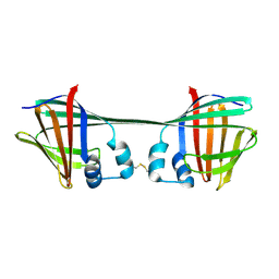
7UCS
DownloadVisualize
BU of 7ucs by Molmil
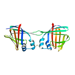
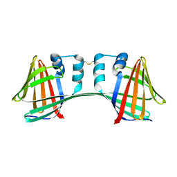
The Crystal Structure of Domain-Swapped Dimer Q108K:T51D:A28C:L36C:F57:H:H:H:R58 Mutant of hCRBPII with Histidine Insertion in the Hinge Loop Region at 1.92 Angstrom Resolution
分子名称: Retinol-binding protein 2
著者Ghanbarpour, A, Geiger, J.
登録日2022-03-17
公開日2023-10-18
実験手法X-RAY DIFFRACTION (1.92 Å)
主引用文献The Crystal Structure of Domain-Sawpped Dimer Q108K:T51D:A28C:L36C:F57:H:R58 Mutant of hCRBPII with a Histidine Insertion in the Hinge Loop Region at 1.96 Angstrom Resolution
To Be Published
7UCT
DownloadVisualize
BU of 7uct by Molmil
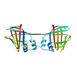
The Crystal Structure of Apo Domain-Swapped Dimer F57:H:H:H:H:H:H:R58 Mutant of HCRBPII with Histidine Insertion in the Hinge Loop Region at 2.5 Angstrom Resolution
分子名称: Retinol-binding protein 2
著者Ghanbarpour, A, Geiger, J.
登録日2022-03-17
公開日2023-10-18
実験手法X-RAY DIFFRACTION (2.52 Å)
主引用文献The Crystal Structure of Domain-Sawpped Dimer Q108K:T51D:A28C:L36C:F57:H:R58 Mutant of hCRBPII with a Histidine Insertion in the Hinge Loop Region at 1.96 Angstrom Resolution
To Be Published
7UD3
DownloadVisualize
BU of 7ud3 by Molmil
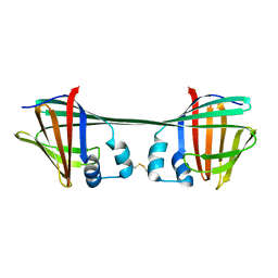
The Crystal Structure of Domain-Swapped Dimer Q108K:T51D:A28C:L36C:F57:W:W:W:R58 Mutant of hCRBPII with a Histidine Insertion in the Hinge Loop Region at 2.36 Angstrom Resolution
分子名称: Retinol-binding protein 2
著者Ghanbarpour, A, Geiger, J.
登録日2022-03-17
公開日2023-10-18
実験手法X-RAY DIFFRACTION (2.37 Å)
主引用文献The Crystal Structure of Domain-Sawpped Dimer Q108K:T51D:A28C:L36C:F57:H:R58 Mutant of hCRBPII with a Histidine Insertion in the Hinge Loop Region at 1.96 Angstrom Resolution
To Be Published
7UCV
DownloadVisualize
BU of 7ucv by Molmil
The Crystal Structure of Apo Domain-Swapped Dimer Q108K:T51D:A28CL36C R58:H:H:H:N59 HCRBPII with Histidine Insertion in the Hinge Loop Region at 2.19 Angstrom Resolution
分子名称: Retinol-binding protein 2
著者Ghanbarpour, A, Geiger, J.
登録日2022-03-17
公開日2023-10-18
実験手法X-RAY DIFFRACTION (2.19 Å)
主引用文献The Crystal Structure of Domain-Sawpped Dimer Q108K:T51D:A28C:L36C:F57:H:R58 Mutant of hCRBPII with a Histidine Insertion in the Hinge Loop Region at 1.96 Angstrom Resolution
To Be Published
7UCZ
DownloadVisualize
BU of 7ucz by Molmil
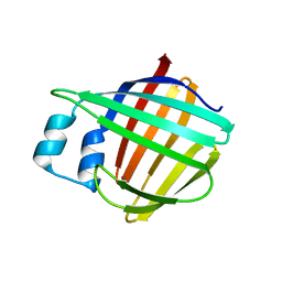
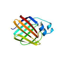
The Crystal Structure of Apo Monomer F57:H:H:H:H:H:H:R58 Mutant of HCRBPII with Histidine Insertion in the Hinge Loop Region at 1.1 Angstrom Resolution
分子名称: Retinol-binding protein 2
著者Ghanbarpour, A, Geiger, J.
登録日2022-03-17
公開日2023-10-18
実験手法X-RAY DIFFRACTION (1.08 Å)
主引用文献The Crystal Structure of Domain-Sawpped Dimer Q108K:T51D:A28C:L36C:F57:H:R58 Mutant of hCRBPII with a Histidine Insertion in the Hinge Loop Region at 1.96 Angstrom Resolution
To Be Published
7UD1
DownloadVisualize
BU of 7ud1 by Molmil
The Crystal Structure of Apo Monomer F57:H:H:H:R58 HCRBPII with Histidine Insertion in the Hinge Loop Region at 1.3 Angstrom Resolution
分子名称: Retinol-binding protein 2
著者Ghanbarpour, A, Geiger, J.
登録日2022-03-17
公開日2023-10-18
実験手法X-RAY DIFFRACTION (1.32 Å)
主引用文献The Crystal Structure of Apo Monomer F57:H:H:H:R58 HCRBPII with Histidine Insertion in the Hinge Loop Region at 1.3 Angstrom Resolution
To Be Published
7UCN
DownloadVisualize
BU of 7ucn by Molmil
The Crystal Structure of Domain-Swapped Dimer Q108K:T51D:A28C:L36C:F57:H:R58 Mutant of hCRBPII with a Histidine Insertion in the Hinge Loop Region at 1.96 Angstrom Resolution
分子名称: Retinol-binding protein 2
著者Ghanbarpour, A, Geiger, J.
登録日2022-03-16
公開日2023-10-18
実験手法X-RAY DIFFRACTION (1.96 Å)
主引用文献The Crystal Structure of Domain-Sawpped Dimer Q108K:T51D:A28C:L36C:F57:H:R58 Mutant of hCRBPII with a Histidine Insertion in the Hinge Loop Region at 1.96 Angstrom Resolution
To Be Published
1I9T
DownloadVisualize
BU of 1i9t by Molmil
CRYSTAL STRUCTURE OF THE OXIDIZED RNA TRIPHOSPHATASE DOMAIN OF MOUSE MRNA CAPPING ENZYME
分子名称: CACODYLATE ION, ISOPROPYL ALCOHOL, MAGNESIUM ION, ...
著者Changela, A, Ho, C.K, Martins, A, Shuman, S, Mondragon, A.
登録日2001-03-20
公開日2001-05-23
最終更新日2023-11-15
実験手法X-RAY DIFFRACTION (1.7 Å)
主引用文献Structure and mechanism of the RNA triphosphatase component of mammalian mRNA capping enzyme.
EMBO J., 20, 2001

222415

件を2024-07-10に公開中

PDB statisticsPDBj update infoContact PDBjnumon