Loading
PDBj
メニューPDBj@FacebookPDBj@TwitterPDBj@YouTubewwPDB FoundationwwPDB
RCSB PDBPDBeBMRBAdv. SearchSearch help
Search by PDB author
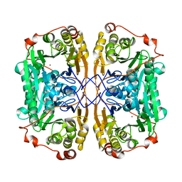
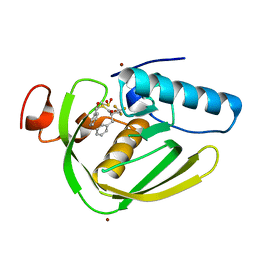
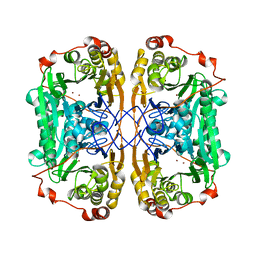
5E5D
DownloadVisualize
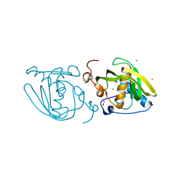
BU of 5e5d by Molmil
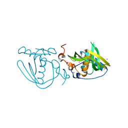
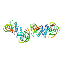
Native structure of Xoo1075, a peptide deformylase from Xanthomonas oryzae pv. oryzae
分子名称: CADMIUM ION, Peptide deformylase
著者Ngo, H.P.T, Kang, L.W.
登録日2015-10-08
公開日2016-10-12
最終更新日2023-11-08
実験手法X-RAY DIFFRACTION (2.6 Å)
主引用文献Native structure of Xoo1075, a peptide deformylase from Xanthomonas oryzae pv. oryzae
To Be Published
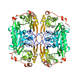
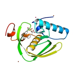
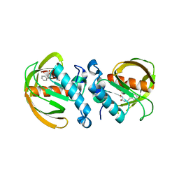
5CY7
DownloadVisualize
BU of 5cy7 by Molmil
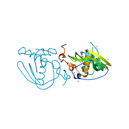
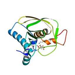
Structure of Xoo1075, a peptide deformylase from Xanthomonas oryze pv oryze, in complex with fragment 275
分子名称: 2-(difluoromethyl)-1H-benzimidazole, ACETATE ION, CADMIUM ION, ...
著者Ngo, H.P.T, Kang, L.W.
登録日2015-07-30
公開日2016-08-03
最終更新日2023-11-08
実験手法X-RAY DIFFRACTION (2.4 Å)
主引用文献Structure of Xoo1075, a peptide deformylase from Xanthomonas oryze pv oryze, in complex with fragment 275
To Be Published
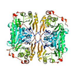
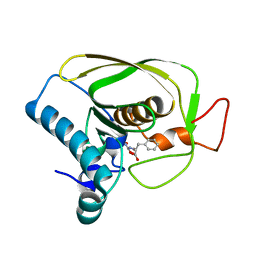
5CX0
DownloadVisualize
BU of 5cx0 by Molmil
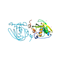
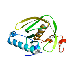
Structure of Xoo1075, a peptide deformylase from Xanthomonas oryzae pv. oryzae, in complex with fragment 322
分子名称: 5-(propan-2-yl)-2-thioxodihydropyrimidine-4,6(1H,5H)-dione, ACETATE ION, CADMIUM ION, ...
著者Ngo, H.P.T, Kang, L.W.
登録日2015-07-28
公開日2016-08-03
最終更新日2024-03-20
実験手法X-RAY DIFFRACTION (2.5 Å)
主引用文献Structure of Xoo1075, a peptide deformylase from Xanthomonas oryzae pv. oryzae, in complex with fragment 322
To Be Published
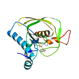
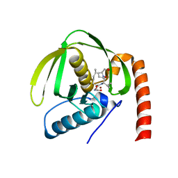
5CXJ
DownloadVisualize
BU of 5cxj by Molmil
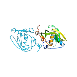
Structure of Xoo1075, a peptide deformylase from Xanthomonas oryzae pv oryzae, in complex with fragment 124
分子名称: 2-(furan-2-yl)ethanamine, ACETATE ION, CADMIUM ION, ...
著者Ngo, H.P.T, Kang, L.W.
登録日2015-07-29
公開日2016-08-03
最終更新日2023-11-08
実験手法X-RAY DIFFRACTION (2.38 Å)
主引用文献Structure of Xoo1075, a peptide deformylase from Xanthomonas oryzae pv oryzae, in complex with fragment 124
To Be Published
5CY8
DownloadVisualize
BU of 5cy8 by Molmil
Structure of Xoo1075, a peptide deformylase from Xanthomonas oryzae pv oryze, in complex with fragment 244
分子名称: (3R)-2,3-dihydro[1,3]thiazolo[3,2-a]benzimidazol-3-ol, ACETATE ION, CADMIUM ION, ...
著者Ngo, H.P.T, Kang, L.W.
登録日2015-07-30
公開日2016-08-03
最終更新日2023-11-08
実験手法X-RAY DIFFRACTION (2.38 Å)
主引用文献Structure of Xoo1075, a peptide deformylase from Xanthomonas oryzae pv oryze, in complex with fragment 244
To Be Published
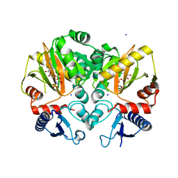
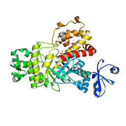
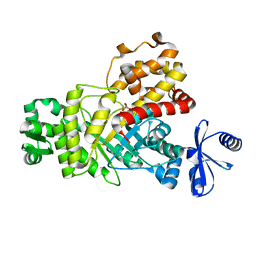
5DMX
DownloadVisualize
BU of 5dmx by Molmil
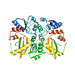
Crystal structure of D-alanine-D-alanine ligase from Acinetobacter baumannii, space group p212121
分子名称: D-alanine--D-alanine ligase
著者Huynh, K.H, Hong, M.K, Kang, L.W.
登録日2015-09-09
公開日2016-08-17
最終更新日2023-11-08
実験手法X-RAY DIFFRACTION (2.81 Å)
主引用文献The crystal structure of the D-alanine-D-alanine ligase from Acinetobacter baumannii suggests a flexible conformational change in the central domain before nucleotide binding
J. Microbiol., 53, 2015
5C1O
DownloadVisualize
BU of 5c1o by Molmil
Crystal structure of AMP-PNP complexed D-alanine-D-alanine ligase(DDL) from Yersinia pestis
分子名称: D-alanine--D-alanine ligase, MAGNESIUM ION, PHOSPHOAMINOPHOSPHONIC ACID-ADENYLATE ESTER, ...
著者Tran, H.T, Kang, L.W, Hong, M.K.
登録日2015-06-15
公開日2016-03-02
最終更新日2024-03-20
実験手法X-RAY DIFFRACTION (2.5 Å)
主引用文献Structure of D-alanine-D-alanine ligase from Yersinia pestis: nucleotide phosphate recognition by the serine loop.
Acta Crystallogr D Struct Biol, 72, 2016
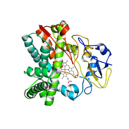
4FXB
DownloadVisualize
BU of 4fxb by Molmil
Crystal structure of CYP105N1 from Streptomyces coelicolor: a cytochrome P450 oxidase in the coelibactin siderophore biosynthetic pathway
分子名称: PROTOPORPHYRIN IX CONTAINING FE, Putative cytochrome P450
著者Hong, M.K, Lim, Y.R, Kim, J.K, Kim, D.H, Kang, L.W.
登録日2012-07-03
公開日2012-11-14
最終更新日2023-11-08
実験手法X-RAY DIFFRACTION (2.9 Å)
主引用文献Crystal structure of cytochrome P450 CYP105N1 from Streptomyces coelicolor, an oxidase in the coelibactin siderophore biosynthetic pathway
Arch.Biochem.Biophys., 528, 2012
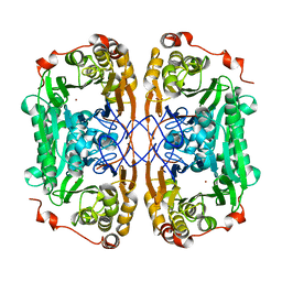
7BZ4
DownloadVisualize
BU of 7bz4 by Molmil
The mutant variant of PNGM-1. H279 was substituted for alanine to study metal coordination.
分子名称: Metallo-beta-lactamase PNGM-1, ZINC ION
著者Park, Y.S, Kang, L.W, Lee, J.H.
登録日2020-04-26
公開日2021-04-28
最終更新日2024-03-27
実験手法X-RAY DIFFRACTION (2.16 Å)
主引用文献Structural Study of Metal Binding and Coordination in Ancient Metallo-beta-Lactamase PNGM-1 Variants.
Int J Mol Sci, 21, 2020
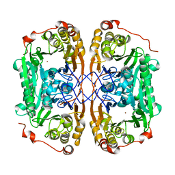
7BYQ
DownloadVisualize
BU of 7byq by Molmil
The mutant variant of PNGM-1. H279A was substituted for alanine to study metal coordination.
分子名称: Metallo-beta-lactamase PNGM-1, ZINC ION
著者Park, Y.S, Kang, L.W, Lee, J.H.
登録日2020-04-24
公開日2021-04-28
最終更新日2023-11-29
実験手法X-RAY DIFFRACTION (1.96 Å)
主引用文献Structural Study of Metal Binding and Coordination in Ancient Metallo-beta-Lactamase PNGM-1 Variants.
Int J Mol Sci, 21, 2020
7BZ1
DownloadVisualize
BU of 7bz1 by Molmil
The mutant variant of PNGM-1. H96 was substituted for alanine to study metal coordination.
分子名称: Metallo-beta-lactamase PNGM-1, ZINC ION
著者Park, Y.S, Kang, L.W, Lee, J.H.
登録日2020-04-26
公開日2021-04-28
最終更新日2023-11-29
実験手法X-RAY DIFFRACTION (2.45 Å)
主引用文献Structural Study of Metal Binding and Coordination in Ancient Metallo-beta-Lactamase PNGM-1 Variants.
Int J Mol Sci, 21, 2020
7BZI
DownloadVisualize
BU of 7bzi by Molmil
The mutant variant of PNGM-1. H91 was substituted for alanine to study metal coordination.
分子名称: Metallo-beta-lactamase PNGM-1, ZINC ION
著者Park, Y.S, Kang, L.W, Lee, J.H.
登録日2020-04-28
公開日2021-04-28
最終更新日2023-11-29
実験手法X-RAY DIFFRACTION (1.94 Å)
主引用文献Structural Study of Metal Binding and Coordination in Ancient Metallo-beta-Lactamase PNGM-1 Variants.
Int J Mol Sci, 21, 2020
7BZ3
DownloadVisualize
BU of 7bz3 by Molmil
The mutant variant of PNGM-1. H257 was substituted for alanine to study substrate binding.
分子名称: Metallo-beta-lactamase PNGM-1, ZINC ION
著者Park, Y.S, Kang, L.W, Lee, J.H.
登録日2020-04-26
公開日2021-04-28
最終更新日2024-03-27
実験手法X-RAY DIFFRACTION (2 Å)
主引用文献Structural Study of Metal Binding and Coordination in Ancient Metallo-beta-Lactamase PNGM-1 Variants.
Int J Mol Sci, 21, 2020
6ILA
DownloadVisualize
BU of 6ila by Molmil
Two Glycerol complexed Crystal structure of fructuronate-tagaturonate epimerase UxaE from Cohnella laeviribosi
分子名称: Fructuronate-tagaturonate epimerase UxaE, GLYCEROL, PHOSPHATE ION, ...
著者Choi, M.Y, Kang, L.W, Ho, T.H, Nguyen, D.Q, Lee, I.H, Lee, J.H, Park, Y.S, Park, H.J.
登録日2018-10-17
公開日2019-10-23
最終更新日2023-11-22
実験手法X-RAY DIFFRACTION (2.69 Å)
主引用文献Crystal structure of fructuronate-tagaturonate epimerase UxaE from Cohnella laeviribosi
To be published
6IKT
DownloadVisualize
BU of 6ikt by Molmil
K1U complex structure of peptide deformylase from Xanthomonas oryzae pv. oryzae
分子名称: (3R)-3-benzyl-4-oxo-4-[(2-oxo-2-phenylethyl)sulfanyl]butanoic acid, CADMIUM ION, NICKEL (II) ION, ...
著者Lee, I.H, Kang, L.W.
登録日2018-10-16
公開日2019-10-23
最終更新日2023-11-22
実験手法X-RAY DIFFRACTION (1.9 Å)
主引用文献K1U complex structure of peptide deformylase from Xanthomonas oryzae pv. oryzae
To be published
6IL0
DownloadVisualize
BU of 6il0 by Molmil
K3U complex structure of peptide deformylase from Xanthomonas oryzae pv. oryzae
分子名称: NICKEL (II) ION, Peptide deformylase, S-(2-oxo-2-phenylethyl) (2R)-2-benzyl-4,4,4-trifluorobutanethioate
著者Lee, I.H, Kang, L.W.
登録日2018-10-16
公開日2019-10-23
最終更新日2023-11-22
実験手法X-RAY DIFFRACTION (1.93 Å)
主引用文献FBIs complex structure of peptide deformylase from Xanthomonas oryzae pv. oryzae
To be published
6IL9
DownloadVisualize
BU of 6il9 by Molmil
One Glycerol complexed Crystal structure of fructuronate-tagaturonate epimerase UxaE from Cohnella laeviribosi
分子名称: Fructuronate-tagaturonate epimerase UxaE from Cohnella laeviribosi in complex with 1 glycerol, GLYCEROL, ZINC ION
著者Choi, M.Y, Kang, L.W, Ho, T.H, Nguyen, D.Q, Lee, I.H, Lee, J.H, Park, Y.S, Park, H.J.
登録日2018-10-17
公開日2019-10-23
最終更新日2023-11-22
実験手法X-RAY DIFFRACTION (1.72005355 Å)
主引用文献Crystal structure of fructuronate-tagaturonate epimerase UxaE from Cohnella laeviribosi
To Be Published
6JFR
DownloadVisualize
BU of 6jfr by Molmil
K3U bound crystal structure of class II peptide deformylase from methicillin resistant Staphylococcus aureus
分子名称: NICKEL (II) ION, Peptide deformylase, S-(2-oxo-2-phenylethyl) (2R)-2-benzyl-4,4,4-trifluorobutanethioate
著者Lee, I.H, Ho, T.H, Kang, L.W.
登録日2019-02-11
公開日2020-02-12
最終更新日2023-11-22
実験手法X-RAY DIFFRACTION (2.4 Å)
主引用文献K3U bound crystal structure of class II peptide deformylase from methicillin resistant Staphylococcus aureus
To be published
6J4N
DownloadVisualize
BU of 6j4n by Molmil
Structure of papua new guinea MBL-1(PNGM-1) native
分子名称: Metallo-beta-lactamases PNGM-1, ZINC ION
著者Hong, M.K, Park, K.S, Jeon, J.H, Lee, J.H, Park, Y.S, Lee, S.H, Kang, L.W.
登録日2019-01-10
公開日2019-02-27
最終更新日2024-03-27
実験手法X-RAY DIFFRACTION (2.1 Å)
主引用文献The novel metallo-beta-lactamase PNGM-1 from a deep-sea sediment metagenome: crystallization and X-ray crystallographic analysis.
Acta Crystallogr F Struct Biol Commun, 74, 2018
6JF8
DownloadVisualize
BU of 6jf8 by Molmil
K4U bound crystal structure of class I type b peptide deformylase from Acinetobacter baumannii
分子名称: L-[(N-HYDROXYAMINO)CARBONYL]PHENYLALANINE, Peptide deformylase, ZINC ION
著者Lee, I.H, Ho, T.H, Kang, L.W.
登録日2019-02-08
公開日2020-02-12
最終更新日2023-11-22
実験手法X-RAY DIFFRACTION (1.7 Å)
主引用文献K4U bound crystal structure of class I type b peptide deformylase from Acinetobacter baumannii
To be published
6JFS
DownloadVisualize
BU of 6jfs by Molmil
K4U bound crystal structure of class II peptide deformylase from methicillin resistant Staphylococcus aureus
分子名称: L-[(N-HYDROXYAMINO)CARBONYL]PHENYLALANINE, NICKEL (II) ION, Peptide deformylase
著者Lee, I.H, Ho, T.H, Kang, L.W.
登録日2019-02-11
公開日2020-02-12
最終更新日2023-11-22
実験手法X-RAY DIFFRACTION (2.25 Å)
主引用文献K4U bound crystal structure of class II peptide deformylase from methicillin resistant Staphylococcus aureus
To be published
6JET
DownloadVisualize
BU of 6jet by Molmil
Actinonin bound crystal structure of class I type a peptide deformylase from Acinetobacter baumannii
分子名称: ACTINONIN, Peptide deformylase, ZINC ION
著者Ho, T.H, Lee, I.H, Kang, L.W.
登録日2019-02-07
公開日2020-02-12
最終更新日2023-11-22
実験手法X-RAY DIFFRACTION (2.6 Å)
主引用文献Actinonin bound crystal structure of class I type a peptide deformylase from Acinetobacter baumannii
To be published
6JF6
DownloadVisualize
BU of 6jf6 by Molmil
Met-ala-ser bound crystal structure of class I type b peptide deformylase from Acinetobacter baumannii
分子名称: MET-ALA-SER, Peptide deformylase, ZINC ION
著者Jung, K.H, Ho, T.H, Lee, I.H, Kang, L.W.
登録日2019-02-08
公開日2020-02-12
最終更新日2023-11-22
実験手法X-RAY DIFFRACTION (2.35 Å)
主引用文献Met-ala-ser bound crystal structure of class I type b peptide deformylase from Acinetobacter baumannii
To be published
6JFO
DownloadVisualize
BU of 6jfo by Molmil
Formyl-Met-Ala-Ser bound crystal structure of class II peptide deformylase from methicillin resistant Staphylococcus aureus
分子名称: FME-ALA-SER, MAGNESIUM ION, Peptide deformylase
著者Lee, I.H, Ho, T.H, Kang, L.W.
登録日2019-02-11
公開日2020-02-12
最終更新日2023-11-22
実験手法X-RAY DIFFRACTION (1.6 Å)
主引用文献Formyl-Met-Ala-Ser bound crystal structure of class II peptide deformylase from methicillin resistant Staphylococcus aureus
To be published
6JF9
DownloadVisualize
BU of 6jf9 by Molmil
Apo crystal structure of class I type b peptide deformylase from Pseudomonas aeruginosa
分子名称: GLYCEROL, NICKEL (II) ION, Peptide deformylase
著者Lee, I.H, Ho, T.H, Kang, L.W.
登録日2019-02-08
公開日2020-02-12
最終更新日2023-11-22
実験手法X-RAY DIFFRACTION (1.7 Å)
主引用文献Apo crystal structure of class I type b peptide deformylase from Pseudomonas aeruginosa
To be published

221051

件を2024-06-12に公開中

PDB statisticsPDBj update infoContact PDBjnumon