1VH8
 
 | |
1VI3
 | |
1VIA
 | |
1VR2
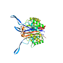
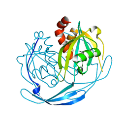
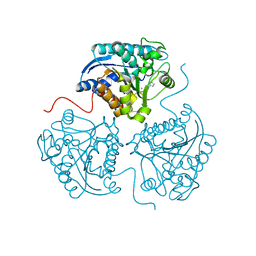
 | | HUMAN VASCULAR ENDOTHELIAL GROWTH FACTOR RECEPTOR 2 (KDR) KINASE DOMAIN | | 分子名称: | PROTEIN (VASCULAR ENDOTHELIAL GROWTH FACTOR RECEPTOR KINASE) | | 著者 | Mctigue, M, Wickersham, J, Pinko, C, Showalter, R, Parast, C, Tempczyk-Russell, A, Gehring, M, Mroczkowski, B, Kan, C, Villafranca, J, Appelt, K. | | 登録日 | 1998-12-03 | | 公開日 | 2000-03-15 | | 最終更新日 | 2023-11-15 | | 実験手法 | X-RAY DIFFRACTION (2.4 Å) | | 主引用文献 | Crystal structure of the kinase domain of human vascular endothelial growth factor receptor 2: a key enzyme in angiogenesis.
Structure Fold.Des., 7, 1999
|
|
1VIQ
 | |
3OHH
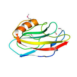
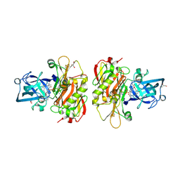 | | Crystal structure of beta-site app-cleaving enzyme 1 (bace-wt) complex with bms-681889 aka n~1~-butyl-5-cyano- n~3~-((1s,2r)-1-(3,5-difluorobenzyl)-2-hydroxy-3-((3- methoxybenzyl)amino)propyl)-n~1~-methyl-1h-indole-1,3- dicarboxamide | | 分子名称: | Beta-secretase 1, GLYCEROL, N~1~-butyl-5-cyano-N~3~-{(1S,2R)-1-(3,5-difluorobenzyl)-2-hydroxy-3-[(3-methoxybenzyl)amino]propyl}-N~1~-methyl-1H-indole-1,3-dicarboxamide, ... | | 著者 | Muckelbauer, J.K. | | 登録日 | 2010-08-17 | | 公開日 | 2011-04-06 | | 最終更新日 | 2017-11-08 | | 実験手法 | X-RAY DIFFRACTION (2.01 Å) | | 主引用文献 | Synthesis and SAR of indole-and 7-azaindole-1,3-dicarboxamide hydroxyethylamine inhibitors of BACE-1.
Bioorg.Med.Chem.Lett., 21, 2011
|
|
1HTI
 | |
1VH4
 | |
1VHO
 | |
1VHY
 | |
1VIY
 | |
1VER
 | |
1VH7
 | |
1VHF
 | |
1VHZ
 | |
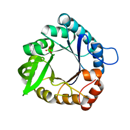
1VIS
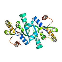
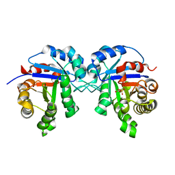
 | | Crystal structure of mevalonate kinase | | 分子名称: | 1,4-DIETHYLENE DIOXIDE, Mevalonate kinase | | 著者 | Structural GenomiX | | 登録日 | 2003-12-01 | | 公開日 | 2003-12-30 | | 最終更新日 | 2023-12-27 | | 実験手法 | X-RAY DIFFRACTION (2.69 Å) | | 主引用文献 | Structural analysis of a set of proteins resulting from a bacterial genomics project
Proteins, 60, 2005
|
|
1VIZ
 | |
1VHA
 | |
1VHU
 | |
1VI5
 | |
1GP2
 | |
3M0C
 | |
3MJL
 | |
1VHG
 | |
1VHT
 | |


