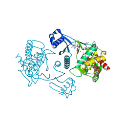
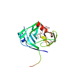
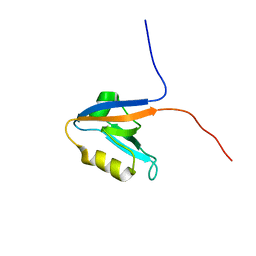
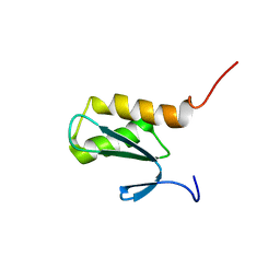
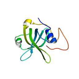
3EO3
 
 | | Crystal structure of the N-acetylmannosamine kinase domain of human GNE protein | | 分子名称: | Bifunctional UDP-N-acetylglucosamine 2-epimerase/N-acetylmannosamine kinase, UNKNOWN ATOM OR ION, ZINC ION | | 著者 | Nedyalkova, L, Tong, Y, Rabeh, W.M, Hong, B, Tempel, W, MacKenzie, F, Arrowsmith, C.H, Edwards, A.M, Bountra, C, Weigelt, J, Bochkarev, A, Park, H, Structural Genomics Consortium (SGC) | | 登録日 | 2008-09-26 | | 公開日 | 2008-10-07 | | 最終更新日 | 2024-02-21 | | 実験手法 | X-RAY DIFFRACTION (2.84 Å) | | 主引用文献 | Crystal structure of the N-acetylmannosamine kinase domain of GNE.
Plos One, 4, 2009
|
|
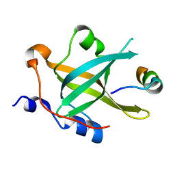
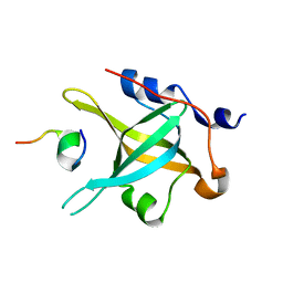
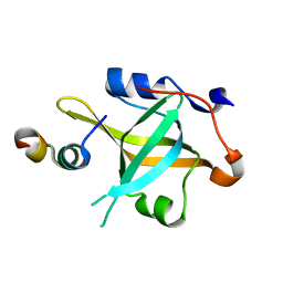
3FEG
 | | Crystal structure of human choline kinase beta in complex with phosphorylated hemicholinium-3 and adenosine nucleotide | | 分子名称: | (2S)-2-[4'-({dimethyl[2-(phosphonooxy)ethyl]ammonio}acetyl)biphenyl-4-yl]-2-hydroxy-4,4-dimethylmorpholin-4-ium, ADENOSINE MONOPHOSPHATE, ADENOSINE-5'-DIPHOSPHATE, ... | | 著者 | Hong, B.S, Tempel, W, Rabeh, W.M, MacKenzie, F, Arrowsmith, C.H, Edwards, A.M, Bountra, C, Weigelt, J, Bochkarev, A, Park, H.W, Structural Genomics Consortium (SGC) | | 登録日 | 2008-11-29 | | 公開日 | 2008-12-23 | | 最終更新日 | 2023-09-06 | | 実験手法 | X-RAY DIFFRACTION (1.302 Å) | | 主引用文献 | Crystal structures of human choline kinase isoforms in complex with hemicholinium-3: single amino acid near the active site influences inhibitor sensitivity.
J.Biol.Chem., 285, 2010
|
|
6KE4
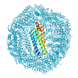 | | ABloop reengineered Ferritin Nanocage | | 分子名称: | CALCIUM ION, CHLORIDE ION, FE (III) ION, ... | | 著者 | Wang, W.M, Wang, H.F. | | 登録日 | 2019-07-03 | | 公開日 | 2019-10-23 | | 最終更新日 | 2023-11-22 | | 実験手法 | X-RAY DIFFRACTION (2.3 Å) | | 主引用文献 | AB loop engineered ferritin nanocages for drug loading under benign experimental conditions.
Chem.Commun.(Camb.), 55, 2019
|
|
6KE2
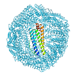 | | ABloop reengineered Ferritin Nanocage | | 分子名称: | CALCIUM ION, CHLORIDE ION, FE (III) ION, ... | | 著者 | Wang, W.M, Wang, H.F. | | 登録日 | 2019-07-03 | | 公開日 | 2019-10-23 | | 最終更新日 | 2023-11-22 | | 実験手法 | X-RAY DIFFRACTION (1.798 Å) | | 主引用文献 | AB loop engineered ferritin nanocages for drug loading under benign experimental conditions.
Chem.Commun.(Camb.), 55, 2019
|
|
6IVE
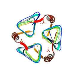 | |
2GVF
 | | HCV NS3-4A protease domain complexed with a macrocyclic ketoamide inhibitor, SCH419021 | | 分子名称: | (6R,8S,11S)-11-CYCLOHEXYL-N-(1-{[(2-{[(1S)-2-(DIMETHYLAMINO)-2-OXO-1-PHENYLETHYL]AMINO}-2-OXOETHYL)AMINO](OXO)ACETYL}BUTYL)-10,13-DIOXO-2,5-DIOXA-9,12-DIAZATRICYCLO[14.3.1.1~6,9~]HENICOSA-1(20),16,18-TRIENE-8-CARBOXAMIDE, Polyprotein, ZINC ION, ... | | 著者 | Arasappan, A, Njoroge, F.G, Chen, K.X, Venkatraman, S, Parekh, T.N, Gu, H, Pichardo, J, Butkiewicz, N, Prongay, A, Madison, V, Girijavallabhan, V. | | 登録日 | 2006-05-02 | | 公開日 | 2007-01-23 | | 最終更新日 | 2024-11-20 | | 実験手法 | X-RAY DIFFRACTION (2.5 Å) | | 主引用文献 | P2-P4 macrocyclic inhibitors of hepatitis C virus NS3-4A serine protease.
Bioorg.Med.Chem.Lett., 16, 2006
|
|
2H6E
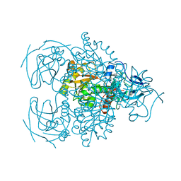 | | Crystal structure of the D-arabinose dehydrogenase from Sulfolobus solfataricus | | 分子名称: | D-arabinose 1-dehydrogenase, ZINC ION | | 著者 | Brouns, S.J.J, Turnbull, A.P, Akerboom, J, Willemen, H.L.D.M, De Vos, W.M, Van der Oost, J. | | 登録日 | 2006-05-31 | | 公開日 | 2007-06-05 | | 最終更新日 | 2024-02-14 | | 実験手法 | X-RAY DIFFRACTION (1.8 Å) | | 主引用文献 | Crystal Structure and Biochemical Properties of the d-Arabinose Dehydrogenase from Sulfolobus solfataricus
J.Mol.Biol., 371, 2007
|
|
2HBS
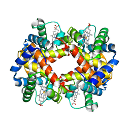 | | THE HIGH RESOLUTION CRYSTAL STRUCTURE OF DEOXYHEMOGLOBIN S | | 分子名称: | HEMOGLOBIN S (DEOXY), ALPHA CHAIN, BETA CHAIN, ... | | 著者 | Harrington, D.J, Adachi, K, Royer Junior, W.E. | | 登録日 | 1997-05-06 | | 公開日 | 1997-07-23 | | 最終更新日 | 2024-02-14 | | 実験手法 | X-RAY DIFFRACTION (2.05 Å) | | 主引用文献 | The high resolution crystal structure of deoxyhemoglobin S.
J.Mol.Biol., 272, 1997
|
|
2K8C
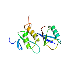 | | Solution structure of PLAA family ubiquitin binding domain (PFUC) trans isomer in complex with ubiquitin | | 分子名称: | Phospholipase A-2-activating protein, Ubiquitin | | 著者 | Fu, Q.S, Zhou, C.J, Gao, H.C, Lin, D.H, Hu, H.Y. | | 登録日 | 2008-09-04 | | 公開日 | 2009-05-05 | | 最終更新日 | 2024-05-22 | | 実験手法 | SOLUTION NMR | | 主引用文献 | Structural Basis for Ubiquitin Recognition by a Novel Domain from Human Phospholipase A2-activating Protein.
J.Biol.Chem., 284, 2009
|
|
2KOJ
 | |
7XV4
 | | Crystal structure of RPA70N-ATRIP fusion | | 分子名称: | ATR-interacting protein, Replication protein A 70 kDa DNA-binding subunit | | 著者 | Wu, Y.Y, Zang, N, Fu, W.M, Zhou, C. | | 登録日 | 2022-05-20 | | 公開日 | 2023-06-14 | | 最終更新日 | 2024-05-08 | | 実験手法 | X-RAY DIFFRACTION (1.6 Å) | | 主引用文献 | Structural characterization of human RPA70N association with DNA damage response proteins.
Elife, 12, 2023
|
|
7XV1
 | | Crystal structure of RPA70N-HelB fusion | | 分子名称: | DNA helicase B, Replication protein A 70 kDa DNA-binding subunit | | 著者 | Wu, Y.Y, Zang, N, Fu, W.M, Zhou, C. | | 登録日 | 2022-05-20 | | 公開日 | 2023-06-14 | | 最終更新日 | 2024-05-08 | | 実験手法 | X-RAY DIFFRACTION (1.8 Å) | | 主引用文献 | Structural characterization of human RPA70N association with DNA damage response proteins.
Elife, 12, 2023
|
|
7XUT
 | | Crystal structure of RPA70N-WRN fusion | | 分子名称: | fusion protein of Replication protein A 70 kDa DNA-binding subunit and Werner syndrome ATP-dependent helicase | | 著者 | Wu, Y.Y, Zang, N, Fu, W.M, Zhou, C. | | 登録日 | 2022-05-19 | | 公開日 | 2023-06-14 | | 最終更新日 | 2024-05-08 | | 実験手法 | X-RAY DIFFRACTION (1.6 Å) | | 主引用文献 | Structural characterization of human RPA70N association with DNA damage response proteins.
Elife, 12, 2023
|
|
2K89
 | | Solution structure of a novel Ubiquitin-binding domain from Human PLAA (PFUC, Gly76-Pro77 cis isomer) | | 分子名称: | Phospholipase A-2-activating protein | | 著者 | Fu, Q.S, Zhou, C.J, Gao, H.C, Lin, D.H, Hu, H.Y. | | 登録日 | 2008-09-04 | | 公開日 | 2009-05-05 | | 最終更新日 | 2024-05-22 | | 実験手法 | SOLUTION NMR | | 主引用文献 | Structural Basis for Ubiquitin Recognition by a Novel Domain from Human Phospholipase A2-activating Protein.
J.Biol.Chem., 284, 2009
|
|
7XV0
 | | Crystal structure of RPA70N-BLMp1 fusion | | 分子名称: | Bloom syndrome protein, Replication protein A 70 kDa DNA-binding subunit | | 著者 | Wu, Y.Y, Zang, N, Fu, W.M, Zhou, C. | | 登録日 | 2022-05-20 | | 公開日 | 2023-06-14 | | 最終更新日 | 2024-05-08 | | 実験手法 | X-RAY DIFFRACTION (1.5 Å) | | 主引用文献 | Structural characterization of human RPA70N association with DNA damage response proteins.
Elife, 12, 2023
|
|
2K8B
 | | Solution structure of PLAA family ubiquitin binding domain (PFUC) cis isomer in complex with ubiquitin | | 分子名称: | Phospholipase A-2-activating protein, Ubiquitin | | 著者 | Fu, Q.S, Zhou, C.J, Gao, H.C, Lin, D.H, Hu, H.Y. | | 登録日 | 2008-09-04 | | 公開日 | 2009-05-05 | | 最終更新日 | 2024-05-22 | | 実験手法 | SOLUTION NMR | | 主引用文献 | Structural Basis for Ubiquitin Recognition by a Novel Domain from Human Phospholipase A2-activating Protein.
J.Biol.Chem., 284, 2009
|
|
5DNK
 | | The structure of PKMT1 from Rickettsia prowazekii in complex with AdoHcy | | 分子名称: | S-ADENOSYL-L-HOMOCYSTEINE, protein lysine methyltransferase 1 | | 著者 | Noinaj, N, Abeykoon, A, He, Y, Yang, D.C, Buchanan, S.K. | | 登録日 | 2015-09-10 | | 公開日 | 2016-08-10 | | 最終更新日 | 2024-03-06 | | 実験手法 | X-RAY DIFFRACTION (1.9 Å) | | 主引用文献 | Structural Insights into Substrate Recognition and Catalysis in Outer Membrane Protein B (OmpB) by Protein-lysine Methyltransferases from Rickettsia.
J.Biol.Chem., 291, 2016
|
|
7XUW
 | | Crystal structure of RPA70N-BLMp2 fusion | | 分子名称: | fusion protein of Replication protein A 70 kDa DNA-binding subunit and Bloom syndrome protein | | 著者 | Wu, Y.Y, Zang, N, Fu, W.M, Zhou, C. | | 登録日 | 2022-05-20 | | 公開日 | 2023-06-14 | | 最終更新日 | 2024-05-08 | | 実験手法 | X-RAY DIFFRACTION (1.8 Å) | | 主引用文献 | Structural characterization of human RPA70N association with DNA damage response proteins.
Elife, 12, 2023
|
|
7XUV
 | | Crystal structure of RPA70N-RMI1 fusion | | 分子名称: | RecQ-mediated genome instability protein 1, Replication protein A 70 kDa DNA-binding subunit | | 著者 | Wu, Y.Y, Zang, N, Fu, W.M, Zhou, C. | | 登録日 | 2022-05-20 | | 公開日 | 2023-06-14 | | 最終更新日 | 2024-05-08 | | 実験手法 | X-RAY DIFFRACTION (1.6 Å) | | 主引用文献 | Structural characterization of human RPA70N association with DNA damage response proteins.
Elife, 12, 2023
|
|
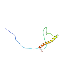
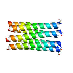
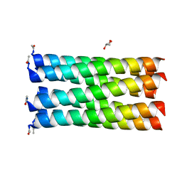
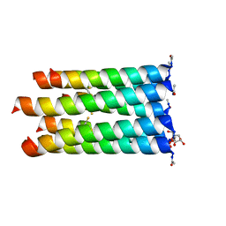
2LNQ
 | | 40-residue D23N beta amyloid fibril | | 分子名称: | P3(40) | | 著者 | Qiang, W, Yau, W, Luo, Y, Mattson, M.P, Tycko, R. | | 登録日 | 2012-01-03 | | 公開日 | 2012-02-08 | | 最終更新日 | 2024-05-15 | | 実験手法 | SOLID-STATE NMR | | 主引用文献 | Antiparallel beta-sheet architecture in Iowa-mutant beta-amyloid fibrils.
Proc.Natl.Acad.Sci.USA, 109, 2012
|
|
2LMQ
 | |
2LJL
 | |
5EZ8
 | |
5EZA
 | |
5EZE
 | |


