3EI1
 
 | |
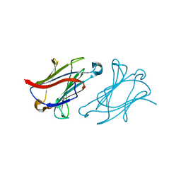
2DQE
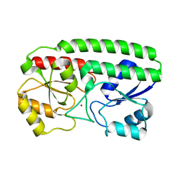
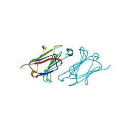
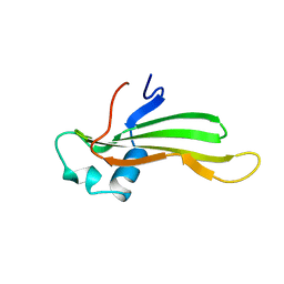
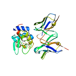 | | Crystal structure of hyhel-10 FV mutant (Hy53a) complexed with hen egg lysozyme | | 分子名称: | Ig VH,anti-lysozyme, Lysozyme C, lysozyme binding Ig kappa chain V23-J2 region | | 著者 | Shiroishi, M, Kondo, H, Tsumoto, K, Kumagai, I. | | 登録日 | 2006-05-25 | | 公開日 | 2007-01-23 | | 最終更新日 | 2023-10-25 | | 実験手法 | X-RAY DIFFRACTION (1.9 Å) | | 主引用文献 | Structural consequences of mutations in interfacial Tyr residues of a protein antigen-antibody complex. The case of HyHEL-10-HEL
J.Biol.Chem., 282, 2007
|
|
2DQJ
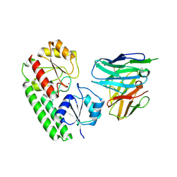
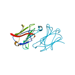
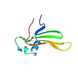
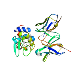 | | Crystal structure of hyhel-10 FV (wild-type) complexed with hen egg lysozyme at 1.8A resolution | | 分子名称: | Ig VH,anti-lysozyme, Lysozyme C, lysozyme binding Ig kappa chain V23-J2 region | | 著者 | Shiroishi, M, Kondo, H, Tsumoto, K, Kumagai, I. | | 登録日 | 2006-05-26 | | 公開日 | 2007-01-23 | | 最終更新日 | 2023-10-25 | | 実験手法 | X-RAY DIFFRACTION (1.8 Å) | | 主引用文献 | Structural consequences of mutations in interfacial Tyr residues of a protein antigen-antibody complex. The case of HyHEL-10-HEL
J.Biol.Chem., 282, 2007
|
|
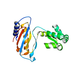
8YJ8
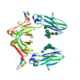
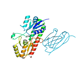 | | Characerization of a novel format scFvXVHH single-chain Biparatopic antibody against a metal binding protein, MtsA | | 分子名称: | Iron ABC transporter substrate-binding lipoprotein MtsA, VHH43, ZINC ION | | 著者 | Ito, S, Nagatoishi, S, Nakakido, M, Tsumoto, K. | | 登録日 | 2024-03-01 | | 公開日 | 2024-06-19 | | 最終更新日 | 2024-06-26 | | 実験手法 | X-RAY DIFFRACTION (1.65 Å) | | 主引用文献 | Characterization of a novel format scFv×VHH single-chain biparatopic antibody against metal binding protein MtsA.
Protein Sci., 33, 2024
|
|
8YJ5
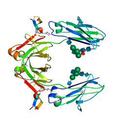
 | | Characerization of a novel format scFvXVHH single-chain Biparatopic antibody against a metal binding protein, MtsA | | 分子名称: | Iron ABC transporter substrate-binding lipoprotein MtsA, VHH43 | | 著者 | Ito, S, Nagatoishi, S, Nakakido, M, Tsumoto, K. | | 登録日 | 2024-03-01 | | 公開日 | 2024-06-19 | | 実験手法 | X-RAY DIFFRACTION (3.66 Å) | | 主引用文献 | Characterization of a novel format scFv×VHH single-chain biparatopic antibody against metal binding protein MtsA.
Protein Sci., 33, 2024
|
|
8YJ6
 | | Characerization of a novel format scFvXVHH single-chain Biparatopic antibody against a metal binding protein, MtsA | | 分子名称: | Iron ABC transporter substrate-binding lipoprotein MtsA, ZINC ION | | 著者 | Ito, S, Nagatoishi, S, Nakakido, M, Tsumoto, K. | | 登録日 | 2024-03-01 | | 公開日 | 2024-06-19 | | 実験手法 | X-RAY DIFFRACTION (1.37 Å) | | 主引用文献 | Characterization of a novel format scFv×VHH single-chain biparatopic antibody against metal binding protein MtsA.
Protein Sci., 33, 2024
|
|
8YJ7
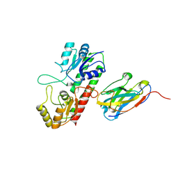
 | | Characerization of a novel format scFvXVHH single-chain Biparatopic antibody against a metal binding protein, MtsA | | 分子名称: | Iron ABC transporter substrate-binding lipoprotein MtsA, ZINC ION, scFv13 | | 著者 | Ito, S, Nagatoishi, S, Nakakido, M, Tsumoto, K. | | 登録日 | 2024-03-01 | | 公開日 | 2024-06-19 | | 最終更新日 | 2024-06-26 | | 実験手法 | X-RAY DIFFRACTION (2.8 Å) | | 主引用文献 | Characterization of a novel format scFv×VHH single-chain biparatopic antibody against metal binding protein MtsA.
Protein Sci., 33, 2024
|
|
6IQG
 | | X-ray crystal structure of Fc and peptide complex | | 分子名称: | 18-mer peptide G(HCS)DCAYHRGELVWCT(HCS)H(NH2), 2-acetamido-2-deoxy-beta-D-glucopyranose-(1-2)-alpha-D-mannopyranose-(1-3)-[2-acetamido-2-deoxy-beta-D-glucopyranose-(1-2)-alpha-D-mannopyranose-(1-6)]beta-D-mannopyranose-(1-4)-2-acetamido-2-deoxy-beta-D-glucopyranose-(1-4)-[alpha-L-fucopyranose-(1-6)]2-acetamido-2-deoxy-beta-D-glucopyranose, CHLORIDE ION, ... | | 著者 | Adachi, M, Ito, Y. | | 登録日 | 2018-11-08 | | 公開日 | 2019-02-13 | | 最終更新日 | 2024-03-13 | | 実験手法 | X-RAY DIFFRACTION (2.998 Å) | | 主引用文献 | Site-Specific Chemical Conjugation of Antibodies by Using Affinity Peptide for the Development of Therapeutic Antibody Format.
Bioconjug. Chem., 30, 2019
|
|
6IQH
 | | X-ray crystal structure of covalent-bonded complex of Fc and peptide | | 分子名称: | 17-mer peptide (GPDCAYHKGELVWCTFH), 2-acetamido-2-deoxy-beta-D-glucopyranose-(1-2)-alpha-D-mannopyranose-(1-3)-[2-acetamido-2-deoxy-beta-D-glucopyranose-(1-2)-alpha-D-mannopyranose-(1-6)]beta-D-mannopyranose-(1-4)-2-acetamido-2-deoxy-beta-D-glucopyranose-(1-4)-[alpha-L-fucopyranose-(1-6)]2-acetamido-2-deoxy-beta-D-glucopyranose, Immunoglobulin gamma-1 heavy chain | | 著者 | Adachi, M, Ito, Y. | | 登録日 | 2018-11-08 | | 公開日 | 2019-02-13 | | 最終更新日 | 2024-01-24 | | 実験手法 | X-RAY DIFFRACTION (2.999 Å) | | 主引用文献 | Site-Specific Chemical Conjugation of Antibodies by Using Affinity Peptide for the Development of Therapeutic Antibody Format.
Bioconjug. Chem., 30, 2019
|
|
7C24
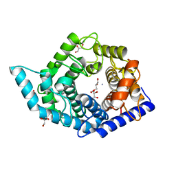 | | Glycosidase F290Y at pH8.0 | | 分子名称: | AMMONIUM ION, GLYCEROL, Isomaltose glucohydrolase | | 著者 | Tagami, T, Kikuchi, A, Okuyama, M, Kimura, A. | | 登録日 | 2020-05-07 | | 公開日 | 2021-05-12 | | 最終更新日 | 2023-11-29 | | 実験手法 | X-RAY DIFFRACTION (1.71 Å) | | 主引用文献 | Structural insights reveal the second base catalyst of isomaltose glucohydrolase.
Febs J., 289, 2022
|
|
7C25
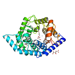 | | Glycosidase Wild Type at pH8.0 | | 分子名称: | AMMONIUM ION, CITRIC ACID, GLYCEROL, ... | | 著者 | Tagami, T, Kikuchi, A, Okuyama, M, Kimura, A. | | 登録日 | 2020-05-07 | | 公開日 | 2021-05-12 | | 最終更新日 | 2023-11-29 | | 実験手法 | X-RAY DIFFRACTION (1.505 Å) | | 主引用文献 | Structural insights reveal the second base catalyst of isomaltose glucohydrolase.
Febs J., 289, 2022
|
|
7C26
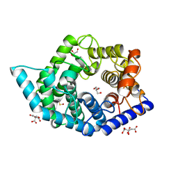 | | Glycosidase Wild Type at pH4.5 | | 分子名称: | AMMONIUM ION, CITRIC ACID, GLYCEROL, ... | | 著者 | Tagami, T, Kikuchi, A, Okuyama, M, Kimura, A. | | 登録日 | 2020-05-07 | | 公開日 | 2021-05-12 | | 最終更新日 | 2023-11-29 | | 実験手法 | X-RAY DIFFRACTION (1.803 Å) | | 主引用文献 | Structural insights reveal the second base catalyst of isomaltose glucohydrolase.
Febs J., 289, 2022
|
|
7C27
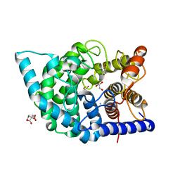 | | Glycosidase F290Y at pH4.5 | | 分子名称: | AMMONIUM ION, CITRIC ACID, GLYCEROL, ... | | 著者 | Tagami, T, Kikuchi, A, Okuyama, M, Kimura, A. | | 登録日 | 2020-05-07 | | 公開日 | 2021-05-12 | | 最終更新日 | 2023-11-29 | | 実験手法 | X-RAY DIFFRACTION (1.91 Å) | | 主引用文献 | Structural insights reveal the second base catalyst of isomaltose glucohydrolase.
Febs J., 289, 2022
|
|
6L67
 | |
6L6D
 | |
6L6B
 | | X-ray structure of human galectin-10 in complex with L-fucose | | 分子名称: | Galectin-10, beta-L-fucopyranose | | 著者 | Kamitori, S. | | 登録日 | 2019-10-28 | | 公開日 | 2020-03-04 | | 最終更新日 | 2023-11-22 | | 実験手法 | X-RAY DIFFRACTION (1.802 Å) | | 主引用文献 | Structures of human galectin-10/monosaccharide complexes demonstrate potential of monosaccharides as effectors in forming Charcot-Leyden crystals.
Biochem.Biophys.Res.Commun., 2020
|
|
6L6C
 | |
6L64
 | | X-ray structure of human galectin-10 in complex with D-glucose | | 分子名称: | Galectin-10, beta-D-glucopyranose | | 著者 | Kamitori, S. | | 登録日 | 2019-10-28 | | 公開日 | 2020-03-04 | | 最終更新日 | 2023-11-22 | | 実験手法 | X-RAY DIFFRACTION (2.08 Å) | | 主引用文献 | Structures of human galectin-10/monosaccharide complexes demonstrate potential of monosaccharides as effectors in forming Charcot-Leyden crystals.
Biochem.Biophys.Res.Commun., 2020
|
|
6L6A
 | | X-ray structure of human galectin-10 in complex with D-mannose | | 分子名称: | Galectin-10, beta-D-mannopyranose | | 著者 | Kamitori, S. | | 登録日 | 2019-10-28 | | 公開日 | 2020-03-04 | | 最終更新日 | 2023-11-22 | | 実験手法 | X-RAY DIFFRACTION (1.81 Å) | | 主引用文献 | Structures of human galectin-10/monosaccharide complexes demonstrate potential of monosaccharides as effectors in forming Charcot-Leyden crystals.
Biochem.Biophys.Res.Commun., 2020
|
|
1DWU
 | | Ribosomal protein L1 | | 分子名称: | RIBOSOMAL PROTEIN L1 | | 著者 | Tishchenko, S.V, Nevskaya, N.A, Pavelyev, M.N, Nikonov, S.V, Garber, M.B, Piendl, W. | | 登録日 | 1999-12-13 | | 公開日 | 2000-12-07 | | 最終更新日 | 2023-12-06 | | 実験手法 | X-RAY DIFFRACTION (2.8 Å) | | 主引用文献 | Structure of Ribosomal Protein L1 from Methanococcus Thermolithotrophicus. Functionally Important Structural Invariants on the L1 Surface
Acta Crystallogr.,Sect.D, 58, 2002
|
|
1CYV
 | | SOLUTION NMR STRUCTURE OF RECOMBINANT HUMAN CYSTATIN A UNDER THE CONDITION OF PH 3.8 AND 310K | | 分子名称: | CYSTATIN A | | 著者 | Tate, S, Tate, N.U, Ushioda, T, Samejima, T, Kainosho, M. | | 登録日 | 1995-08-24 | | 公開日 | 1995-12-07 | | 最終更新日 | 2024-05-22 | | 実験手法 | SOLUTION NMR | | 主引用文献 | Solution structure of a human cystatin A variant, cystatin A2-98 M65L, by NMR spectroscopy. A possible role of the interactions between the N- and C-termini to maintain the inhibitory active form of cystatin A.
Biochemistry, 34, 1995
|
|
1CYU
 | | SOLUTION NMR STRUCTURE OF RECOMBINANT HUMAN CYSTATIN A UNDER THE CONDITION OF PH 3.8 AND 310K | | 分子名称: | CYSTATIN A | | 著者 | Tate, S, Tate, N.U, Ushioda, T, Samejima, T, Kainosho, M. | | 登録日 | 1995-08-24 | | 公開日 | 1995-12-07 | | 最終更新日 | 2024-05-22 | | 実験手法 | SOLUTION NMR | | 主引用文献 | Solution structure of a human cystatin A variant, cystatin A2-98 M65L, by NMR spectroscopy. A possible role of the interactions between the N- and C-termini to maintain the inhibitory active form of cystatin A.
Biochemistry, 34, 1995
|
|
6LM0
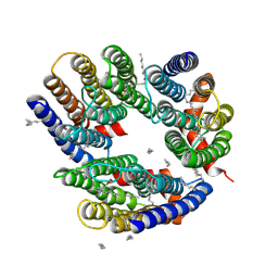 | | The crystal structure of cyanorhodopsin (CyR) N2098R from cyanobacteria Calothrix sp. NIES-2098 | | 分子名称: | DECANE, HEXANE, N-OCTANE, ... | | 著者 | Hosaka, T, Kimura-Someya, T, Shirouzu, M. | | 登録日 | 2019-12-24 | | 公開日 | 2020-10-21 | | 最終更新日 | 2023-11-22 | | 実験手法 | X-RAY DIFFRACTION (2.65 Å) | | 主引用文献 | A unique clade of light-driven proton-pumping rhodopsins evolved in the cyanobacterial lineage.
Sci Rep, 10, 2020
|
|
5F9E
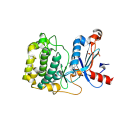 | | Structure of Protein Kinase C theta with compound 10: 2,2-dimethyl-7-(2-oxidanylidene-3~{H}-imidazo[4,5-b]pyridin-1-yl)-1-(phenylmethyl)-3~{H}-quinazolin-4-one | | 分子名称: | 2,2-dimethyl-7-(2-oxidanylidene-3~{H}-imidazo[4,5-b]pyridin-1-yl)-1-(phenylmethyl)-3~{H}-quinazolin-4-one, Protein kinase C theta type | | 著者 | Klein, M. | | 登録日 | 2015-12-09 | | 公開日 | 2016-05-11 | | 実験手法 | X-RAY DIFFRACTION (2 Å) | | 主引用文献 | Discovery and optimization of 1,7-disubstituted-2,2-dimethyl-2,3-dihydroquinazolin-4(1H)-ones as potent and selective PKC theta inhibitors.
Bioorg.Med.Chem., 24, 2016
|
|
6LM1
 | | The crystal structure of cyanorhodopsin (CyR) N4075R from cyanobacteria Tolypothrix sp. NIES-4075 | | 分子名称: | DECANE, DODECANE, HEXADECANE, ... | | 著者 | Hosaka, T, Kimura-Someya, T, Shirouzu, M. | | 登録日 | 2019-12-24 | | 公開日 | 2020-10-21 | | 最終更新日 | 2023-11-22 | | 実験手法 | X-RAY DIFFRACTION (1.9 Å) | | 主引用文献 | A unique clade of light-driven proton-pumping rhodopsins evolved in the cyanobacterial lineage.
Sci Rep, 10, 2020
|
|


