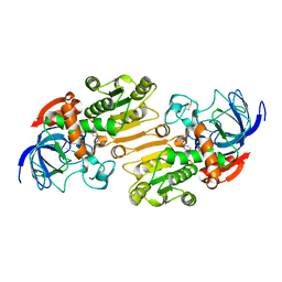
3HT1
 
 | |
3HT2
 | |
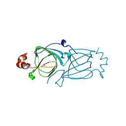
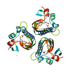
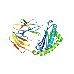
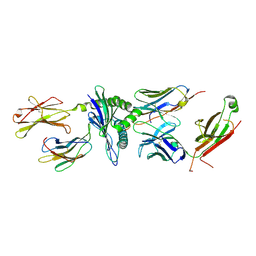
2OYA
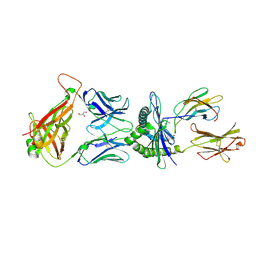
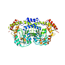
 | | Crystal structure analysis of the dimeric form of the SRCR domain of mouse MARCO | | 分子名称: | Macrophage receptor MARCO, SULFATE ION | | 著者 | Ojala, J.R.M, Pikkarainen, T, Tuuttila, A, Sandalova, T, Tryggvason, K. | | 登録日 | 2007-02-21 | | 公開日 | 2007-04-17 | | 最終更新日 | 2023-08-30 | | 実験手法 | X-RAY DIFFRACTION (1.77 Å) | | 主引用文献 | Crystal structure of the cysteine-rich domain of scavenger receptor MARCO reveals the presence of a basic and an acidic cluster that both contribute to ligand recognition.
J.Biol.Chem., 282, 2007
|
|
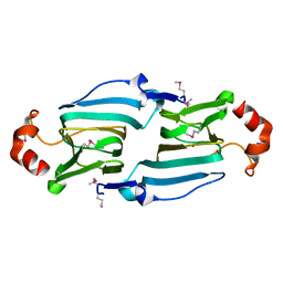
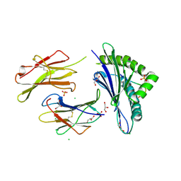
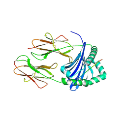
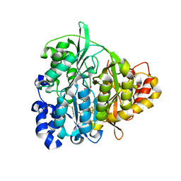
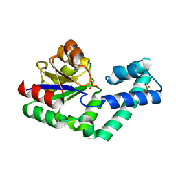
2OY3
 | | Crystal structure analysis of the monomeric SRCR domain of mouse MARCO | | 分子名称: | MAGNESIUM ION, Macrophage receptor MARCO | | 著者 | Ojala, J.R.M, Pikkarainen, T, Tuuttila, A, Sandalova, T, Tryggvason, K. | | 登録日 | 2007-02-21 | | 公開日 | 2007-04-17 | | 最終更新日 | 2023-08-30 | | 実験手法 | X-RAY DIFFRACTION (1.78 Å) | | 主引用文献 | Crystal structure of the cysteine-rich domain of scavenger receptor MARCO reveals the presence of a basic and an acidic cluster that both contribute to ligand recognition.
J.Biol.Chem., 282, 2007
|
|
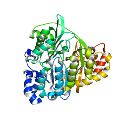
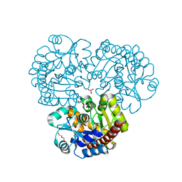
5STD
 | | SCYTALONE DEHYDRATASE PLUS INHIBITOR 2 | | 分子名称: | (6,7-DIFLUORO-QUINAZOLIN-4-YL)-(1-METHYL-2,2-DIPHENYL-ETHYL)-AMINE, CALCIUM ION, Scytalone dehydratase | | 著者 | Wawrzak, Z, Sandalova, T, Steffens, J.J, Basarab, G.S, Lundqvist, T, Lindqvist, Y, Jordan, D.B. | | 登録日 | 1999-02-10 | | 公開日 | 1999-12-29 | | 最終更新日 | 2023-09-20 | | 実験手法 | X-RAY DIFFRACTION (1.95 Å) | | 主引用文献 | High-resolution structures of scytalone dehydratase-inhibitor complexes crystallized at physiological pH.
Proteins, 35, 1999
|
|
6H6D
 | |
5JLZ
 | | Crystal structure of HLA-DRB1*04:01 in complex with modified alpha-enolase peptide 26-40 with citrulline at the position 32 | | 分子名称: | Alpha-enolase, HLA class II histocompatibility antigen, DR alpha chain, ... | | 著者 | Dubnovitsky, A, Kozhukh, G, Sandalova, T, Achour, A. | | 登録日 | 2016-04-28 | | 公開日 | 2016-12-07 | | 最終更新日 | 2024-01-10 | | 実験手法 | X-RAY DIFFRACTION (1.99 Å) | | 主引用文献 | Functional and Structural Characterization of a Novel HLA-DRB1*04:01-Restricted alpha-Enolase T Cell Epitope in Rheumatoid Arthritis.
Front Immunol, 7, 2016
|
|
5JWE
 | | Crystal structure of H-2Db in complex with the LCMV-derived GP92-101 peptide | | 分子名称: | Beta-2-microglobulin, GLYCEROL, H-2 class I histocompatibility antigen, ... | | 著者 | Buratto, J, Badia-Martinez, D, Norstrom, M, Sandalova, T, Achour, A. | | 登録日 | 2016-05-12 | | 公開日 | 2017-05-24 | | 最終更新日 | 2024-01-10 | | 実験手法 | X-RAY DIFFRACTION (2.4 Å) | | 主引用文献 | Crystal structures of H-2Db in complex with the LCMV-derived peptides GP92 and GP392 explain pleiotropic effects of glycosylation on antigen presentation and immunogenicity.
PLoS ONE, 12, 2017
|
|
5JWD
 | | Crystal structure of H-2Db in complex with the LCMV-derived GP392-401 peptide | | 分子名称: | Beta-2-microglobulin, GLYCEROL, H-2 class I histocompatibility antigen, ... | | 著者 | Buratto, J, Badia-Martinez, D, Norstrom, M, Sandalova, T, Achour, A. | | 登録日 | 2016-05-12 | | 公開日 | 2017-05-24 | | 最終更新日 | 2024-01-10 | | 実験手法 | X-RAY DIFFRACTION (2.5 Å) | | 主引用文献 | Crystal structures of H-2Db in complex with the LCMV-derived peptides GP92 and GP392 explain pleiotropic effects of glycosylation on antigen presentation and immunogenicity.
PLoS ONE, 12, 2017
|
|
5LAX
 | | Crystal structure of HLA_DRB1*04:01 in complex with alpha-enolase peptide 26-40 | | 分子名称: | HLA class II histocompatibility antigen, DR alpha chain, DRB1-4 beta chain, ... | | 著者 | Dubnovitsky, A, Kozhukh, G, Sandalova, T, Achour, A. | | 登録日 | 2016-06-15 | | 公開日 | 2016-12-07 | | 最終更新日 | 2024-01-10 | | 実験手法 | X-RAY DIFFRACTION (2.6 Å) | | 主引用文献 | Functional and Structural Characterization of a Novel HLA-DRB1*04:01-Restricted alpha-Enolase T Cell Epitope in Rheumatoid Arthritis.
Front Immunol, 7, 2016
|
|
5M00
 | | Crystal structure of murine P14 TCR complex with H-2Db and Y4A, modified gp33 peptide from LCMV | | 分子名称: | Beta-2-microglobulin, H-2 class I histocompatibility antigen, D-B alpha chain, ... | | 著者 | Achour, A, Sandalova, T, Sun, R, Han, X. | | 登録日 | 2016-10-03 | | 公開日 | 2017-12-20 | | 最終更新日 | 2024-01-17 | | 実験手法 | X-RAY DIFFRACTION (1.95 Å) | | 主引用文献 | Thernary complexes of TCR P14 give insights into the mechanisms behind reestablishment of CTL responses against a viral escape mutant
To Be Published
|
|
5M01
 | | Crystal structure of murine P14 TCR/ H-2Db complex with PA, modified gp33 peptide from LCMV | | 分子名称: | Beta-2-microglobulin, GLYCEROL, H-2 class I histocompatibility antigen, ... | | 著者 | Achour, A, Sandalova, T, Sun, R, Han, X. | | 登録日 | 2016-10-03 | | 公開日 | 2017-10-25 | | 最終更新日 | 2024-01-17 | | 実験手法 | X-RAY DIFFRACTION (1.95 Å) | | 主引用文献 | Thernary complexes of TCR P14 give insights into the mechanisms behind reestablishment of CTL responses against a viral escape mutant
To Be Published
|
|
5M02
 | |
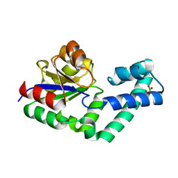
1ZJ8
 | | Structure of Mycobacterium tuberculosis NirA protein | | 分子名称: | CHLORIDE ION, IRON/SULFUR CLUSTER, Probable ferredoxin-dependent nitrite reductase NirA, ... | | 著者 | Schnell, R, Sandalova, T, Hellman, U, Lindqvist, Y, Schneider, G, Structural Proteomics in Europe (SPINE) | | 登録日 | 2005-04-28 | | 公開日 | 2005-05-31 | | 最終更新日 | 2023-12-27 | | 実験手法 | X-RAY DIFFRACTION (2.8 Å) | | 主引用文献 | Siroheme- and [Fe4-S4]-dependent NirA from Mycobacterium tuberculosis Is a Sulfite Reductase with a Covalent Cys-Tyr Bond in the Active Site
J.Biol.Chem., 280, 2005
|
|
1ZJ9
 | | Structure of Mycobacterium tuberculosis NirA protein | | 分子名称: | CHLORIDE ION, IRON/SULFUR CLUSTER, Probable ferredoxin-dependent nitrite reductase NirA, ... | | 著者 | Schnell, R, Sandalova, T, Hellman, U, Lindqvist, Y, Schneider, G. | | 登録日 | 2005-04-28 | | 公開日 | 2005-05-31 | | 最終更新日 | 2023-12-27 | | 実験手法 | X-RAY DIFFRACTION (2.9 Å) | | 主引用文献 | Siroheme- and [Fe4-S4]-dependent NirA from Mycobacterium tuberculosis Is a Sulfite Reductase with a Covalent Cys-Tyr Bond in the Active Site
J.Biol.Chem., 280, 2005
|
|
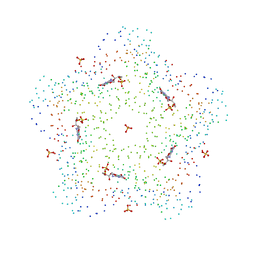
1C2Y
 | | CRYSTAL STRUCTURES OF A PENTAMERIC FUNGAL AND AN ICOSAHEDRAL PLANT LUMAZINE SYNTHASE REVEALS THE STRUCTURAL BASIS FOR DIFFERENCES IN ASSEMBLY | | 分子名称: | 5-NITROSO-6-RIBITYL-AMINO-2,4(1H,3H)-PYRIMIDINEDIONE, PROTEIN (LUMAZINE SYNTHASE) | | 著者 | Persson, K, Schneider, G, Jordan, D.B, Viitanen, P.V, Sandalova, T. | | 登録日 | 1999-07-27 | | 公開日 | 2000-07-30 | | 最終更新日 | 2023-08-09 | | 実験手法 | X-RAY DIFFRACTION (3.3 Å) | | 主引用文献 | Crystal structure analysis of a pentameric fungal and an icosahedral plant lumazine synthase reveals the structural basis for differences in assembly.
Protein Sci., 8, 1999
|
|
1C41
 | | CRYSTAL STRUCTURES OF A PENTAMERIC FUNGAL AND AN ICOSAHEDRAL PLANT LUMAZINE SYNTHASE REVEALS THE STRUCTURAL BASIS FOR DIFFERENCES IN ASSEMBLY | | 分子名称: | 5-NITROSO-6-RIBITYL-AMINO-2,4(1H,3H)-PYRIMIDINEDIONE, LUMAZINE SYNTHASE, SULFATE ION | | 著者 | Persson, K, Schneider, G, Jordan, D.B, Viitanen, P.V, Sandalova, T. | | 登録日 | 1999-08-03 | | 公開日 | 2000-08-06 | | 最終更新日 | 2023-08-09 | | 実験手法 | X-RAY DIFFRACTION (3.1 Å) | | 主引用文献 | Crystal structure analysis of a pentameric fungal and an icosahedral plant lumazine synthase reveals the structural basis for differences in assembly
Protein Sci., 8, 1999
|
|
4EX6
 | | Crystal structure of the alnumycin P phosphatase AlnB | | 分子名称: | AlnB, BORIC ACID, MAGNESIUM ION | | 著者 | Oja, T, Niiranen, L, Sandalova, T, Klika, K.D, Niemi, J, Mantsala, P, Schneider, G, Metsa-Ketela, M. | | 登録日 | 2012-04-30 | | 公開日 | 2013-01-16 | | 最終更新日 | 2023-09-13 | | 実験手法 | X-RAY DIFFRACTION (1.25 Å) | | 主引用文献 | Structural basis for C-ribosylation in the alnumycin A biosynthetic pathway.
Proc.Natl.Acad.Sci.USA, 110, 2013
|
|
1E3I
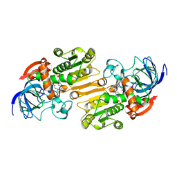 | | Mouse class II alcohol dehydrogenase complex with NADH and inhibitor | | 分子名称: | 1,4-DIHYDRONICOTINAMIDE ADENINE DINUCLEOTIDE, ALCOHOL DEHYDROGENASE, CLASS II, ... | | 著者 | Svensson, S, Hoog, J.O, Schneider, G, Sandalova, T. | | 登録日 | 2000-06-16 | | 公開日 | 2000-09-12 | | 最終更新日 | 2023-12-13 | | 実験手法 | X-RAY DIFFRACTION (2.08 Å) | | 主引用文献 | Crystal Structure of Mouse Class II Alcohol Dehydrogenase Reveal Determinants of Substrate Specificity and Catalytic Efficiency
J.Mol.Biol., 302, 2000
|
|
1E3E
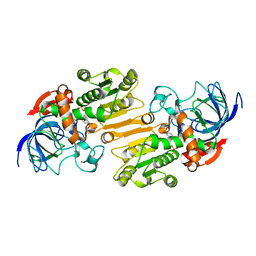 | | Mouse class II alcohol dehydrogenase complex with NADH | | 分子名称: | 1,4-DIHYDRONICOTINAMIDE ADENINE DINUCLEOTIDE, ALCOHOL DEHYDROGENASE, CLASS II, ... | | 著者 | Svensson, S, Hoeoeg, J.O, Schneider, G, Sandalova, T. | | 登録日 | 2000-06-14 | | 公開日 | 2000-09-12 | | 最終更新日 | 2023-12-13 | | 実験手法 | X-RAY DIFFRACTION (2.12 Å) | | 主引用文献 | Crystal Structure of Mouse Class II Alcohol Dehydrogenase Reveal Determinants of Substrate Specificity and Catalytic Efficiency
J.Mol.Biol., 302, 2000
|
|
1E3L
 | | P47H mutant of mouse class II alcohol dehydrogenase complex with NADH | | 分子名称: | ALCOHOL DEHYDROGENASE, CLASS II, NICOTINAMIDE-ADENINE-DINUCLEOTIDE, ... | | 著者 | Svensson, S, Hoog, J.O, Schneider, G, Sandalova, T. | | 登録日 | 2000-06-19 | | 公開日 | 2000-09-12 | | 最終更新日 | 2023-12-13 | | 実験手法 | X-RAY DIFFRACTION (2.5 Å) | | 主引用文献 | Crystal Structure of Mouse Class II Alcohol Dehydrogenase Reveal Determinants of Substrate Specificity and Catalytic Efficiency
J.Mol.Biol., 302, 2000
|
|
5TIL
 | |
4EX7
 | | Crystal structure of the alnumycin P phosphatase in complex with free phosphate | | 分子名称: | AlnB, BORIC ACID, MAGNESIUM ION, ... | | 著者 | Oja, T, Niiranen, L, Sandalova, T, Klika, K.D, Niemi, J, Mantsala, P, Schneider, G, Metsa-Ketela, M. | | 登録日 | 2012-04-30 | | 公開日 | 2013-01-16 | | 最終更新日 | 2024-04-03 | | 実験手法 | X-RAY DIFFRACTION (1.5 Å) | | 主引用文献 | Structural basis for C-ribosylation in the alnumycin A biosynthetic pathway.
Proc.Natl.Acad.Sci.USA, 110, 2013
|
|
4EX8
 | | Crystal structure of the prealnumycin C-glycosynthase AlnA | | 分子名称: | AlnA, CALCIUM ION, CHLORIDE ION, ... | | 著者 | Oja, T, Niiranen, L, Sandalova, T, Klika, K.D, Niemi, J, Mantsala, P, Schneider, G, Metsa-Ketela, M. | | 登録日 | 2012-04-30 | | 公開日 | 2013-01-16 | | 最終更新日 | 2023-09-13 | | 実験手法 | X-RAY DIFFRACTION (2.1 Å) | | 主引用文献 | Structural basis for C-ribosylation in the alnumycin A biosynthetic pathway.
Proc.Natl.Acad.Sci.USA, 110, 2013
|
|
6EWQ
 | | Putative sugar aminotransferase Spr1654 from Streptococcus pneumoniae, PLP-form | | 分子名称: | PYRIDOXAL-5'-PHOSPHATE, Putative capsular polysaccharide biosynthesis protein | | 著者 | Achour, A, Sun, R, Sandalova, T, Han, X. | | 登録日 | 2017-11-06 | | 公開日 | 2018-05-02 | | 最終更新日 | 2024-01-17 | | 実験手法 | X-RAY DIFFRACTION (2.2 Å) | | 主引用文献 | Structural and functional studies of Spr1654: an essential aminotransferase in teichoic acid biosynthesis inStreptococcus pneumoniae.
Open Biol, 8, 2018
|
|


