Loading
PDBj
メニューPDBj@FacebookPDBj@TwitterPDBj@YouTubewwPDB FoundationwwPDB
RCSB PDBPDBeBMRBAdv. SearchSearch help
Search by PDB author
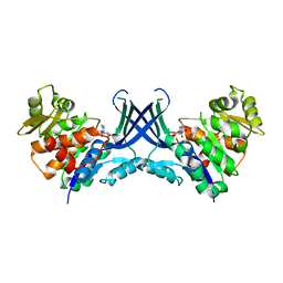
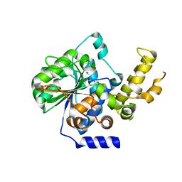
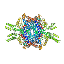
5E9G
DownloadVisualize
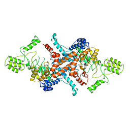
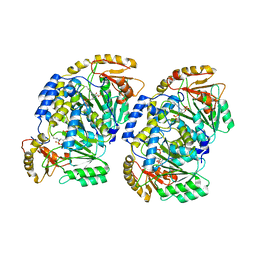
BU of 5e9g by Molmil
Structural insights of isocitrate lyases from Magnaporthe oryzae
分子名称: GLYCEROL, GLYOXYLIC ACID, Isocitrate lyase, ...
著者Park, Y, Cho, Y, Lee, Y.-H, Lee, Y.-W, Rhee, S.
登録日2015-10-15
公開日2016-04-27
最終更新日2023-11-08
実験手法X-RAY DIFFRACTION (2.1 Å)
主引用文献Crystal structure and functional analysis of isocitrate lyases from Magnaporthe oryzae and Fusarium graminearum
J.Struct.Biol., 194, 2016
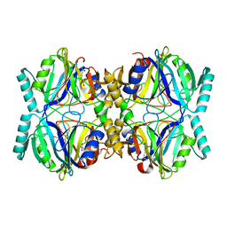
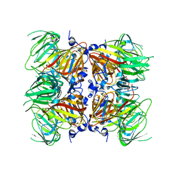
5E9H
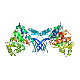
DownloadVisualize
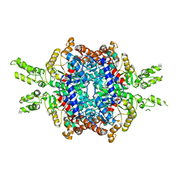
BU of 5e9h by Molmil
Structural insights of isocitrate lyases from Fusarium graminearum
分子名称: Isocitrate lyase, MALONATE ION, MANGANESE (II) ION
著者Park, Y, Cho, Y, Lee, Y.-H, Lee, Y.-W, Rhee, S.
登録日2015-10-15
公開日2016-04-27
最終更新日2023-11-08
実験手法X-RAY DIFFRACTION (2.3 Å)
主引用文献Crystal structure and functional analysis of isocitrate lyases from Magnaporthe oryzae and Fusarium graminearum
J.Struct.Biol., 194, 2016
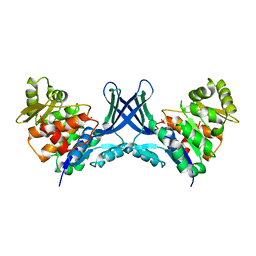
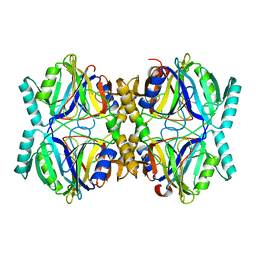
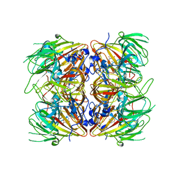
5E9F
DownloadVisualize
BU of 5e9f by Molmil
Structural insights of isocitrate lyases from Magnaporthe oryzae
分子名称: Isocitrate lyase, MAGNESIUM ION
著者Park, Y, Cho, Y, Lee, Y.-H, Lee, Y.-W, Rhee, S.
登録日2015-10-15
公開日2016-04-27
最終更新日2023-11-08
実験手法X-RAY DIFFRACTION (2.8 Å)
主引用文献Crystal structure and functional analysis of isocitrate lyases from Magnaporthe oryzae and Fusarium graminearum
J.Struct.Biol., 194, 2016
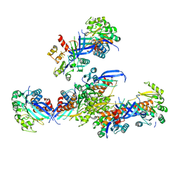
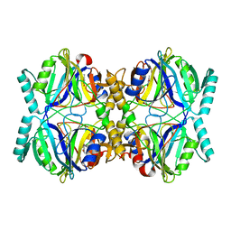
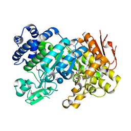
7EF6
DownloadVisualize
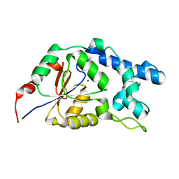
BU of 7ef6 by Molmil
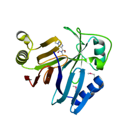
Crystal Structure of Xanthosine monophosphate phosphatase in the unliganded state
分子名称: MAGNESIUM ION, Xanthosine monophosphate phosphatase
著者Yang, S, Rhee, S.
登録日2021-03-21
公開日2021-11-03
最終更新日2023-11-29
実験手法X-RAY DIFFRACTION (1.34 Å)
主引用文献Initiation of cytosolic plant purine nucleotide catabolism involves a monospecific xanthosine monophosphate phosphatase.
Nat Commun, 12, 2021
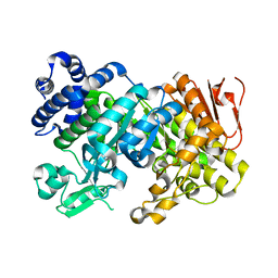
7EF7
DownloadVisualize
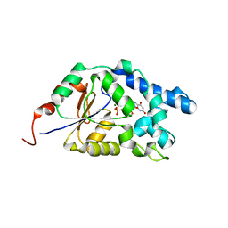
BU of 7ef7 by Molmil
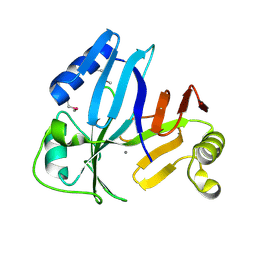
Crystal Structure of Xanthosine monophosphate phosphatase complex with XMP
分子名称: At2g32150/F22D22.10, MAGNESIUM ION, XANTHOSINE-5'-MONOPHOSPHATE
著者Yang, S, Rhee, S.
登録日2021-03-21
公開日2021-11-03
最終更新日2023-11-29
実験手法X-RAY DIFFRACTION (1.5 Å)
主引用文献Initiation of cytosolic plant purine nucleotide catabolism involves a monospecific xanthosine monophosphate phosphatase.
Nat Commun, 12, 2021
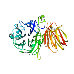
4FFF
DownloadVisualize
BU of 4fff by Molmil
Crystal Structure of Levan Fructotransferase from Arthrobacter ureafaciens
分子名称: Levan fructotransferase
著者Park, J, Rhee, S.
登録日2012-06-01
公開日2012-07-18
最終更新日2024-02-28
実験手法X-RAY DIFFRACTION (2.57 Å)
主引用文献Structural and functional basis for substrate specificity and catalysis of levan fructotransferase.
J.Biol.Chem., 287, 2012
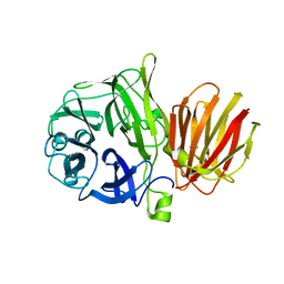
4FFI
DownloadVisualize
BU of 4ffi by Molmil
Crystal Structure of Levan Fructotransferase D54N mutant from Arthrobacter ureafaciens in complex with levanbiose
分子名称: Levan fructotransferase, beta-D-fructofuranose-(2-6)-beta-D-fructofuranose, beta-D-fructofuranose-(2-6)-beta-D-fructofuranose-(2-6)-beta-D-fructofuranose
著者Park, J, Rhee, S.
登録日2012-06-01
公開日2012-07-18
最終更新日2024-02-28
実験手法X-RAY DIFFRACTION (2.3 Å)
主引用文献Structural and functional basis for substrate specificity and catalysis of levan fructotransferase.
J.Biol.Chem., 287, 2012
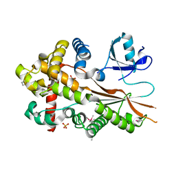
5XU6
DownloadVisualize
BU of 5xu6 by Molmil
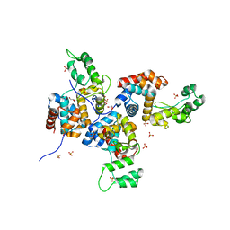
Crystal structure of inositol 1,3,4,5,6-pentakisphosphate 2-kinase (IPK1) from Cryptococcus neoformans
分子名称: Inositol-pentakisphosphate 2-kinase, SULFATE ION
著者Oh, J, Rhee, S.
登録日2017-06-22
公開日2017-10-04
最終更新日2017-11-29
実験手法X-RAY DIFFRACTION (2.35 Å)
主引用文献Crystal structure of inositol 1,3,4,5,6-pentakisphosphate 2-kinase from Cryptococcus neoformans.
J. Struct. Biol., 200, 2017
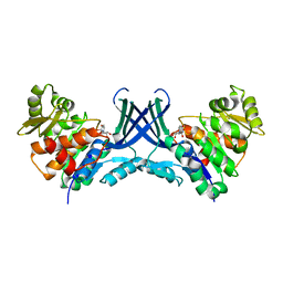
7VTE
DownloadVisualize
BU of 7vte by Molmil
uridine bound structure of Pseudouridine kinase (PUKI) from Escherichia coli strain B
分子名称: POTASSIUM ION, Pseudouridine kinase, URIDINE
著者Kim, S.H, Rhee, S.
登録日2021-10-29
公開日2022-04-06
最終更新日2023-11-29
実験手法X-RAY DIFFRACTION (2.15296578 Å)
主引用文献Substrate-binding loop interactions with pseudouridine trigger conformational changes that promote catalytic efficiency of pseudouridine kinase PUKI.
J.Biol.Chem., 298, 2022
7VTG
DownloadVisualize
BU of 7vtg by Molmil
Pseudouridine bound structure of Pseudouridine kinase (PUKI) S30A mutant from Escherichia coli strain B
分子名称: 5-[(2~{S},3~{R},4~{S},5~{R})-5-(hydroxymethyl)-3,4-bis(oxidanyl)oxolan-2-yl]-1~{H}-pyrimidine-2,4-dione, Pseudouridine kinase
著者Kim, S.H, Rhee, S.
登録日2021-10-29
公開日2022-04-06
最終更新日2023-11-29
実験手法X-RAY DIFFRACTION (1.89859128 Å)
主引用文献Substrate-binding loop interactions with pseudouridine trigger conformational changes that promote catalytic efficiency of pseudouridine kinase PUKI.
J.Biol.Chem., 298, 2022
7VTD
DownloadVisualize
BU of 7vtd by Molmil
Unliganded structure of Pseudouridine kinase (PUKI) from Escherichia coli strain B
分子名称: POTASSIUM ION, Pseudouridine kinase
著者Kim, S.H, Rhee, S.
登録日2021-10-29
公開日2022-04-06
最終更新日2023-11-29
実験手法X-RAY DIFFRACTION (2.1505487 Å)
主引用文献Substrate-binding loop interactions with pseudouridine trigger conformational changes that promote catalytic efficiency of pseudouridine kinase PUKI.
J.Biol.Chem., 298, 2022
7VTF
DownloadVisualize
BU of 7vtf by Molmil
cytidine bound structure of Pseudouridine kinase (PUKI) from Escherichia coli strain B
分子名称: 4-AMINO-1-BETA-D-RIBOFURANOSYL-2(1H)-PYRIMIDINONE, Pseudouridine kinase
著者Kim, S.H, Rhee, S.
登録日2021-10-29
公開日2022-04-06
最終更新日2023-11-29
実験手法X-RAY DIFFRACTION (2.20203447 Å)
主引用文献Substrate-binding loop interactions with pseudouridine trigger conformational changes that promote catalytic efficiency of pseudouridine kinase PUKI.
J.Biol.Chem., 298, 2022
7VVA
DownloadVisualize
BU of 7vva by Molmil
Pseudouridine bound structure of Pseudouridine kinase (PUKI) from Escherichia coli strain B
分子名称: 5-[(2~{S},3~{R},4~{S},5~{R})-5-(hydroxymethyl)-3,4-bis(oxidanyl)oxolan-2-yl]-1~{H}-pyrimidine-2,4-dione, Pseudouridine kinase
著者Kim, S.H, Rhee, S.
登録日2021-11-05
公開日2022-04-06
最終更新日2023-11-29
実験手法X-RAY DIFFRACTION (2.75029182 Å)
主引用文献Substrate-binding loop interactions with pseudouridine trigger conformational changes that promote catalytic efficiency of pseudouridine kinase PUKI.
J.Biol.Chem., 298, 2022
7VRX
DownloadVisualize
BU of 7vrx by Molmil
Pad-1 in the absence of substrate
分子名称: Aminotransferase, SULFATE ION
著者Choi, M, Rhee, S.
登録日2021-10-25
公開日2022-04-20
最終更新日2023-11-29
実験手法X-RAY DIFFRACTION (1.96634674 Å)
主引用文献Structural and biochemical basis for the substrate specificity of Pad-1, an indole-3-pyruvic acid aminotransferase in auxin homeostasis.
J.Struct.Biol., 214, 2022
3OUM
DownloadVisualize
BU of 3oum by Molmil
Crystal Structure of toxoflavin-degrading enzyme in complex with toxoflavin
分子名称: 1,6-dimethylpyrimido[5,4-e][1,2,4]triazine-5,7(1H,6H)-dione, MANGANESE (II) ION, toxoflavin-degrading enzyme
著者Kim, M.I, Rhee, S.
登録日2010-09-15
公開日2011-08-10
最終更新日2024-04-03
実験手法X-RAY DIFFRACTION (2 Å)
主引用文献Structural and functional analysis of phytotoxin toxoflavin-degrading enzyme
Plos One, 6, 2011
3OUL
DownloadVisualize
BU of 3oul by Molmil
Crystal Structure of toxoflavin-degrading enzyme in a substrate-free form
分子名称: MANGANESE (II) ION, Toxoflavin-degrading enzyme
著者Kim, M.I, Rhee, S.
登録日2010-09-15
公開日2011-08-10
最終更新日2024-04-03
実験手法X-RAY DIFFRACTION (1.6 Å)
主引用文献Structural and functional analysis of phytotoxin toxoflavin-degrading enzyme
Plos One, 6, 2011
4OK7
DownloadVisualize
BU of 4ok7 by Molmil
Structure of bacteriophage SPN1S endolysin from Salmonella typhimurium
分子名称: Endolysin, GLYCEROL, SULFATE ION
著者Park, Y, Lim, J, Kong, M, Ryu, S, Rhee, S.
登録日2014-01-22
公開日2014-03-19
最終更新日2024-02-28
実験手法X-RAY DIFFRACTION (1.9 Å)
主引用文献Structure of bacteriophage SPN1S endolysin reveals an unusual two-module fold for the peptidoglycan lytic and binding activity.
Mol.Microbiol., 92, 2014
4P5F
DownloadVisualize
BU of 4p5f by Molmil
The crystal structure of type III effector protein XopQ complexed with adenosine diphosphate ribose
分子名称: CALCIUM ION, Inosine-uridine nucleoside N-ribohydrolase, [(2R,3S,4R,5R)-5-(6-AMINOPURIN-9-YL)-3,4-DIHYDROXY-OXOLAN-2-YL]METHYL [HYDROXY-[[(2R,3S,4R,5S)-3,4,5-TRIHYDROXYOXOLAN-2-YL]METHOXY]PHOSPHORYL] HYDROGEN PHOSPHATE
著者Yu, S, Hwang, I, Rhee, S.
登録日2014-03-17
公開日2014-08-13
最終更新日2023-12-27
実験手法X-RAY DIFFRACTION (2.1 Å)
主引用文献The crystal structure of type III effector protein XopQ from Xanthomonas oryzae complexed with adenosine diphosphate ribose.
Proteins, 82, 2014
3HQ0
DownloadVisualize
BU of 3hq0 by Molmil
Crystal Structure Analysis of the 2,3-dioxygenase LapB from Pseudomonas in complex with a product
分子名称: (2E,4E)-2-hydroxy-6-oxohepta-2,4-dienoic acid, Catechol 2,3-dioxygenase, FE (III) ION
著者Cho, J.-H, Rhee, S.
登録日2009-06-05
公開日2009-10-20
最終更新日2024-04-03
実験手法X-RAY DIFFRACTION (2 Å)
主引用文献Crystal structure and functional analysis of the extradiol dioxygenase LapB from a long-chain alkylphenol degradation pathway in Pseudomonas
J.Biol.Chem., 284, 2009
3HPV
DownloadVisualize
BU of 3hpv by Molmil
Crystal Structure Analysis of the 2,3-dioxygenase LapB from Pseudomonas sp. KL28
分子名称: Catechol 2,3-dioxygenase, FE (II) ION
著者Cho, J.-H, Rhee, S.
登録日2009-06-05
公開日2009-10-20
最終更新日2023-11-01
実験手法X-RAY DIFFRACTION (2.3 Å)
主引用文献Crystal structure and functional analysis of the extradiol dioxygenase LapB from a long-chain alkylphenol degradation pathway in Pseudomonas
J.Biol.Chem., 284, 2009
3HPY
DownloadVisualize
BU of 3hpy by Molmil
Crystal Structure Analysis of the 2,3-dioxygenase LapB from Pseudomonas in the complex with 4-methylcatechol
分子名称: 4-METHYLCATECHOL, Catechol 2,3-dioxygenase, FE (III) ION
著者Cho, J.-H, Rhee, S.
登録日2009-06-05
公開日2009-10-20
最終更新日2024-04-03
実験手法X-RAY DIFFRACTION (1.94 Å)
主引用文献Crystal structure and functional analysis of the extradiol dioxygenase LapB from a long-chain alkylphenol degradation pathway in Pseudomonas
J.Biol.Chem., 284, 2009
4E2Q
DownloadVisualize
BU of 4e2q by Molmil
Crystal Structure of (S)-Ureidoglycine Aminohydrolase from Arabidopsis thaliana
分子名称: MANGANESE (II) ION, Ureidoglycine aminohydrolase
著者Shin, I, Rhee, S.
登録日2012-03-09
公開日2012-04-18
最終更新日2024-03-20
実験手法X-RAY DIFFRACTION (2.5 Å)
主引用文献Structural and functional insights into (S)-ureidoglycine aminohydrolase, key enzyme of purine catabolism in Arabidopsis thaliana
J.Biol.Chem., 287, 2012
4E2S
DownloadVisualize
BU of 4e2s by Molmil
Crystal structure of (S)-Ureidoglycine Aminohydrolase from Arabidopsis thaliana in complex with its substrate, (S)-Ureidoglycine
分子名称: (2S)-amino(carbamoylamino)ethanoic acid, MANGANESE (II) ION, Ureidoglycine aminohydrolase
著者Shin, I, Rhee, S.
登録日2012-03-09
公開日2012-04-18
最終更新日2024-03-20
実験手法X-RAY DIFFRACTION (2.59 Å)
主引用文献Structural and functional insights into (S)-ureidoglycine aminohydrolase, key enzyme of purine catabolism in Arabidopsis thaliana
J.Biol.Chem., 287, 2012
3CZG
DownloadVisualize
BU of 3czg by Molmil
Crystal Structure Analysis of Sucrose hydrolase (SUH)-glucose complex
分子名称: Sucrose hydrolase, alpha-D-glucopyranose
著者Kim, M.I, Rhee, S.
登録日2008-04-29
公開日2008-07-15
最終更新日2020-07-29
実験手法X-RAY DIFFRACTION (1.8 Å)
主引用文献Crystal structures and mutagenesis of sucrose hydrolase from Xanthomonas axonopodis pv. glycines: insight into the exclusively hydrolytic amylosucrase fold.
J.Mol.Biol., 380, 2008
3CZL
DownloadVisualize
BU of 3czl by Molmil
Crystal Structure Analysis of Sucrose hydrolase(SUH) E322Q-glucose complex
分子名称: alpha-D-glucopyranose, sucrose hydrolase
著者Kim, M.I, Rhee, S.
登録日2008-04-29
公開日2008-07-15
最終更新日2023-11-01
実験手法X-RAY DIFFRACTION (2 Å)
主引用文献Crystal structures and mutagenesis of sucrose hydrolase from Xanthomonas axonopodis pv. glycines: insight into the exclusively hydrolytic amylosucrase fold.
J.Mol.Biol., 380, 2008

221051

件を2024-06-12に公開中

PDB statisticsPDBj update infoContact PDBjnumon