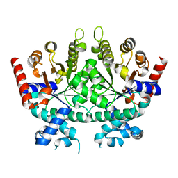
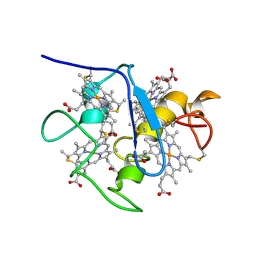
2DDR
 
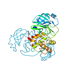
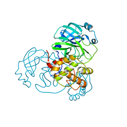
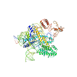
 | | Crystal structure of sphingomyelinase from Bacillus cereus with calcium ion | | 分子名称: | CALCIUM ION, Sphingomyelin phosphodiesterase | | 著者 | Ago, H, Oda, M, Takahashi, M, Tsuge, H, Ochi, S, Katunuma, N, Miyano, M, Sakurai, J, RIKEN Structural Genomics/Proteomics Initiative (RSGI) | | 登録日 | 2006-02-02 | | 公開日 | 2006-05-02 | | 最終更新日 | 2011-07-13 | | 実験手法 | X-RAY DIFFRACTION (1.4 Å) | | 主引用文献 | Structural Basis of the Sphingomyelin Phosphodiesterase Activity in Neutral Sphingomyelinase from Bacillus cereus.
J.Biol.Chem., 281, 2006
|
|
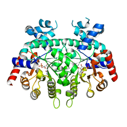
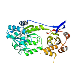
2MNS
 | |
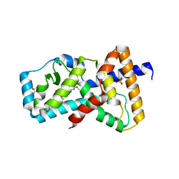
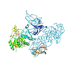
1WAD
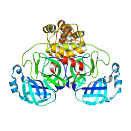
 | | CYTOCHROME C3 WITH 4 HEME GROUPS AND ONE CALCIUM ION | | 分子名称: | CALCIUM ION, CYTOCHROME C3, PROTOPORPHYRIN IX CONTAINING FE | | 著者 | Matias, P.M, Morais, J, Coelho, R, Carrondo, M.A, Wilson, K, Dauter, Z, Sieker, L. | | 登録日 | 1996-01-10 | | 公開日 | 1997-01-27 | | 最終更新日 | 2024-06-05 | | 実験手法 | X-RAY DIFFRACTION (1.8 Å) | | 主引用文献 | Cytochrome c3 from Desulfovibrio gigas: crystal structure at 1.8 A resolution and evidence for a specific calcium-binding site.
Protein Sci., 5, 1996
|
|
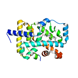
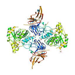
6B30
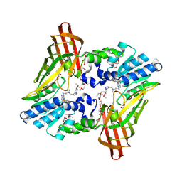
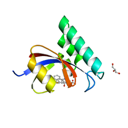
 | | Structure of RORgt in complex with a novel inverse agonist 1 | | 分子名称: | N-[(1R)-1-(4-methoxyphenyl)-2-oxo-2-{[4-(trimethylsilyl)phenyl]amino}ethyl]-N-methyl-3-oxo-2,3-dihydro-1,2-oxazole-5-carboxamide, Nuclear receptor ROR-gamma | | 著者 | Skene, R.J, Hoffman, I. | | 登録日 | 2017-09-20 | | 公開日 | 2018-01-03 | | 最終更新日 | 2024-03-13 | | 実験手法 | X-RAY DIFFRACTION (2.69 Å) | | 主引用文献 | Discovery of orally efficacious ROR gamma t inverse agonists, part 1: Identification of novel phenylglycinamides as lead scaffolds.
Bioorg. Med. Chem., 26, 2018
|
|
2X9H
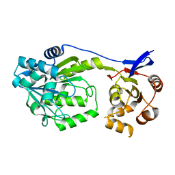
 | | CRYSTAL STRUCTURE OF MYOSIN-2 MOTOR DOMAIN IN COMPLEX WITH ADP- METAVANADATE AND PENTACHLOROCARBAZOLE | | 分子名称: | 2,3,4,6,8-PENTACHLORO-9H-CARBAZOL-1-OL, ADP METAVANADATE, MAGNESIUM ION, ... | | 著者 | Selvadurai, J, Kirst, J, Knoelker, H.J, Manstein, D.J. | | 登録日 | 2010-03-19 | | 公開日 | 2011-05-11 | | 最終更新日 | 2023-12-20 | | 実験手法 | X-RAY DIFFRACTION (2.7 Å) | | 主引用文献 | Crystal Structure of Myosin-2 Motor Domain in Complex with Adp-Metavanadate and Pentachlorocarbazole
To be Published
|
|
2OXB
 | | Crystal structure of a cell-wall invertase (E203Q) from Arabidopsis thaliana in complex with sucrose | | 分子名称: | 2-acetamido-2-deoxy-beta-D-glucopyranose, Beta-fructofuranosidase, beta-D-fructofuranose-(2-1)-alpha-D-glucopyranose | | 著者 | Lammens, W, Le Roy, K, Van Laere, A, Van den Ende, W, Rabijns, A. | | 登録日 | 2007-02-20 | | 公開日 | 2008-01-22 | | 最終更新日 | 2023-08-30 | | 実験手法 | X-RAY DIFFRACTION (2.6 Å) | | 主引用文献 | An alternate sucrose binding mode in the E203Q Arabidopsis invertase mutant: An X-ray crystallography and docking study.
Proteins, 71, 2007
|
|
6DSQ
 | |
6DSR
 | |
8SMQ
 | | Crystal Structure of the N-terminal Domain of the Cryptic Surface Protein (CD630_25440) from Clostridium difficile. | | 分子名称: | 1,2-ETHANEDIOL, CHLORIDE ION, GLYCEROL, ... | | 著者 | Minasov, G, Shuvalova, L, Brunzelle, J.S, Kiryukhina, O, Wawrzak, Z, Satchell, K.J.F, Center for Structural Biology of Infectious Diseases (CSBID), Center for Structural Genomics of Infectious Diseases (CSGID) | | 登録日 | 2023-04-26 | | 公開日 | 2023-05-10 | | 最終更新日 | 2023-12-06 | | 実験手法 | X-RAY DIFFRACTION (2 Å) | | 主引用文献 | Protein target highlights in CASP15: Analysis of models by structure providers.
Proteins, 91, 2023
|
|
6E3E
 | | Structure of RORgt in complex with a novel inverse agonist. | | 分子名称: | (4S)-2-METHYL-2,4-PENTANEDIOL, 5-[(5R)-5-[(7-fluoro-1,1-dimethyl-2,3-dihydro-1H-inden-5-yl)carbamoyl]-2-methoxy-7,8-dihydro-1,6-naphthyridin-6(5H)-yl]-5-oxopentanoic acid, Nuclear receptor ROR-gamma, ... | | 著者 | Skene, R.J, Hoffman, I. | | 登録日 | 2018-07-13 | | 公開日 | 2019-07-17 | | 最終更新日 | 2024-03-13 | | 実験手法 | X-RAY DIFFRACTION (2.47 Å) | | 主引用文献 | Design, Synthesis, and Biological Evaluation of Retinoic Acid-Related Orphan Receptor gamma t (ROR gamma t) Agonist Structure-Based Functionality Switching Approach from In House ROR gamma t Inverse Agonist to ROR gamma t Agonist.
J.Med.Chem., 62, 2019
|
|
6E3G
 | | Structure of RORgt in complex with a novel agonist. | | 分子名称: | (5R)-6-acetyl-2-methoxy-N-{4-[(2-methoxyphenyl)methoxy]phenyl}-5,6,7,8-tetrahydro-1,6-naphthyridine-5-carboxamide, 1,2-ETHANEDIOL, Nuclear receptor ROR-gamma, ... | | 著者 | Skene, R.J, Hoffman, I. | | 登録日 | 2018-07-13 | | 公開日 | 2019-06-12 | | 最終更新日 | 2024-03-13 | | 実験手法 | X-RAY DIFFRACTION (2.1 Å) | | 主引用文献 | Design, Synthesis, and Biological Evaluation of Retinoic Acid-Related Orphan Receptor gamma t (ROR gamma t) Agonist Structure-Based Functionality Switching Approach from In House ROR gamma t Inverse Agonist to ROR gamma t Agonist.
J.Med.Chem., 62, 2019
|
|
8DMN
 | |
8EJ9
 | |
8EJ7
 | |
6NT2
 | | type 1 PRMT in complex with the inhibitor GSK3368715 | | 分子名称: | 2,3-DIHYDROXY-1,4-DITHIOBUTANE, GLYCEROL, N~1~-({5-[4,4-bis(ethoxymethyl)cyclohexyl]-1H-pyrazol-4-yl}methyl)-N~1~,N~2~-dimethylethane-1,2-diamine, ... | | 著者 | Concha, N.O. | | 登録日 | 2019-01-28 | | 公開日 | 2019-07-10 | | 最終更新日 | 2019-07-24 | | 実験手法 | X-RAY DIFFRACTION (2.48 Å) | | 主引用文献 | Anti-tumor Activity of the Type I PRMT Inhibitor, GSK3368715, Synergizes with PRMT5 Inhibition through MTAP Loss.
Cancer Cell, 36, 2019
|
|
1A2I
 | | SOLUTION STRUCTURE OF DESULFOVIBRIO VULGARIS (HILDENBOROUGH) FERROCYTOCHROME C3, NMR, 20 STRUCTURES | | 分子名称: | CYTOCHROME C3, HEME C | | 著者 | Messias, A.C, Kastrau, D.H.K, Costa, H.S, Legall, J, Turner, D.L, Santos, H, Xavier, A.V. | | 登録日 | 1998-01-05 | | 公開日 | 1998-07-08 | | 最終更新日 | 2022-02-16 | | 実験手法 | SOLUTION NMR | | 主引用文献 | Solution structure of Desulfovibrio vulgaris (Hildenborough) ferrocytochrome c3: structural basis for functional cooperativity.
J.Mol.Biol., 281, 1998
|
|
7S91
 | |
6XFI
 | | Crystal Structures of beta-1,4-N-Acetylglucosaminyltransferase 2 (POMGNT2): Structural Basis for Inherited Muscular Dystrophies | | 分子名称: | 2-acetamido-2-deoxy-beta-D-glucopyranose, PHOSPHATE ION, Protein O-linked-mannose beta-1,4-N-acetylglucosaminyltransferase 2, ... | | 著者 | Halmo, S.M, Yeh, J, Wells, L, Moremen, K.W, Lanzilotta, W.N. | | 登録日 | 2020-06-15 | | 公開日 | 2021-04-21 | | 実験手法 | X-RAY DIFFRACTION (2 Å) | | 主引用文献 | Crystal structures of beta-1,4-N-acetylglucosaminyltransferase 2: structural basis for inherited muscular dystrophies.
Acta Crystallogr D Struct Biol, 77, 2021
|
|
6XI2
 | | Apo form of POMGNT2 | | 分子名称: | 2-acetamido-2-deoxy-beta-D-glucopyranose, ALA-ALA-ALA-ALA-ALA-ALA-ALA-ALA-ALA-ALA, ALA-GLY-ALA-GLY-ALA-ALA-ALA-ALA-ALA-ALA, ... | | 著者 | Halmo, S.M, Yeh, J, Wells, L, Moremen, K.W, Lanzilotta, W.N. | | 登録日 | 2020-06-19 | | 公開日 | 2021-04-21 | | 実験手法 | X-RAY DIFFRACTION (2.57 Å) | | 主引用文献 | Crystal structures of beta-1,4-N-acetylglucosaminyltransferase 2: structural basis for inherited muscular dystrophies.
Acta Crystallogr D Struct Biol, 77, 2021
|
|
4TYO
 | | PPIase in complex with a non-phosphate small molecule inhibitor. | | 分子名称: | 3-(6-fluoro-1H-benzimidazol-2-yl)-N-(naphthalen-2-ylcarbonyl)-D-alanine, GLYCEROL, Peptidyl-prolyl cis-trans isomerase NIMA-interacting 1 | | 著者 | Greasley, S.E, Ferre, R.A. | | 登録日 | 2014-07-08 | | 公開日 | 2014-08-20 | | 最終更新日 | 2023-12-27 | | 実験手法 | X-RAY DIFFRACTION (1.75 Å) | | 主引用文献 | Structure-based design of novel human Pin1 inhibitors (III): Optimizing affinity beyond the phosphate recognition pocket.
Bioorg.Med.Chem.Lett., 24, 2014
|
|
1A9U
 | | THE COMPLEX STRUCTURE OF THE MAP KINASE P38/SB203580 | | 分子名称: | 4-[5-(4-FLUORO-PHENYL)-2-(4-METHANESULFINYL-PHENYL)-3H-IMIDAZOL-4-YL]-PYRIDINE, MAP KINASE P38 | | 著者 | Wang, Z, Canagarajah, B, Boehm, J.C, Kassis, S, Cobb, M.H, Young, P.R, Abdel-Meguid, S, Adams, J.L, Goldsmith, E.J. | | 登録日 | 1998-04-10 | | 公開日 | 1999-04-20 | | 最終更新日 | 2024-04-03 | | 実験手法 | X-RAY DIFFRACTION (2.5 Å) | | 主引用文献 | Structural basis of inhibitor selectivity in MAP kinases.
Structure, 6, 1998
|
|
6D6Z
 | |
6NY3
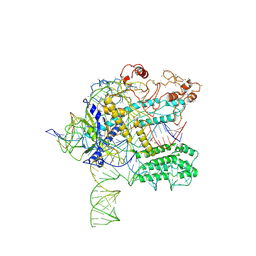 | | CasX ternary complex with 30bp target DNA | | 分子名称: | CasX, DNA Non-target strand, DNA target strand, ... | | 著者 | Liu, J.J, Orlova, N, Nogales, E, Doudna, J.A. | | 登録日 | 2019-02-10 | | 公開日 | 2019-02-27 | | 最終更新日 | 2019-12-25 | | 実験手法 | ELECTRON MICROSCOPY (3.7 Å) | | 主引用文献 | CasX enzymes comprise a distinct family of RNA-guided genome editors.
Nature, 566, 2019
|
|
6NY2
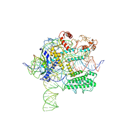 | | CasX-gRNA-DNA(45bp) state I | | 分子名称: | CasX, DNA Non-target strand, DNA target strand, ... | | 著者 | Liu, J.J, Orlova, N, Nogales, E, Doudna, J.A. | | 登録日 | 2019-02-10 | | 公開日 | 2019-02-27 | | 最終更新日 | 2019-12-25 | | 実験手法 | ELECTRON MICROSCOPY (3.2 Å) | | 主引用文献 | CasX enzymes comprise a distinct family of RNA-guided genome editors.
Nature, 566, 2019
|
|
6NY1
 | | CasX-gRNA-DNA(30bp) State II | | 分子名称: | CasX, DNA Non-target strand, DNA target strand, ... | | 著者 | Liu, J.J, Orlova, N, Nogales, E, Doudna, J.A. | | 登録日 | 2019-02-10 | | 公開日 | 2019-02-27 | | 最終更新日 | 2024-03-13 | | 実験手法 | ELECTRON MICROSCOPY (4.2 Å) | | 主引用文献 | CasX enzymes comprise a distinct family of RNA-guided genome editors.
Nature, 566, 2019
|
|


